. 2004 Feb;72(2):1043-56.
doi: 10.1128/IAI.72.2.1043-1056.2004.

Functional analysis of the cag pathogenicity island in Helicobacter pylori isolates from patients with gastritis, peptic ulcer, and gastric cancer

Affiliations

Functional analysis of the cag pathogenicity island in Helicobacter pylori isolates from patients with gastritis, peptic ulcer, and gastric cancer

Steffen Backert et al. Infect Immun. 2004 Feb.

Abstract

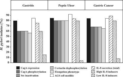
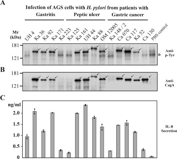
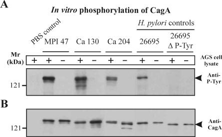
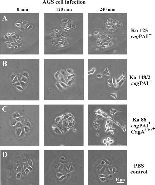
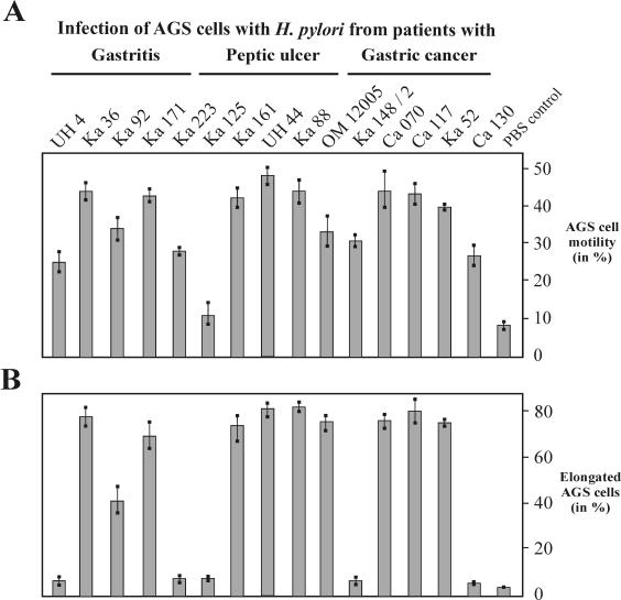
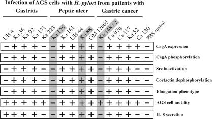
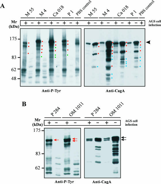
Helicobacter pylori is the causative agent of a variety of gastric diseases, but the clinical relevance of bacterial virulence factors is still controversial. Virulent strains carrying the cag pathogenicity island (cagPAI) are thought to be key players in disease development. Here, we have compared cagPAI-dependent in vitro responses in H. pylori isolates obtained from 75 patients with gastritis, peptic ulcer, and gastric cancer (n = 25 in each group). AGS gastric epithelial cells were infected with each strain and assayed for (i) CagA expression, (ii) translocation and tyrosine phosphorylation of CagA, (iii) c-Src inactivation, (iv) cortactin dephosphorylation, (v) induction of actin cytoskeletal rearrangements associated with cell elongation, (vi) induction of cellular motility, and (vii) secretion of interleukin-8. Interestingly, we found high but similar prevalences of all of these cagPAI-dependent host cell responses (ranging from 56 to 80%) among the various groups of patients. This study revealed CagA proteins with unique features, CagA subspecies of various sizes, and new functional properties for the phenotypic outcomes. We further showed that induction of AGS cell motility and elongation are two independent processes. Our data corroborate epidemiological studies, which indicate a significant association of cagPAI presence and functionality with histopathological findings in gastritis, peptic ulcer, and gastric cancer patients, thus emphasizing the importance of the cagPAI for the pathogenicity of H. pylori. Nevertheless, we found no significant association of the specific H. pylori-induced responses with any particular patient group. This may indicate that the determination of disease development is highly complex and involves multiple bacterial and/or host factors.


Figures

FIG. 1.
FIG. 1.
Comparison of cagPAI-dependent host cell responses in AGS cells after infection with H. pylori isolates obtained from 75 patients with gastritis, peptic ulcer, or gastric cancer disease (n = 25 in each group). Among the patients, we detected 64 cagPAI+ strains and 12 cagPAI strains. The percentages of H. pylori isolates corresponding to the various parameters are shown. CagA expression, CagA phosphorylation, Src inactivation, and cortactin dephosphorylation were determined in Western blot analyses using specific antibodies. The elongation phenotype and cellular motility were quantified as described in Materials and Methods. IL-8 secretion was measured by standard ELISA. IL-8-inducing strains can be divided into high inducers (1.0 to 4.0 ng/ml) and low inducers (0.3 to 0.9 ng/ml). IL-8 induction of <0.3 ng/ml was in the range of PBS control levels. The numerical data and their respective percentages and P values are given in Table 2. Further details are provided in the text.
FIG. 2.
FIG. 2.
Phosphorylation of the CagA protein and induction of IL-8 during infection of AGS cells with 15 representative H. pylori isolates. (A) CagA tyrosine phosphorylation was analyzed in Western blots with a phosphotyrosine-specific antibody, PY-99 (arrows). The asterisk indicates the position of an unknown 125-kDa host cell protein that is phosphorylated in the PBS control. This protein changed its phosphorylation status during infection. (B) Stripping and reprobing of the blot with an anti-CagA antibody (Ab-1) indicated the positions of the different CagA protein species on the gel (arrows). UH4, Ka223, Ka125, and Ka148/2 are cagPAI strains and did not express CagA. CagA of strain Ca130 was expressed but not phosphorylated. Infection was for 4 h at an MOI of 100. (C) In a parallel experiment, AGS cells were infected under the same conditions for 24 h and IL-8 release into the culture supernatant was measured by ELISA. The results shown are representative of three independent experiments. The error bars indicate standard deviations.
FIG. 3.
FIG. 3.
In vitro phosphorylation of CagA. (A) We observed nine nonphosphorylated CagA protein species during infection of AGS cells with the following H. pylori strains: M15, M27, M33, and M64 (gastritis group); Ka61 (peptic ulcer group); and MPI47, Ca115, Ca130, and Ca204 (gastric cancer group). These CagA proteins could be phosphorylated in an in vitro phosphorylation assay. The results are shown for three strains. Incubation of the H. pylori lysate with lysate of AGS cells or recombinant human c-Src (data not shown) resulted in specific CagA phosphorylation (arrowhead). The lysates of the TIGR H. pylori strain 26695 and a phosphorylation-deficient deletion mutant (ΔP-Tyr) were used as positive and negative controls, respectively. CagA phosphorylation was not detected either in the lysate of wild-type H. pylori incubated without c-Src or in the lysate of the CagAΔP-Tyr mutant incubated with c-Src. (B) An anti-CagA blot with antibody Ab-1 was performed as a control. +, present; −, absent.
FIG. 4.
FIG. 4.
Induction of c-Src inactivation and cortactin dephosphorylation. (A) CagAP-Tyr-specific inactivation of c-Src. Western blotting with a phosphospecific anti-c-Src antibody revealed that some, but not all, H. pylori strains induce c-Src inactivation by dephosphorylation of residue Y-418 (top) in the catalytic kinase domain. Densitometric measurements of band intensities revealed the Src activity in comparison to uninfected AGS cells in the PBS control (middle). Stripping and reprobing of the blot with a monoclonal anti-Src antibody revealed that equal amounts of Src are present in all lanes (bottom). (B) Cortactin is a major phosphorylated protein in uninfected AGS cells and is specifically dephosphorylated during infection with some H. pylori strains (top). Cortactin was immunoprecipitated from infected AGS cells with a monoclonal anti-cortactin antibody, and the blots were probed with an anti-phosphotyrosine antibody (66). The phosphotyrosine pattern of cell lysates at 80 kDa shows dephosphorylation of cortactin in infections with those H. pylori isolates that induce CagA phosphorylation and Src inactivation (middle). Stripping and reprobing of the blot with a monoclonal anti-cortactin antibody revealed that similar amounts of cortactin were present in all lanes (bottom).
FIG. 5.
FIG. 5.
Induction of two phenotypes associated with the stimulation of migratory behavior of H. pylori-infected host cells. Time lapse phase-contrast micrographs of AGS cells infected with wild-type strain Ka125 (A), Ka148/2 (B), and Ka88 (C) are shown as examples. Ka125 had no stimulatory effect on motility and elongation. In contrast, Ka148/2 induced a strong motility phenotype (B) and Ka88 induced motility and the elongation phenotype of AGS cells (C). (D) PBS as a control had no effect. In each experiment, H. pylori infection was for 4 h at an MOI of 100.
FIG. 6.
FIG. 6.
Quantification of motility (A) and elongation (B) phenotypes during infection with H. pylori. AGS cells were infected for 4 h at an MOI of 100. A single field of the coverslip was labeled and photographed before and after infection. One hundred cells from each photograph were counted and evaluated. The results are the means of three independent experiments. The error bars indicate standard deviations.
FIG. 7.
FIG. 7.
Summary of host cell responses during infection with 15 representative H. pylori isolates from patients with gastritis, peptic ulcer, or gastric cancer. The capacity of each wild-type strain to induce a certain response in infected AGS cells is represented as efficient induction (+) or no or drastically reduced induction (−). The chart demonstrates that translocation and phosphorylation of CagA correlates in each infection with Src inactivation, cortactin dephosphorylation, and induction of the elongation phenotype. AGS cell motility and IL-8 induction were observed with both the sets of H. pylori that induced the aforementioned responses and some cagPAI strains. The phenotypic outcomes of the shaded strains are shown in Fig. 5. See the text for more details.
FIG. 8.
FIG. 8.
Detection of unique and multiple CagA protein species. (A) Anti-phosphotyrosine patterns of AGS infected with different H. pylori strains revealed unique phosphorylated protein species in each lane (left). The blot was stripped and reprobed with an anti-CagA antibody (right). This exposure revealed 5 to 13 CagA protein species with masses between 40 and 120 kDa, depending on the strain. The 25- to 35-kDa fragmentation products (10, 47, 53) were also observed but ran out of the gels shown. Full-length CagA is indicated by the arrowhead. The red dots indicate phosphorylated proteins corresponding in size to CagA protein species, the green dots indicate phosphorylated proteins not corresponding in size to CagA protein species, and the blue dots label CagA protein species with no detectable phosphorylated form during infection. H. pylori strain P1 served as a control because the fragmentation of 135-kDa wild-type CagA into an amino-terminal p100CagA fragment and a carboxy-terminal p35CagA fragment has been described (10). +, present; −, absent. (B) H. pylori strains P284 and OM1011 are unique and showed a double band of full-length CagA. This double band had nearly identical intensities in the anti-CagA blot (black arrows) but showed a different phosphorylation pattern during infection (red arrows).

References

    1. Akanuma, M., S. Maeda, K. Ogura, Y. Mitsuno, Y. Hirata, T. Ikenoue, M. Otsuka, T. Watanabe, Y. Yamaji, H. Yoshida, T. Kawabe, Y. Shiratori, and M. Omata. 2002. The evaluation of putative virulence factors of Helicobacter pylori for gastroduodenal disease by use of a short-term Mongolian gerbil infection model. J. Infect. Dis. 185:341-347. - PubMed
    1. Akopyants, N. S., S. W. Clifton, D. Kersulyte, J. E. Crabtree, B. E. Youree, C. A. Reece, N. O. Bukanov, E. S. Drazek, B. A. Roe, and D. E. Berg. 1998. Analyses of the cag pathogenicity island of Helicobacter pylori. Mol. Microbiol. 28:37-53. - PubMed
    1. Amieva, M. R., R. Vogelmann, A. Covacci, L. S. Tompkins, W. J. Nelson, and S. Falkow. 2003. Disruption of the epithelial apical-junctional complex by Helicobacter pylori CagA. Science 300:1430-1434. - PMC - PubMed
    1. Ando, T., K. Kusugami, M. Ohsuga, M. Shinoda, M. Sakakibara, H. Saito, A. Fukatsu, S. Ichiyama, and M. Ohta. 1996. Interleukin-8 activity correlates with histological severity in Helicobacter pylori-associated antral gastritis. Am. J. Gastroenterol. 91:1150-1156. - PubMed
    1. Ando, T., R. M. Peek, Jr., Y. C. Lee, U. Krishna, K. Kusugami, and M. J. Blaser. 2002. Host cell responses to genotypically similar Helicobacter pylori isolates from United States and Japan. Clin. Diagn. Lab. Immunol. 9:167-175. - PMC - PubMed

Publication types

MeSH terms