. 2004 Apr;15(4):1816-32.
doi: 10.1091/mbc.e03-08-0623. Epub 2004 Jan 23.

NO66, a highly conserved dual location protein in the nucleolus and in a special type of synchronously replicating chromatin

Affiliations

NO66, a highly conserved dual location protein in the nucleolus and in a special type of synchronously replicating chromatin

Jens Eilbracht et al. Mol Biol Cell. 2004 Apr.

Abstract

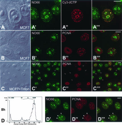
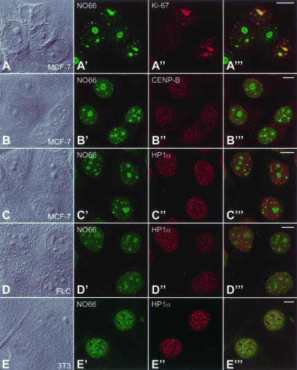
It has recently become clear that the nucleolus, the most prominent nuclear subcompartment, harbors diverse functions beyond its classic role in ribosome biogenesis. To gain insight into nucleolar functions, we have purified amplified nucleoli from Xenopus laevis oocytes using a novel approach involving fluorescence-activated cell sorting techniques. The resulting protein fraction was analyzed by mass spectrometry and used for the generation of monoclonal antibodies directed against nucleolar components. Here, we report the identification and molecular characterization of a novel, ubiquitous protein, which in most cell types appears to be a constitutive nucleolar component. Immunolocalization studies have revealed that this protein, termed NO66, is highly conserved during evolution and shows in most cells analyzed a dual localization pattern, i.e., a strong enrichment in the granular part of nucleoli and in distinct nucleoplasmic entities. Colocalizations with proteins Ki-67, HP1alpha, and PCNA, respectively, have further shown that the staining pattern of NO66 overlaps with certain clusters of late replicating chromatin. Biochemical experiments have revealed that protein NO66 cofractionates with large preribosomal particles but is absent from cytoplasmic ribosomes. We propose that in addition to its role in ribosome biogenesis protein NO66 has functions in the replication or remodeling of certain heterochromatic regions.


Figures

Figure 1.
Figure 1.
Purification of nucleoli from mass-isolated Xenopus laevis oocyte nuclei by fluorescence labeling and particle sorting. Amplified nucleoli were labeled by incubating nuclear homogenates with FITC-coupled mAb No-185 (No-185-FITC) directed against the major nucleolar protein NO38. (A) Two-parameter frequency profile obtained by flow cytometry. Besides FITC fluorescence, light scattering depending on the particle size was measured. Strongly fluorescent large particles, i.e., nucleoli, appearing in the indicated rectangle were separated from other particles, including yolk platelets and debris of nuclear membranes. (B and C) Light microscopy of ruptured oocyte nuclei labeled with No-185-FITC before (B and B′) and after (C and C′) particle sorting. (B and C) Phase contrast. (B′ and C′) FITC fluorescence. Bar, 20 μm. (D) Electron microscopy of amplified nucleoli of Xenopus enriched by particle sorting. Bar, 5 μm. (E) Mass-isolated total nuclei (lane 1) and purified nucleoli (lane 2) were analyzed by immunoblotting using anti-NO38, anti-xNopp180, anti-SF3b155, and antilamin LIII. The 50-kDa signal appearing in the nucleoli fraction after long exposure time results from the secondary antibodies to mouse-IgG detecting the heavy chain of the mAb No-185-FITC antibody originally used as fluorescence marker for particle sorting. Reference proteins (lane R) represent, from top to bottom: 205, 116, 97, 66, 45, and 29 kDa.
Figure 2.
Figure 2.
Immunological characterization of mAb No-5-1-1 and identification of protein NO66. (A and A′) Immunolocalization of the protein recognized by mAb No-5-1-1 in cultured Xenopus laevis kidney epithelial cells, line A6 (XLKE-A6). (A) Phase contrast. (A′) Corresponding immunofluorescence micrograph. Bar, 10 μm. (B) Immunoprecipitation experiments using mAb No-5-1-1. Proteins of the following fractions were separated by SDS-PAGE and stained with Coomassie blue: XLKE-A6 cell extract before immunoprecipitation (lane 1); cell lysate proteins nonspecifically bound to protein G-Sepharose in the absence of antibody (lane 2); immunoprecipitate obtained from XLKE-A6 cell lysate (lane 3); and immunoprecipitate after incubation with extraction buffer as a negative control (lane 4). Reference proteins (lane R) are the same as in Figure 1. A protein of ∼66 kDa (termed NO66, indicated by an arrowhead in lane 3) is specifically immunoprecipitated by mAb No-5-1-1. (C) Peptide sequences of Xenopus laevis protein NO66 obtained after excision of the specifically immunoprecipitated protein band from gels as shown in B, lane 3 and “in-gel” digestion with trypsin, followed by chromatographic separation and Edman sequencing of the tryptic fragments.
Figure 5.
Figure 5.
Identification and immunolocalization of protein NO66 and its homologues in different cultured cell lines from diverse species. (A) Coomassie blue-stained total cellular proteins of different human cell lines. Reference proteins (lane R) are the same as in Figure 1. (A′) Corresponding autoradiogram showing ECL detection of the antigenic polypeptide detected by antibody NO66-2. (B) Separation of total cellular proteins from cultured cells of different species by SDS-PAGE and Coomassie blue staining. (B′) Corresponding immunoblot, using antibody NO66-2, reveals the presence of homologous proteins in all the species analyzed. Note that only one polypeptide is recognized in A′ and B′, but that the Mr of the protein exhibits some variation in the diverse species. (C and C′-H and H′) The intracellular distribution of protein NO66 was analyzed by immunofluorescence microscopy in cultured cells from different species (as indicated). Shown here are examples of human (C-F), Xenopus (G), and murine (H) origin. Phase-contrast micrographs are shown in C-H, and the corresponding immunofluorescence micrographs are shown in C′-H′. Bar, 10 μm. (I) Immunoelectron microscopy of protein NO66 within the nucleolus of XLKE-A6 cells showing segregation of nucleolar components upon AMD-treatment. Antibody NO66-2 was detected by secondary antibodies coupled to nanogold particles. The granular component (GC) of the segregated nucleolus is specifically labeled, whereas the dense fibrillar component (DFC) is almost devoid of silver-enhanced gold particles. Bar, 0.5 μm.
Figure 9.
Figure 9.
Relationship of nuclear bodies containing protein NO66 to replication foci. (A) Differential interference contrast (DIC) micrograph of MCF-7 cells microinjected with Cy3-dCTP. (A′) Immunolocalization of protein NO66 with antiserum NO66-2. (A″) Fluorescence of microinjected Cy3-dCTP, which was subsequently incorporated into newly synthesized DNA during replication. (A‴) Corresponding merged image. (B and C) DIC micrographs of MCF-7 cells. (B′ and C′) Immunolocalization of protein NO66 with antiserum NO66-2. (B″ and C″) Immunolocalization of PCNA using mAb PC10. (B‴ and C‴) Corresponding merged images. The cells presented in C and C‴ had been treated with Triton X-100 before fixation to remove PCNA from non-S-phase cells (indicated by arrowheads). (D) Flow cytometric analysis of the relative DNA content of synchronized HeLa cells during late S-phase, 8 h after release from a double-thymidine block. The DNA content according to the DAPI fluorescence intensity of the cells corresponds to G1-, S-, or G2/M-phase as indicated. (D′-D‴) Immunofluorescence analysis of the same HeLa cell population with antiserum NO66-2 against protein hsNO66 (D′) and with mAb PC10 against PCNA (D″). The corresponding merged image is given in D‴. NO66 and PCNA colocalize in replication foci during late S-phase (arrowheads) The micrographs in A-D‴ have been obtained with a confocal laser scanning microscope and show single optical sections, with exception of C′-C‴ where maximum intensity projections of multiple optical sections are shown. Bars, 10 μm.
Figure 3.
Figure 3.
Identification of proteins homologous to Xenopus laevis protein NO66. (A) Database searches with NO66 peptides listed in Figure 2 revealed the hypothetical human protein “hsNO66” as a putative homologue of protein NO66. An alignment of hsNO66 with matching NO66 peptides obtained from Xenopus laevis is shown (identical aa residues are indicated in bold letters). (B) The homology of the human hypothetical protein with Xenopus laevis protein NO66 was confirmed by analyses of homologues found in other species. A fragment of the corresponding chicken NO66 homolog encoded by an EST shows high similarity to X. laevis peptide t59, which fits only weakly to the human sequence. (C) Two EST clones were identified that together encoded the carboxy-terminal part of the protein NO66 homolog of Xenopus tropicalis. Peptides t30 and t26 of NO66 could not be directly allocated to the human sequence but showed significant homology to the Xenopus tropicalis sequence. (D) Northern blot analysis of total RNA (lane 1) and poly(A)+ RNA (lane 2) from human MCF-7 cells with a random prime-labeled probe obtained from a cDNA clone encoding hsNO66. RNA size markers (R): 2.4 (top) and 1.4 kb (bottom). (E) Phase-contrast microscopy of human MCF-7 cells used to determine the subcellular localization of the hsNO66 protein carrying an amino-terminal myc-tag in transient transfection experiments. (E′) Corresponding immunofluorescence using mAb 9E10 recognizing the myc-tag. Bar, 10 μm.
Figure 4.
Figure 4.
Characterization of antibodies to protein NO66. (A) Immunoprecipitation of the X. laevis protein NO66 with mAb No-5-1-1. Proteins of the following fractions were separated by SDS-PAGE and stained with Coomassie blue: XLKE-A6 cell extract before immunoprecipitation (lane 1), supernatant after incubation of cell extract with protein G-Sepharose in the absence of antibody (lane 2), supernatant after incubation of cell extract with protein G-Sepha-rose in the presence of mAb No-5-1-1 (lane 3), extract nonspecifically bound to protein G-Sepharose (lane 4), immunoprecipitate obtained from XLKE-A6 cell lysates (lane 5), and immunoprecipitate after incubation with lysis buffer, as a negative control (lane 6). The arrowhead (lane 5) points to the immunoprecipitated Xenopus NO66. (B) Corresponding immunoblot probed with the guinea pig antibodies NO66-2 to protein NO66 reacting specifically with the 66-kDa protein present in the cell extract (lanes 1 and 2) and in the immunoprecipitate (lane 5). Reference proteins (lanes R) are the same as in Figure 1.
Figure 6.
Figure 6.
Identification of protein NO66 in cellular fractions from Xenopus laevis and human cell cultures and biochemical analysis by sucrose gradient centrifugation and gel filtration. (A) Proteins of cell extracts from A-431 cells were fractionated by gel filtration, separated by SDS-PAGE, and stained with Coomassie blue. (A′) Corresponding immunoblot probed with the antiserum NO66-2. Peak positions of reference macromolecules are indicated by arrowheads: dextran blue (d; Mapp 2000,000; fraction 2), thyroglobulin (t; Mapp 669,000; fraction 4), ferritin (f; Mapp 440,000; fraction 6), aldolase (a; Mapp 158,000; fraction 10), bovine serum albumin (b; Mapp 66,000; fraction 13), and ovalbumin (o; Mapp 43,000; fraction 15). Protein NO66 is mainly detected in fractions 4 and 5, corresponding to a mean Mapp 600,000. (B) Cell lysates of human A-431 cells were fractionated after sucrose density centrifugation. The resulting fractions (lanes 1-15 from top to bottom of the gradient) were separated by SDS-PAGE and subjected to Coomassie blue staining. (B′) Corresponding immunoblot analysis using the NO66-2 antiserum. Arrowheads indicate the peak positions of the reference proteins fractionated in parallel: bovine serum albumin (b; 4.3 S, fraction 3), catalase (c; 11.3 S, fraction 7), and thyroglobulin (t; 16.5 S, fraction 10). Protein NO66 is recovered in fractions 7 and 8, with a sedimentation coefficient of ∼12S. (C) Same experiment as presented in B and B′, but after treatment of the cell lysates with RNase A. Under these conditions, the antigenic polypeptide was only weakly detectable by the NO66-specific antibodies and sedimented to the bottom of the gradient (fractions 13-15). (D) Coomassie blue staining of various fractions from MCF-7 cells separated by SDS-PAGE (lys, total cell lysate; cp, cytoplasmic fraction; nu, fraction enriched in nuclei; no, fraction enriched in nucleoli). (D′) Corresponding immunoblot using antibody NO66-2, which reacts specifically with a ∼79 kDa protein (hsNO66) highly enriched in the nuclear and nucleolar fractions. (D″) To ascertain the fractionation procedure, a parallel immunoblot was probed with antibody B2.4-1 directed against the 146-kDa splicing factor SF3b155.(E and E′) Total proteins of mass isolated oocyte nuclei (MI), of the LSP, HSP, and HSS fractions from oocyte nuclei, of a Xenopus egg extract (Egg) and from total Xenopus XLKE-A6 cells (XLKE) were separated by SDS-PAGE, stained with Coomassie blue (E) and analyzed by immunoblotting using antibody NO66-2 (E′). (F-F″) Fractionation of HSP fractions from Xenopus oocyte nuclei by sucrose gradient centrifugation. The resulting protein fractions were analyzed by SDS-PAGE and Coomassie blue staining (F) and further examined by immunoblotting using antibody NO66-2 (F′) or mAb No-185 against the nucleolar protein NO38 (F″). The peak fractions (indicated by arrowheads) of the preribosomal precursor molecules of 40S (fraction 6) and 65S (fraction 10) were determined by their extinction at 260 nm (marked in F′). Although protein NO38 was associated with both the 40S and the 65S preribosomal particles, protein NO66 was detected in fractions 10-15, suggesting an association with the 65S preribosomes and larger particles. (G and G′) RNase treatment of the HSP fractions before centrifugation resulted in the expected shift of the protein NO38 position to lower density fractions, where it peaked in fraction 3 (G′). By contrast, only trace amounts of protein NO66, sedimenting to the bottom of the gradient, were detected (G). Reference proteins (lanes R) are the same as in Figure 1.
Figure 7.
Figure 7.
Identification of constitutive nucleolar proteins associated with protein NO66 in immunoprecipitation experiments. (A-A″) Immunoprecipitation of proteins from XLKE-A6 cell extract with mAb No-5-1-1 directed against NO66. The analyzed fractions are the same as shown in Figure 2B. The arrowhead (A) points to immunoprecipitated protein NO66 seen after Commassie blue staining. Corresponding immunoblot analysis with mAb P7-1A4 against nucleolin (A′) and mAb No-185 against protein NO38 (A″) shows that the 90- and 95-kDa homologues of nucleolin (arrowheads in A′) and the two isoforms of NO38 (arrowheads in A″) were coimmunoprecipitated by mAb No-5-1-1. Strong signals in the range of 25 and 50 kDa are due to binding of the secondary antibody used for immunodetection to the light and heavy chain of the NO66-specific antibody. (B and B′) In a reciprocal experiment, immunoprecipitation from extracts of XLKE-A6 cells was performed with mAb No-185. (B) SDS-PAGE-separated immunoprecipitation fractions after staining with Coomassie blue. Arrowheads mark the immunoprecipitated protein NO38 variants. (B′) Coimmunoprecipitated protein NO66 (arrowhead) was detected by immunoblotting with antiserum NO66-2. (C and C′) When immunoprecipitation from XLKE-A6 cell extracts was performed with mAb P7-1A4, which precipitated both forms of nucleolin (arrowheads in Coomassie blue-stained gel in C), significant amounts of protein NO66 were detected with NO66-2 antibodies (arrowhead in C′).
Figure 8.
Figure 8.
Double label immunolocalization studies of protein NO66 in comparison with various marker proteins characteristic for distinct nuclear subcompartments. Micrographs were taken with a confocal laser scanning microscope and single optical sections are shown. (A-G) Differential interference contrast (DIC) micrographs. (A′-G') Immunolocalization of protein NO66 with antiserum NO66-2. (A″-G″) Immunolocalization of constituents of different nuclear domains: nucleolar RNA helicase NOH61 (A″), p80-coilin as a marker for Cajal bodies (B″), survival of motor neuron (SMN) protein present in the cytoplasm and in nucleoplasmic SMN bodies, also termed gems (C″), promyelotic leukemia (PML) protein as a marker for PML bodies (D″), splicing factor SF3b155 accumulating in nuclear speckles (E″), protein PSP1 defining paraspeckles (F″), and PTB protein decorating the perinucleolar compartment (PNC; G″). (A‴-G‴) Corresponding overlay images. Note that the extranucleolar structures stained by the antibodies to protein NO66 do not show significant colocalization with any of the other defined nuclear domains. Bars, 10 μm.
Figure 10.
Figure 10.
Double immunolocalization of protein NO66 in comparison with proteins Ki-67, CENP-B, and HP1α. Micrographs were taken with a confocal laser scanning microscope and show single optical sections. (A-E) Differential interference contrast (DIC) micrographs of cells of lines MCF-7, PLC and 3T3 as indicated. (A′-E′) Immunolocalization of protein NO66 with antiserum NO66-2. (A″) Immunolocalization of the proliferation marker protein Ki-67 with mAb MB67, (B″) of centromeric protein CENP-B with antiserum anti-CENP-B and (C″-E″) of heterochromatin protein HP1α with mAb 2HP 1H5. (A‴-E‴) Corresponding merged immunofluorescence images. Bars, 10 μm.

Similar articles

Cited by

References

    1. Adams, C.C., Jakovljevic, J., Roman, J., Harnipicharnchai, P., and Woolford, J.L., Jr. (2002). Saccharomyces cerevisiae nucleolar protein Nop7p is necessary for biogenesis of 60S ribosomal subunits. RNA 8, 150-165. - PMC - PubMed
    1. Andersen, J.S., Lyon, C.E., Fox, A.H., Leung, A.K., Lam, Y.W., Stehen, H., Mann, M., and Lamond, A.I. (2002). Directed proteomic analysis of the human nucleolus. Curr. Biol. 12, 1-11. - PubMed
    1. Ayoub, N., Noma, K., Isaac, S., Kahan, T., Grewal, S.I., and Cohen, A. (2003). A novel jmjC domain protein modulates heterochromatization in fission yeast. Mol. Cell. Biol. 23, 4356-4370. - PMC - PubMed
    1. Azum-Gélade, M.C., Noaillac-Depeyre, J., Caizergues-Ferrer, M., and Gas, N. (1994). Cell cycle redistribution of U3 snRNA and fibrillarin. Presence in the cytoplasmic nucleolus remnant and in the prenucleolar bodies at telophase. J. Cell Sci. 107, 463-475. - PubMed
    1. Baynton, K., Otterlei, M., Bjoras, M., von Kobbe, C., Bohr, V.A., and Seeberg, E. (2003). WRN interacts physically and functionally with the recombination mediator protein RAD52. J. Biol. Chem. 278, 36476-36486. - PubMed

Publication types

MeSH terms