. 2004 Apr;15(4):1760-75.
doi: 10.1091/mbc.e03-07-0527. Epub 2004 Jan 23.

Identification of a novel sequence in PDZ-RhoGEF that mediates interaction with the actin cytoskeleton

Affiliations

Identification of a novel sequence in PDZ-RhoGEF that mediates interaction with the actin cytoskeleton

Jayashree Banerjee et al. Mol Biol Cell. 2004 Apr.

Abstract

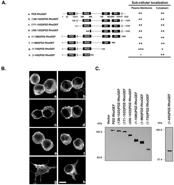
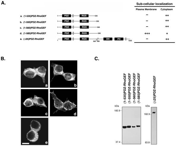
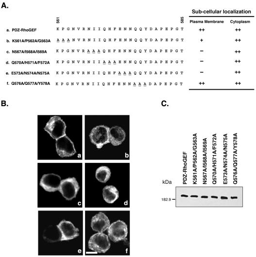
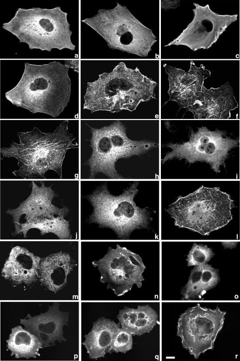
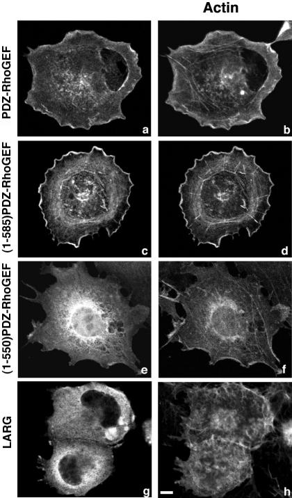
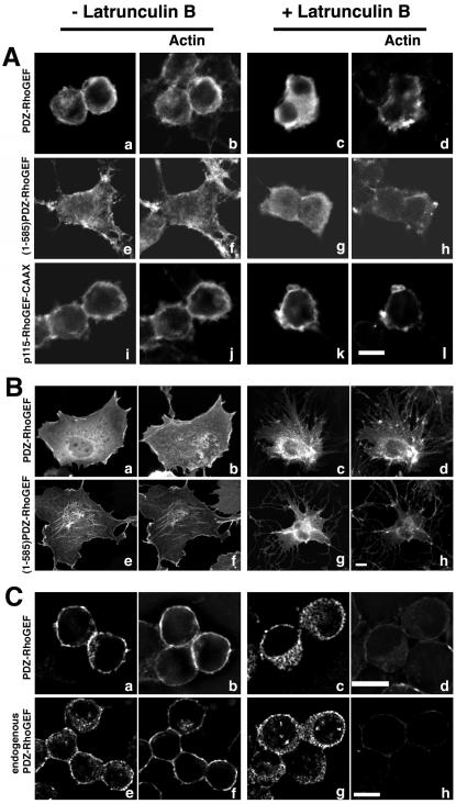
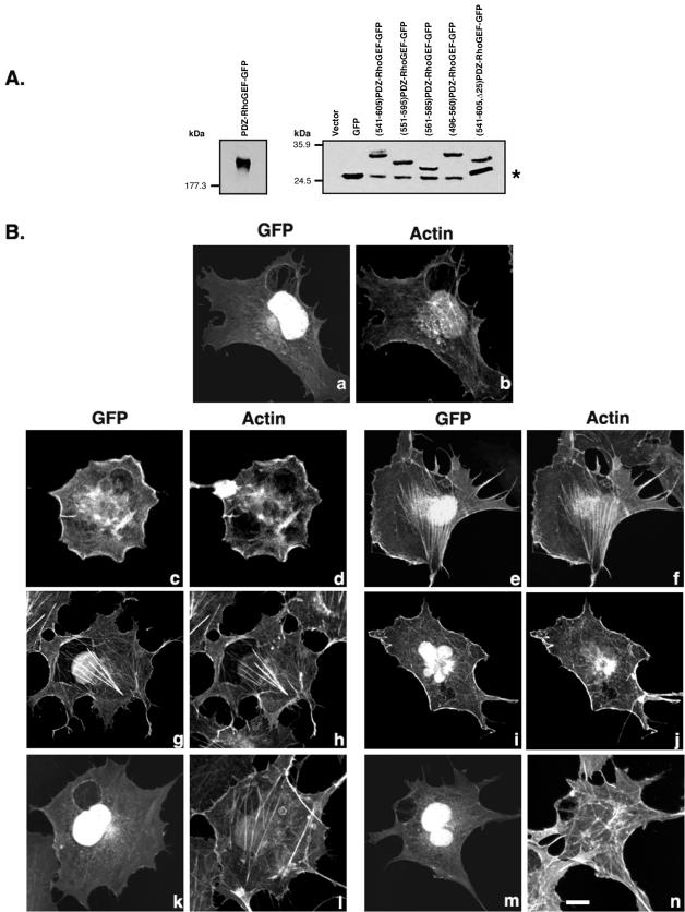
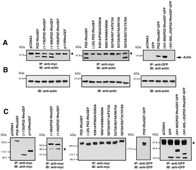
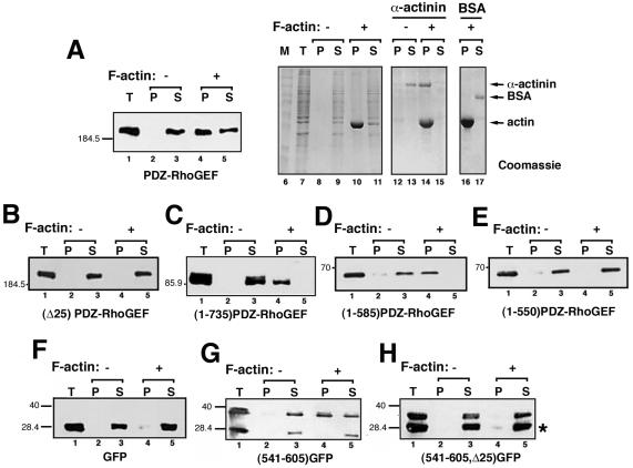
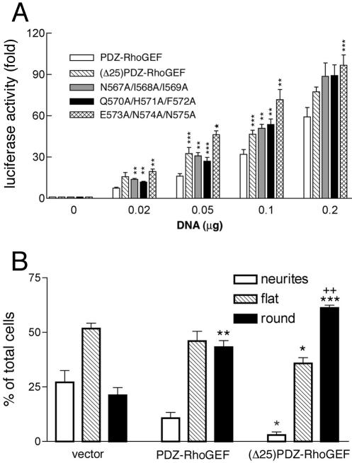
Small GTPases of the Rho family are crucial regulators of actin cytoskeleton rearrangements. Rho is activated by members of the Rho guanine-nucleotide exchange factor (GEF) family; however, mechanisms that regulate RhoGEFs are not well understood. This report demonstrates that PDZ-RhoGEF, a member of a subfamily of RhoGEFs that contain regulator of G protein signaling domains, is partially localized at or near the plasma membranes in 293T, COS-7, and Neuro2a cells, and this localization is coincident with cortical actin. Disruption of the cortical actin cytoskeleton in cells by using latrunculin B prevents the peri-plasma membrane localization of PDZ-RhoGEF. Coimmunoprecipitation and F-actin cosedimentation assays demonstrate that PDZ-RhoGEF binds to actin. Extensive deletion mutagenesis revealed the presence of a novel 25-amino acid sequence in PDZ-RhoGEF, located at amino acids 561-585, that is necessary and sufficient for localization to the actin cytoskeleton and interaction with actin. Last, PDZ-RhoGEF mutants that fail to interact with the actin cytoskeleton display enhanced Rho-dependent signaling compared with wild-type PDZ-RhoGEF. These results identify interaction with the actin cytoskeleton as a novel function for PDZ-RhoGEF, thus implicating actin interaction in organizing PDZ-RhoGEF signaling.


Figures

Figure 1.
Figure 1.
Localization of N- and C-terminal deletion mutants of PDZ-RhoGEF. (A) Domain structure of PDZ-RhoGEF is presented (Fukuhara et al., 1999; Longenecker et al., 2001), and the location of the indicated domains and proline-rich regions (P) are shown. N-Terminal and C-terminal deletion mutants of PDZ-RhoGEF are depicted (left), and include full-length PDZ-RhoGEF (a), (128-1522)PDZ-RhoGEF (b), (171-1522)PDZ-RhoGEF (c), (435-1522)PDZ-RhoGEF (d), (1-1085)PDZ-RhoGEF (e), (1-960)PDZ-RhoGEF (f), (1-735)PDZ-RhoGEF (g), and (1-435)PDZ-RhoGEF (h). All constructs have an N-terminal Myc epitope tag. The subcellular localization of these mutants is summarized (right), as described in MATERIALS AND METHODS. (B) 293T cells were transiently transfected with expression vectors (1 μg) encoding PDZ-RhoGEF deletion mutants (a–h, as described in A). Twenty-four hours after transfection, cells were fixed and processed for immunofluorescence, and images were recorded using confocal microscopy as described in MATERIALS AND METHODS. More than 100 cells were examined in at least three separate experiments, and a representative image is shown. Bar, 10 μm. (C) Lysates from cells expressing the indicated PDZ-RhoGEF proteins were immunoblotted with an anti-Myc antibody to compare expression levels.
Figure 2.
Figure 2.
Role for amino acids 561–585 in localization of PDZ-RhoGEF. (A) Deletion mutants of PDZ-RhoGEF analyzed are depicted (left) and include (1-535)PDZ-RhoGEF (a), (1-550)PDZ-RhoGEF (b), (1-560)PDZ-RhoGEF (c), (1-585)PDZ-RhoGEF (d), and (Δ25)PDZ-RhoGEF (e). All constructs have an N-terminal Myc epitope tag. The subcellular localization of these mutants is summarized (right), as described in MATERIALS AND METHODS. (B) 293T cells were transiently transfected with expression vectors (1 μg) encoding PDZ-RhoGEF deletion mutants (a–e, as described in A). Twenty-four hours after transfection, cells were fixed and processed for immunofluorescence, and images were recorded using confocal microscopy as described in MATERIALS AND METHODS. More than 100 cells were examined in at least three separate experiments, and a representative image is shown. Bar, 10 μm. (C) Lysates from cells expressing the indicated PDZ-RhoGEF proteins were immunoblotted with an anti-Myc antibody to compare expression levels.
Figure 3.
Figure 3.
Alanine scanning mutagenesis of amino acids 561–585 in PDZ-RhoGEF. (A) Amino acid sequence between 561 and 585 is depicted for PDZ-RhoGEF (a) and the following triple alanine mutants: K561A/P562A/G563A (b), N567A/I568A/I569A (c), Q570A/H571A/F572A (d), E573A/N574A/N575A (e), and Q576A/Q577A/Y578A (f). The alanine mutations are underlined. All constructs have an N-terminal Myc epitope tag. The subcellular localization of these mutants is summarized (right), as described in MATERIALS AND METHODS. (B) 293T cells were transiently transfected with expression vectors (1 μg) encoding full-length PDZ-RhoGEF (a) or the triple alanine mutants (b-f, as described in A). Twenty-four hours after transfection, cells were fixed and processed for immunofluorescence, and images were recorded using confocal microscopy as described in MATERIALS AND METHODS. More than 100 cells were examined in at least three separate experiments, and a representative image is shown. Bar, 10 μm. (C) Lysates from cells expressing the indicated PDZ-RhoGEF proteins were immunoblotted with an anti-Myc antibody to compare expression levels.
Figure 4.
Figure 4.
Subcellular localization of PDZ-RhoGEF and mutants in COS-7 cells. COS-7 cells were transiently transfected with 1 μg of an expression vector encoding Mycepitope–tagged PDZ-RhoGEF (a), (128-1522)PDZ-RhoGEF (b), (171-1522) PDZ-RhoGEF (c), (435-1522)PDZ-RhoGEF (d), (1-1085)PDZ-RhoGEF (e), (1-960)PDZ-RhoGEF (f), (1-735)PDZ-RhoGEF (g), (1-435)PDZ-RhoGEF (h), (1-535) PDZ-RhoGEF (i), (1-550)PDZ-RhoGEF (j), (1-560)PDZ-RhoGEF (k), (1-585)PDZ-RhoGEF (l), (Δ25)PDZ-RhoGEF (m), K561A/P562A/G563A (n), N567A/I568A/I569A (o), Q570A/H571A/F572A (p), E573A/N574A/N575A (q), or Q576A/Q577A/Y578A (r). Twenty-four hours after transfection, cells were fixed and processed for immunofluorescence, and images were recorded using confocal microscopy as described in MATERIALS AND METHODS. More than 100 cells were examined in at least three separate experiments, and a representative image is shown. Bar, 10 μm.
Figure 5.
Figure 5.
PDZ-RhoGEF colocalizes with actin. COS-7 cells were transiently transfected with 1 μg of an expression vector encoding Myc-epitope–tagged PDZ-RhoGEF (a and b), (1-585)PDZ-RhoGEF (c and d), (1-550)PDZ-RhoGEF (e and f), or AU1 epitope-tagged LARG (g and h). Expressed proteins were detected with an anti-Myc 9E10 (a, c, and e) or anti-AU1 antibody (g) followed by Alexa 488 conjugated to an anti-mouse antibody. Actin was visualized in the same cells by costaining with Alexa 594 conjugated to phalloidin (b, d, f, and h). Representative images were recorded by confocal microscopy. Bar, 10 μm.
Figure 6.
Figure 6.
Latrunculin B treatment disrupts PDZ-RhoGEF localization. (A) 293T cells were transiently transfected with 1 μg of an expression vector encoding Myc-epitope–tagged PDZ-RhoGEF (a–d), (1-585)PDZ-RhoGEF (e–h), or p115-RhoGEF-CAAX (i–l). Before fixation, cells on coverslips were untreated (a, b, e, f, i, and j) or treated (c, d, g, h, k, and l) with 10 μM latrunculin B for 15 min at 37°C. Cells were fixed and dual stained with anti-Myc antibody 9E10 (a, c, e, g, i, and k) followed by an Alexa 488 conjugated secondary antibody and with Alexa 594 conjugated to phalloidin (b, d, f, h, j, and l). Representative images were recorded by confocal microscopy. Bar, 10 μm. (B) COS cells were transiently transfected with 1 μg of an expression vector encoding Myc-epitope–tagged PDZ-RhoGEF (a–d) or (1-585)PDZ-RhoGEF (e–h). Before fixation, cells on coverslips were untreated (a, b, e, and f) or treated (c, d, g, and h) with 10 μM latrunculin B for 15 min at 37°C. Cells were fixed and dual stained with anti-Myc antibody 9E10 (a, c, e, and g) followed by an Alexa 488-conjugated secondary antibody and with Alexa 594 conjugated to phalloidin (b, d, f, and h). Representative images were recorded by confocal microscopy. Bar, 10 μm. (C) Neuro2a cells were transiently transfected with 1 μg of an expression vector encoding Myc-epitope–tagged PDZ-RhoGEF (a–d) or were not transfected (e–h). Before fixation, cells on coverslips were untreated (a, b, e, and f) or treated (c, d, g, and h) with 10 μM latrunculin B for 15 min at 37°C. Cells were fixed and dual stained with anti-Myc antibody 9E10 (a and c) or anti-PDZ-RhoGEF polyclonal antibody (Jackson et al., 2001) (e and g) followed by appropriate Alexa 594-conjugated secondary antibody and with Alexa 647 conjugated to phalloidin (b, d, f, and h). Representative images were recorded by deconvolution microscopy. Bar, 10 μm. Endogenous PDZ-RhoGEF was more readily visualized in rounded Neuro2a cells compared with cells with very flat morphologies.
Figure 7.
Figure 7.
Amino acids 561–585 of PDZ-RhoGEF are sufficient for colocalization with F-actin. (A) Lysates of cells transfected with 1 μg vector alone or with the indicated expression vector for GFP-tagged forms of PDZ-RhoGEF were subject to Western blotting with an anti-GFP antibody. The asterisk (right) indicates a faster migrating band detected by the anti-GFP antibody and present in all GFP-tagged constructs. This protein comigrates with GFP alone and likely represents degradation or translational initiation from an internal methionine. (B) COS-7 cells were transfected with expression vectors for GFP alone (a and b), full-length PDZRhoGEF-GFP (c and d), (541-605)PDZ-RhoGEF-GFP (e and f), (551-595)PDZ-RhoGEF-GFP (g and h), (561-585)PDZ-RhoGEF-GFP (i and j), (496-560)PDZ-RhoGEF-GFP (k and l), or (541-605, Δ25)PDZ-RhoGEF-GFP (m and n). Twenty-four hours after transfection the cells were fixed and processed for confocal microscopy. Expressed proteins were visualized by GFP fluorescence (a, c, e, g, i, k, and m), and actin was visualized (b, d, f, h, j, l, and n) in the same cells by staining with Alexa 594 conjugated to phalloidin. Bar, 10 μm.
Figure 8.
Figure 8.
PDZ-RhoGEF coimmunoprecipitates with actin. (A) COS-7 cells were transiently transfected with 3 μg of an expression vector encoding PDZ-RhoGEF or the indicated mutants with Myc epitope or GFP tags. Myc-tagged p115RhoGEF was used as a control. Cell lysates were immunoprecipitated with a mouse monoclonal anti-Myc antibody (left and middle) or a polyclonal anti-GFP antibody (right), and immunoprecipitates were analyzed by Western blot by using anti-actin mAb. (B) Presence of actin in the cell lysates was detected by Western blot by using anti-actin mAb. (C) Immunopreciptation of the Myc-tagged or GFP-tagged PDZ-RhoGEF constructs was confirmed by Western blot of the immunoprecipitates using anti-Myc (left and middle) or anti-GFP (right) antibody. Bands marked (*) represent immunoglobulins precipitated from each immunoprecipitation.
Figure 9.
Figure 9.
PDZ-RhoGEF binds to F-actin. COS-7 cells were transfected with an expression vector for full-length PDZ-RhoGEF (A), (Δ25)PDZ-RhoGEF (B), (1-735)PDZ-RhoGEF (C), (1-585)PDZ-RhoGEF (D), (1-550)PDZ-RhoGEF (E), GFP alone (F), (541-605)PDZ-RhoGEF-GFP (G), or (541-605, Δ25)PDZ-RhoGEF-GFP (H). Cell lysates were prepared, and F-actin cosedimentation assays were performed as described in MATERIALS AND METHODS. Lysates were either untreated (lanes 2 and 3) or incubated with F-actin (lanes 4 and 5) and then separated into pellet (P, lanes 2 and 4) and soluble (S, lanes 3 and 5) fractions. Western blots of the P and S fractions (lanes 2–5) along with a sample of the total cell lysates (T) (lane 1) were performed using an anti-Myc (A–E) or anti-GFP (F–H) antibody. The asterisk (*) identifies a faster migrating band detected by the anti-GFP antibody (G and H), and its presence is likely due to degradation or translational initiation from an internal methionine. Note that cosedimentation with F-actin is only observed for the slower migrating species (G, lane 4) that contains the PDZ-RhoGEF 541–605 sequence. Several controls were performed for every F-actin cosedimentation assay, and an example is shown (A, lanes 6–17). An aliquot of the samples corresponding to lanes 1–5 (lanes 7–11) along with marker proteins (lane 6) were resolved by SDS-PAGE and stained with Coomassie Blue to demonstrate sedimentation of F-actin. In addition, purified α-actinin (lanes 12–15) and purified bovine serum albumin (BSA) (lanes 16–17) were incubated without (lanes 12 and 13) or with (lanes 14–17) F-actin, to serve as positive and negative controls, respectively. After sedimentation, these control samples (lanes 12–17) were resolved by SDS-PAGE and stained with Coomassie Blue.
Figure 10.
Figure 10.
Signaling by PDZ-RhoGEF mutants. (A) 293T cells were cotransfected with pSRE, pCMV-βgal, and expression vectors for either PDZ-RhoGEF, (Δ25)PDZ-RhoGEF, N567A/I568A/I569A, Q570A/H571A/F572A, or E573A,N574A,N575A. Cells were transfected with 0.02 to 0.2 μg of the PDZ-RhoGEF mutants, as indicated, and processed for the SRE luciferase assay, as described in MATERIALS AND METHODS. The data represent luciferase activity normalized by β-galactosidase activity present in each cellular lysate, expressed as fold induction with respect to control cells, and are the mean ± S.E. of two experiments each performed in duplicate (n = 4). Asterisks indicate statistical difference between the indicated mutant and wild-type PDZ-RhoGEF (*p < 0.0001, **p < 0.005, ***p < 0.02). (B) Neuro2a cells on coverslips were transiently transfected with 0.5 μg of an expression vector encoding Myc-epitope tagged PDZ-RhoGEF or (Δ25)PDZ-RhoGEF, or 0.1 μg of pEGFP-N1 (vector). Cells were prepared for immunofluorescence microscopy as described in MATERIALS AND METHODS. Expressing cells were scored as containing long extensions (neurites), displaying a flattened morphology (flat), or contracted (round). The data represent the mean ± S.E. for >500 cells from three independent experiments. Asterisks indicate statistical difference between vector alone and wild-type PDZ-RhoGEF or (Δ25)PDZ-RhoGEF–transfected cells (***p < 0.001, **p < 0.01, *p < 0.02). Pluses indicate statistical difference between wild-type PDZ-RhoGEF and (Δ25)PDZ-RhoGEF transfected cells (++, p < 0.005).

Similar articles

Cited by

References

    1. Arai, A., Spencer, J.A., and Olson, E.N. (2002). STARS, a striated muscle activator of Rho signaling and serum response factor-dependent transcription. J. Biol. Chem. 277, 24453-24459. - PubMed
    1. Aurandt, J., Vikis, H.G., Gutkind, J.S., Ahn, N., and Guan, K.L. (2002). The semaphorin receptor plexin-B1 signals through a direct interaction with the Rho-specific nucleotide exchange factor, LARG. Proc. Natl. Acad. Sci. USA 99, 12085-12090. - PMC - PubMed
    1. Ausubel, F.M., Brent, R.E., Kingston, R.E., Moore, D.D., Seidman, J.G., Smith, J.A., and Struhl, K. (1992). Short Protocols in Molecular Biology. John Wiley & Sons: New York.
    1. Bellanger, J.M., Astier, C., Sardet, C., Ohta, Y., Stossel, T.P., and Debant, A. (2000). The Rac1- and RhoG-specific GEF domain of Trio targets filamin to remodel cytoskeletal actin. Nat. Cell Biol. 2, 888-892. - PubMed
    1. Bhattacharyya, R., and Wedegaertner, P.B. (2000). Galpha 13 requires palmitoylation for plasma membrane localization, Rho-dependent signaling, and promotion of p115-RhoGEF membrane binding. J. Biol. Chem. 275, 14992-14999. - PubMed

Publication types

MeSH terms

LinkOut - more resources