. 2004 Apr;15(4):1533-43.
doi: 10.1091/mbc.e03-07-0511. Epub 2004 Jan 23.

O-glycosylation as a sorting determinant for cell surface delivery in yeast

Affiliations

O-glycosylation as a sorting determinant for cell surface delivery in yeast

Tomasz J Proszynski et al. Mol Biol Cell. 2004 Apr.

Abstract

Little is known about the mechanisms that determine localization of proteins to the plasma membrane in Saccharomyces cerevisiae. The length of the transmembrane domains and association of proteins with lipid rafts have been proposed to play a role in sorting to the cell surface. Here, we report that Fus1p, an O-glycosylated integral membrane protein involved in cell fusion during yeast mating, requires O-glycosylation for cell surface delivery. In cells lacking PMT4, encoding a mannosyltransferase involved in the initial step of O-glycosylation, Fus1p was not glycosylated and accumulated in late Golgi structures. A chimeric protein lacking O-glycosylation motif was missorted to the vacuole and accumulated in late Golgi in wild-type cells. Exocytosis of this protein could be restored by addition of a 33-amino acid portion of an O-glycosylated sequence from Fus1p. Our data suggest that O-glycosylation functions as a sorting determinant for cell surface delivery of Fus1p.


Figures

Figure 1.
Figure 1.
Schematic representation of constructs used in this study. The amino acid sequence of the extracellular domain of Fus1p is shown at the bottom. In red are shown potentially O-glycosylated amino acids. Green arrows and numbers indicate the length of the segment from the extracellular domain of Fus1p added to the invertase fusion constructs. Colors represent sequence from different proteins: yellow, Fus1p; red, Mid2p; and white, invertase. All protein construct contain a C-terminal added GFP tag. Additionally, Fus1p was also tagged with the TAP-tag (see Figures 2A and 3).
Figure 2.
Figure 2.
Fus1 processing and its surface delivery is blocked in pmt4Δ cells. (A) Western blot analysis of Fus1-TAP expressed in different pmt mutants. (B) Cellular localization of Fus1-GFP in wild-type and pmt4Δ cells. In wild-type cells, the protein is localized to the plasma membrane and the vacuole (arrow). In pmt4Δ cells, protein accumulates in dot-like structures inside the cell and in the vacuole (arrow). Quantitative analysis of fluorescence from plasma membrane and inside of the cell (see MATERIALS AND METHODS) demonstrated that Fus1-GFP is much more efficiently delivered to the cell surface in wild-type cells than in the pmt4Δ cells (see Figure 9).
Figure 3.
Figure 3.
Unglycosylated Fus1 accumulates in pmt4Δ cells. Western blot analysis of Fus1-TAP expressed in wild-type, pmt4Δ, and different secretory mutant cells. The form of Fus1 that is produced in the pmt4Δ mutant migrates at the level of the unglycosylated protein produced in sec53 cells at 37°C (sec53 form). In sec18 cells at 37°C, the protein accumulated in the ER and migrated as a partially glycosylated precursor (p) form. In sec14 cells at 37°C, the protein accumulated in the Golgi as a fully glycosylated mature form (m1). In the Golgi complex, a second mature form (m2) was generated. The m2 form migrated more rapidly than all the other forms. In pmt4Δ cells, minute amounts of m1, p, and m2 forms were also detected.
Figure 4.
Figure 4.
Processing of Fus1-GFP in wild-type and pmt4Δ cells. Cells were grown in medium containing raffinose as the carbon source and expression of Fus1-GFP was induced for 15 min by addition of galactose. The cells were then pulse labeled with [35S]methionine for 5 min and chased for various times as indicated. In wild-type cells, at the beginning of chase the sec53 and p forms of Fus1 were present. After 5 min, m1 form was visible and after 30 min the m2 form was generated. Throughout the chase period, the p and sec53 forms were also detected. In pmt4Δ cells throughout the chase period Fus1 migrated with the same mobility as the unglycosylated sec53 form.
Figure 5.
Figure 5.
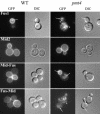
Cellular localization of Fus-GFP, Mid2-GFP, and chimeric proteins in wild-type and pmt4 cells. In wild-type cells, Fus1-GFP was delivered to the cell surface and to the vacuole (arrow). In pmt4Δ cells, protein accumulated in dot-like structures (arrowhead) inside the cell and in the vacuole (arrow). Mid2-GFP was efficiently delivered to the cell surface in wild-type and pmt4Δ mutant. Similarly, Mid-Fus was delivered to the plasma membrane both in wild-type and pmt4Δ cells. In the pmt4Δ mutant, the protein was also detected in the vacuole (arrow). Fus-Mid surface delivery was dependent on PMT4. In the pmt4Δ mutant, the Fus-Mid protein accumulated in dot-like structures (arrowhead) inside the cell and in the vacuole (arrow). Quantitative fluorescence analysis confirmed increased intracellular accumulation of Fus-Mid in pmt4Δ cells compared with wild-type cells (see Figure 9).
Figure 6.
Figure 6.
In pmt4Δ cells Fus1 and Fus-Mid accumulates in the late Golgi structures. In pmt4Δ mutant, Fus1-GFP and Fus-Mid GFP colocalized with Sec7-DsRed, a late Golgi marker.
Figure 7.
Figure 7.
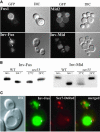
Inv-Fus and Inv-Mid are not delivered to the cell surface in wild-type cells and accumulate in late Golgi compartments. (A) Surface delivery of Fus1-GFP, Mid2-GFP and chimeric invertase constructs in wild-type cells. Both Fus1-GFP and Mid2-GFP were efficiently delivered to the cell surface. GFP fusion constructs of Fus1 and Mid2 carrying a portion of invertase sequence instead of their extracellular domain accumulated in dot-like structures (arrowhead) inside the cell and in vacuole (arrow). Quantitative analysis showed strongly reduced surface delivery of both invertase fusion constructs (Inv-Fus and Inv-Mid) compared to O-glycosylated constructs (Fus-GFP and Fus-Mid in wild-type) (see Figure 9). (B) Inv-Fus and Inv-Mid are N-but not O-glycosylated. Western blot analysis showed that Inv-Fus and Inv-Mid expressed in wild-type cells in presence of tunicamycin (tun) migrated faster than the proteins expressed in the absence of tunicamycin. After inhibition of N-glycosylation (WT + tunicamycin) both proteins migrated with the same mobility as the proteins produced in sec53 cells at the restrictive temperature. (C) Inv-Fus was missorted to the vacuole (double arrowhead) and also accumulated in late Golgi structures (marked by Sec7-DsRed). Note, that two different strains were used in A–C (see MATERIALS AND METHODS).
Figure 8.
Figure 8.
Surface delivery of invertase constructs can be rescued by addition of portion of the Fus1p O-glycosylated extracellular domain and is dependent on PMT4. (A) Cellular localization of invertase rescue constructs containing portions from the extracellular domain of Fus1p. The constructs were expressed in wild-type and pmt4Δ cells as indicated. Quantification showed that the addition of 33 amino acids from the extracellular part of Fus1p could restore the plasma membrane localization of Inv33Mid (see Figure 9). Cells were grown in medium containing a raffinose as a carbon source, and proteins expression was induced overnight by addition of galactose. Western blot analysis of Inv33Mid (B) and Inv33Fus (C) expressed in pmt4Δ mutant, wild-type, and sec53 cells (temperature as indicated) in the presence or absence of tunicamycin (tun-/+). Differences in protein migration due to N- and O-glycosylation are indicated on the right side of the figure.
Figure 9.
Figure 9.
Quantification of the plasma membrane delivery of different protein constructs. Images of cells expressing different constructs were taken using the same conditions and the intensity of the fluorescence were measured for >40 cells in each experiment using the IpLab software (see MATERIALS AND METHODS). The bars (with SEM) represent the ratio of the total fluorescence measured for the plasma membrane area and for the intracellular area of each cell.

Similar articles

Cited by

References

    1. Aalto, M.K., Ronne, H., and Keranen, S. (1993). Yeast syntaxins Sso1p and Sso2p belong to a family of related membrane proteins that function in vesicular transport. EMBO J. 12, 4095-4104. - PMC - PubMed
    1. Alfalah, M., Jacob, R., Preuss, U., Zimmer, K.P., Naim, H., and Naim, H.Y. (1999). O-Linked glycans mediate apical sorting of human intestinal sucraseisomaltase through association with lipid rafts. Curr. Biol. 9, 593-596. - PubMed
    1. Babczinski, P., and Tanner, W. (1973). Involvement of dolicholmonophosphate in the formation of specific mannosyl-linkages in yeast glycoproteins. Biochem. Biophys. Res. Commun. 54, 1119-1124. - PubMed
    1. Bagnat, M., Chang, A., and Simons, K. (2001). Plasma membrane proton ATPase Pma1p requires raft association for surface delivery in yeast. Mol. Biol. Cell 12, 4129-4138. - PMC - PubMed
    1. Bagnat, M., Keranen, S., Shevchenko, A., and Simons, K. (2000). Lipid rafts function in biosynthetic delivery of proteins to the cell surface in yeast. Proc. Natl. Acad. Sci. USA 97, 3254-3259. - PMC - PubMed

Publication types

MeSH terms