. 2004 May 1;556(Pt 3):739-54.
doi: 10.1113/jphysiol.2003.051284. Epub 2004 Jan 23.

Serotonergic facilitation of synaptic activity in the developing rat prefrontal cortex

Affiliations

Serotonergic facilitation of synaptic activity in the developing rat prefrontal cortex

Jean-Claude Béïque et al. J Physiol. .

Abstract

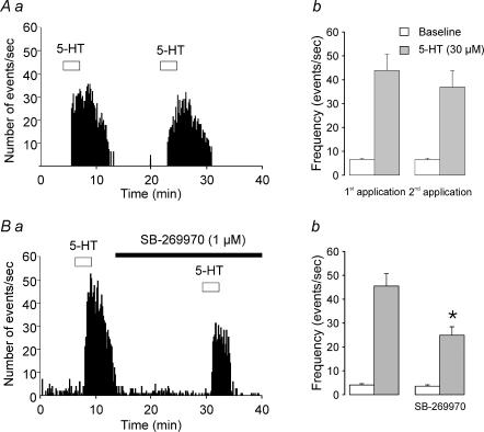
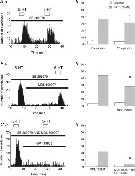
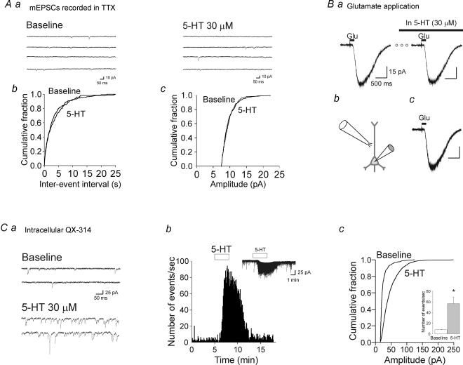
Previous studies have outlined an important role for serotonin (5-HT) in the development of synaptic connectivity and function in the cerebral cortex. In this study, we have examined the effects of 5-HT on synaptic function in prefrontal cortex at a time of intense synapse formation and remodelling. Whole-cell recordings in slices derived from animals aged postnatal (P) days 16-20 showed that administration of 5-HT induced a robust increase in synaptic activity that was blocked by CNQX but not by bicuculline. This 5-HT-induced increase in glutamate-mediated synaptic activity was pharmacologically heterogeneous as it was differentially inhibited by the receptor subtype-selective antagonists SB-269970, MDL 100907 and GR 113808 and thus involved 5-HT(7), 5-HT(2A) and 5-HT(4) receptors. These results, obtained in juvenile cortex, contrast with those seen in adults where the increase in spontaneous excitatory postsynaptic currents (sEPSCs) was mediated solely by 5-HT(2A) receptors. In developing cortex, activation of 5-HT(7), but not 5-HT(2A) or 5-HT(4) receptors, elicited a robust inward current. However, the facilitation of synaptic activity mediated by all three of these receptors involved increases in both the amplitude and frequency of sEPSCs and was blocked by TTX. These results are best interpreted as indicating that all three receptor subtypes increase synaptic activity by exciting neuronal elements within the slice. No evidence was found for a postsynaptic facilitation of synaptic currents by 5-HT. Together, these results show that the repertoire of electrophysiologically active 5-HT receptors in prefrontal cortex is developmentally regulated, and that 5-HT(7) and 5-HT(4) receptors play a previously unsuspected role in regulating synaptic activity in this region.


Figures

Figure 1
Figure 1. Administration of 5-HT induces an inward current and an increase in the frequency and amplitude of spontaneous postsynaptic currents in layer V pyramidal neurones in the developing cortex
Aa, in this recording from a P19 animal, bath administration of 5-HT (30 μm) induced an exceptionally large inward current and a large increase in synaptic activity. The increase in synaptic activity is depicted in more detail using an expanded time scale in the current traces at times 1 and 2. Ab, cumulative distribution of inter-event intervals for the cell illustrated in Aa. 5-HT significantly increased the frequency of synaptic currents (P < 0.05, K-S test). Ac, amplitude distribution of spontaneous synaptic currents during baseline acquisition (open bars) and in the presence of 5-HT (filled bars) for the same cell. Administration of 5-HT increased the frequency of synaptic currents at all amplitudes and shifted their distribution towards larger size classes. The noise distribution is shown as a dashed line. Ad, cumulative distribution of synaptic current amplitudes for the cell illustrated in A. 5-HT significantly shifted to the right the cumulative distribution synaptic current amplitudes (P < 0.05; K-S test). Holding potential, −70 mV; holding current, −5 pA. B, the synaptic events induced by 5-HT reversed at a membrane potential close to 0 mV. Current traces from a layer V pyramidal neurone recording using a caesium-based intracellular solution are shown during baseline acquisition and in the presence of 5-HT (30 μm). Traces are shown at holding potentials of −70, −40 and +40 mV. Note that no or very few synaptic events are readily detected as outward synaptic current in the presence of 5-HT at −40 mV. Inset, using the same caesium-based intracellular solution as for the recording depicted in B, GABAA receptor-mediated IPSCs were evoked with bipolar stimulating electrodes while holding a layer V pyramidal neurone at −80, −70 and −40 mV. These current traces (average of 5 consecutive traces) were obtained in the presence of CNQX (30 μm) and d-AP-5 (30 μm) and the IPSCs were completely blocked by bicuculline (30 μm; not shown). Note that GABAA receptor-mediated IPSCs are readily detected as outward currents at −40 mV.
Figure 2
Figure 2. Predominant role of 5-HT7 receptor activation in the 5-HT-induced inward current in layer V pyramidal neurones
Aa, current trace from a layer V pyramidal neurone illustrating the inward current induced by bath administration of 5-HT (30 μm). This effect is inhibited by the selective 5-HT7 receptor antagonist SB-269970 (1 μm). The three open circles represent a period of 17 min. Holding potential, −70 mV; holding current, −15 pA. Ab, summary plot depicting the amplitude of the 5-HT-induced inward current before and during bath administration of SB-269970 (1 μm; n = 7; P < 0.01, Student's paired t test). Ba, current trace from a layer V pyramidal neurone illustrating the inward current induced by bath administration of 5-HT (30 μm) before and during bath administration of the selective 5-HT2A receptor antagonist MDL 100907 (300 nm). The three open circles represent a period of 15 min. Bb, summary plot depicting the amplitude of the 5-HT-induced inward current before and during bath administration of MDL 100907 (300 nm to 1 μm; n = 7; P < 0.05; Student's paired t test).
Figure 3
Figure 3. The 5-HT-induced increase in synaptic activity reflects action potential-dependent release of glutamate
Aa, histogram depicting the frequency of synaptic currents as a function of time. Prolonged administration of 5-HT induces a persistent increase in the frequency of synaptic potentials. Ab, sample current traces for the experiment illustrated in Aa. Ba, administration of TTX (1 μm) induces a rapid and complete suppression of the synaptic activity induced by 5-HT. Bb, sample current traces for the experiment depicted in Ba. Ca, administration of the AMPA/kainate antagonist CNQX (30 μm) blocks the 5-HT-induced increase in synaptic activity, and thus identifies the synaptic currents as sEPSCs. Cb, sample current traces for the experiment depicted in Ca.
Figure 4
Figure 4. Activation of 5-HT7 receptors mediates, in part, the 5-HT-induced increase in sEPSCs in layer V pyramidal neurones of developing prefrontal cortex
Aa, histogram depicting the increase in sEPSC frequency induced by two successive applications of 5-HT (30 μm). Ab, plot comparing the response elicited by 5-HT (30 μm; n = 4) on consecutive administrations. Although the second 5-HT administration tended to elicit somewhat smaller responses, this difference did not reach statistical significance (P = 0.2, Student's paired t test). Ba, histogram illustrating the antagonism of the 5-HT-induced increase in sEPSCs by SB-269970 (1 μm). Bb, plot summarizing the effect of SB-269970 (1 μm). SB-269970 significantly inhibited the effect of 5-HT on sEPSCs (n = 6; P < 0.01, Student's paired t test).
Figure 5
Figure 5. Activation of 5-HT2A and 5-HT4 receptors also contribute to the 5-HT-induced increase in sEPSCs recorded from layer V pyramidal neurones
These experiments were all conducted in the presence of SB-269970 (1 μm) to block 5-HT7 receptors. Aa, histogram depicting the increase in sEPSC frequency induced by two successive applications of 5-HT (30 μm). Ab, plot comparing the response to successive applications of 5-HT in the presence of SB-269970 (1 μm, n = 4, P = 0.36). Ba, histogram illustrating the effect of the 5-HT2A receptor antagonist MDL 100907 (300 nm) on the 5-HT-induced increase in sEPSC frequency. Bb, plot summarizing the effect of MDL 100907 (1 μm; n = 8). MDL 100907 partially inhibited the effect of 5-HT (P < 0.01; Student's paired t test). Ca, histogram illustrating the effect of GR 113808 (1 μm) co-administered with MDL 100907 (300 nm) on the increase in synaptic activity elicited by 5-HT. Cb, plot summarizing the effect of GR 113808 + MDL 100907 (300 nm; n = 4). Co-administration of these antagonists almost completely suppressed the effect of 5-HT (P < 0.01; Student's paired t test).
Figure 6
Figure 6. Selective activation of 5-HT4 or 5-HT2A receptors increases the frequency and amplitude of sEPSCs
A, effect of 5-HT in the presence of 1 μm MDL 100907 and 1 μm SB-269970 (selective activation of 5-HT4 receptors). Aa, current trace from a layer V pyramidal neurone showing the effect of bath administration of 30 μm 5-HT. Holding potential, −70 mV; holding current, −25 pA. Ab, administration of 5-HT significantly shifts to the left the cumulative distribution of inter-event intervals indicating that 5-HT significantly increased the frequency of sEPSCs (P < 0.05, K-S test). Ac, amplitude distribution of sEPSCs during baseline acquisition (open bars) and in the presence of 5-HT (filled bars). 5-HT shifts the amplitude distribution of synaptic currents towards larger size classes. The noise distribution is shown as a dashed line. Ad, the cumulative distribution of sEPSC amplitudes (P < 0.05; K-S test) indicates that 5-HT increased the amplitude of sEPSCs. B, effect of 5-HT in the presence of 1 μm GR 113808 and 1 μm SB-269970 (selective activation of 5-HT2A receptors). Ba, current trace from a layer V pyramidal neurone showing the effect of bath administration of 30 μm 5-HT. Bb, administration of 5-HT significantly shifted to the left the cumulative distribution of inter-events intervals (P < 0.05, K-S test) indicating that 5-HT significantly increased the frequency of sEPSCs. Bc, amplitude distribution of sEPSCs during baseline (open bars) and in the presence of 5-HT (filled bars). 5-HT shifted the amplitude distribution of synaptic currents towards larger size classes. The noise distribution is shown as a dashed line. Bd, the cumulative distribution of sEPSC amplitudes shows that 5-HT increases the amplitude of sEPSCs (P < 0.05; K-S test).
Figure 7
Figure 7. The increase in synaptic activity induced by 5-HT does not involve a postsynaptic facilitation of glutamate-mediated synaptic currents
A, administration of 5-HT does not increase the frequency or the amplitude of mEPSCs. Aa, current traces recorded from a layer V pyramidal neurones recorded in the presence of TTX (1 μm) and (−)bicuculline (30 μm), before and during bath administration of 5-HT (30 μm). Ab, cumulative probability plot for mEPSC inter-event intervals recorded from the cell in Aa before and during the application of 30 μm 5-HT (P > 0.05; K-S test). Ac, cumulative probability plot for mEPSC amplitudes recorded from the same cell, before and during the application of 30 μm 5-HT (P > 0.05; K-S test). B, administration of 5-HT does not alter the amplitude of the inward current induced by exogenous glutamate. Ba, current traces (averaged from 10 consecutive traces) recorded from a layer V pyramidal neurone showing the rapid inward current induced by a 200 ms long application of glutamate (1 μm; from a perfusion tube located above the apical dendrite of the recorded neurone, as depicted in Bb) before, and during, bath administration of 30 μm 5-HT. Recordings were carried out in the continuous presence of TTX. Bc, the traces in Ba are shown here superimposed. Holding potential, −70 mV; holding current, −10 pA. C, intracellular administration of the sodium channel blocker QX-314 did not prevent the increase in frequency and amplitude of sEPSCs induced by bath administration of 5-HT. Ca, current traces of a layer V pyramidal neurone recorded with an intracellular solution containing QX-314 (5 mm) before and after bath administration of 30 μm 5-HT. Holding potential, −70 mV; holding current, +20 pA. Cb, histogram depicting the increase in sEPSC frequency elicited by 5-HT under these conditions. Inset, current trace for this neurone depicted using a slower time scale. Cc, cumulative distribution of sEPSC amplitudes for this same cell. 5-HT significantly increased the amplitude of sEPSCs (P < 0.05, K-S test). Inset, plot depicting the average increase in sEPSC frequency induced by 5-HT in whole-cell recordings with intracellular QX-314 (n = 11, P < 0.01).
Figure 8
Figure 8. The 5-HT-induced increase in sEPSC frequency and amplitude in the adult rat prefrontal cortex is entirely dependent on the activation of 5-HT2A receptors
A, current traces depicting the effect of 30 μm 5-HT on a layer V pyramidal neurone. Traces 1 and 2 depict current traces plotted using an expanded time. Ba, 5-HT significantly shifted to the left the cumulative distribution of inter-event intervals (P < 0.05, K-S test) indicating that 5-HT significantly increased the frequency of sEPSCs. Bb, amplitude distribution of sEPSCs during baseline acquisition (open bars) and in the presence of 5-HT (filled bars). 5-HT increased the frequency of sEPSCs at all amplitudes and shifted the distribution towards larger size classes. The noise distribution is shown as a dashed line. Bc, the cumulative distribution for sEPSC amplitudes is significantly shifted to the right by 5-HT (P < 0.05; K-S test. Holding potential, −70 mV. Ca, the increase in sEPSC frequency induced by bath administration of 5-HT is blocked by the selective 5-HT2A antagonist MDL 100907 (300 nm). Cb, plot comparing the effect of two consecutive applications of 5-HT on sEPSC frequency. The increase in frequency of sEPSCs induced by two successive applications of 5-HT did not show significant desensitization (n = 5; Student's paired t test). Cc, plot summarizing the effect of MDL 100907 (300 nm; n = 3). MDL 100907 completely blocked the effect of 5-HT on sEPSCs (P < 0.01; Student's paired t test).

References

    1. Aghajanian GK, Marek GJ. Serotonin induces excitatory postsynaptic potentials in apical dendrites of neocortical pyramidal cells. Neuropharmacology. 1997;36:589–599. - PubMed
    1. Andrade R, Chaput Y. 5-Hydroxytryptamine4-like receptors mediate the slow excitatory response to serotonin in the rat hippocampus. J Pharmacol Exp Ther. 1991;257:930–937. - PubMed
    1. Ansanay H, Dumuis A, Sebben M, Bockaert J, Fagni L. cAMP-dependent, long-lasting inhibition of a K+ current in mammalian neurons. Proc Natl Acad Sci U S A. 1995;92:6635–6639. - PMC - PubMed
    1. Araneda R, Andrade R. 5-Hydroxytryptamine2 and 5-hydroxytryptamine1A receptors mediate opposing responses on membrane excitability in rat association cortex. Neuroscience. 1991;40:399–412. - PubMed
    1. Bennett-Clarke CA, Lane RD, Rhoades RW. Fenfluramine depletes serotonin from the developing cortex and alters thalamocortical organization. Brain Res. 1995;702:255–260. - PubMed

Publication types

MeSH terms

Substances

LinkOut - more resources