. 2004 Feb;202(2):157-63.
doi: 10.1002/path.1510.

Tissue and cellular tropism of the coronavirus associated with severe acute respiratory syndrome: an in-situ hybridization study of fatal cases

Affiliations

Tissue and cellular tropism of the coronavirus associated with severe acute respiratory syndrome: an in-situ hybridization study of fatal cases

K F To et al. J Pathol. 2004 Feb.

Abstract

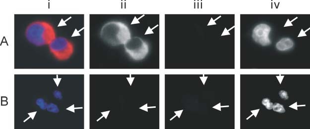
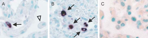
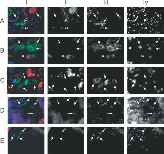
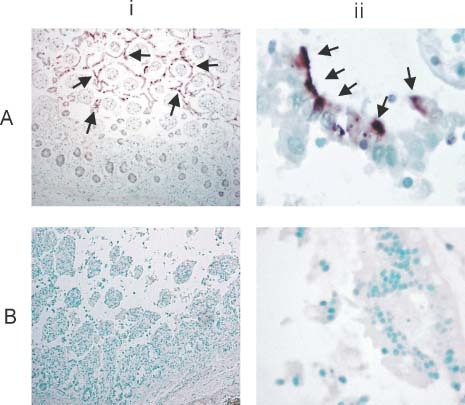
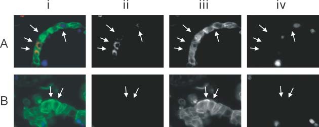
Severe acute respiratory syndrome (SARS) is a new human infectious disease with significant morbidity and mortality. The disease has been shown to be associated with a new coronavirus (SARS-CoV). The clinical and epidemiological aspects of SARS have been described. Moreover, the viral genome of SARS-CoV has been fully sequenced. However, much of the biological behaviour of the virus is not known and data on the tissue and cellular tropism of SARS-CoV are limited. In this study, six fatal cases of SARS were investigated for the tissue and cellular tropism of SARS-CoV using an in-situ hybridization (ISH) technique. Among all the tissues studied, positive signals were seen in pneumocytes in the lungs and surface enterocytes in the small bowel. Infected pneumocytes were further confirmed by immunofluorescence-fluorescence in-situ hybridization (FISH) analysis. These results provide important information concerning the tissue tropism of SARS-CoV, which is distinct from previously identified human coronaviruses, and suggest the possible involvement of novel receptors in this infection. Whereas the lung pathology was dominated by diffuse alveolar damage, the gut was relatively intact. These findings indicated that tissue responses to SARS-CoV infection are distinct in different organs.


Figures

Figure 1
Figure 1
Fluorescence in‐situ hybridization (FISH) of SARS‐CoV in infected (A) and non‐infected (B) Vero cells. The Vero cells are indicated by arrows. The composite picture is shown in i. Images captured under the red, green, and blue filters are shown as grey scale images in ii, iii, and iv, respectively. FISH signals of SARS‐CoV were detected in the cytoplasm of infected, but not in uninfected, Vero cells (ii). No bleaching of filters or autofluorescence was detected (iii). The nuclei were counterstained with DAPI (iv)
Figure 2
Figure 2
In situ hybridization (ISH) of SARS‐CoV in the lung. (A) Infected pneumocytes (arrow) with positive cytoplasmic signals are seen lining the alveolar septa. The other cellular components in the alveolar septa and histiocytes (arrow‐head) are negative (×400). (B) Scattered infected cells show a diffuse cytoplasmic distribution of the viral genome in infected cells (indicated by arrows; ×400). The infected cells, as suggested by the immunofluorescence–FISH study (Figure 3), were pneumocytes. (C) Control non‐SARS lung shows no specific ISH signals (×400)
Figure 3
Figure 3
Immunofluorescence–FISH with cytokeratin (AE1/AE3) and macrophage (CD68) markers in lung tissue. The panels show (i) the composite picture; (ii) FISH signals of SARS‐CoV in the red channel; (iii) immunofluorescence in the green channel; and (iv) counterstaining of the nuclei in the blue channel. Panels ii–iv are shown as grey scale captured images. (A) Low‐power view of the lung showing a SARS‐CoV‐infected pneumocyte (double arrow) among other uninfected pneumocytes (arrows) (×400). Pneumocytes were highlighted by immunofluorescence using a cytokeratin marker (AE1/AE3). (B) High‐power view of A showing clearly the cytoplasmic signals (red and in ii) in the infected pneumocytes (double arrows) (×1000). Among 100 infected cells (double arrow) counted in each positive case, all were pneumocytes. (C) High‐power view of the no‐probe control in the same lung specimen. Arrows indicate some of the detached pneumocytes that stained positive for cytokeratin with minimal cytoplasmic signals when viewed under the red filter. The oval background red signals represent autofluorescence from red blood cells. (D) Macrophages were seen among the floating cells and were recognized by their CD68 immunoreactivity (arrows). Among 100 infected cells (double arrow) counted in each positive case, none were macrophages (×400). (E) High‐power view of the no‐probe control in the same lung specimen. Arrows indicate macrophages in the alveolar space. No autofluorescence signal is seen in these cells
Figure 4
Figure 4
In situ hybridization (ISH) of SARS‐CoV in the small intestine. The panels show (i) a low‐power view (×40) and (ii) a high‐power view (×400). (A) Despite the autolytic changes, positive signals were seen and mainly detected in the surface enterocytes (arrows). High‐power view showing the positive cytoplasmic signals detected in the partially detached surface enterocytes. The positive signals appear polarized towards the apical part of the enterocytes. (B) Control non‐SARS intestine section reveals no specific ISH signals. High‐power view shows no cytoplasmic signals in the detached surface enterocytes
Figure 5
Figure 5
Immunofluorescence–FISH with cytokeratin (AE1/AE3) in the small intestine. The panels show (i) the composite picture; (ii) FISH signals of SARS‐CoV in the red channel; (iii) immunofluorescence in the green channel; and (iv) counterstaining of the nuclei in the blue channel. Panels ii–iv are shown as grey scale captured images. (A) High‐power view of the small intestine showing SARS‐CoV‐infected surface enterocytes (arrows) that have become detached (×1000). Surface enterocytes are highlighted by immunofluorescence using the cytokeratin marker (AE1/AE3). (B) High‐power view of the no‐probe control showing minimal autofluorescence signals in the detached enterocytes (arrows)

Comment in

References

    1. World Health Organization . Severe acute respiratory syndrome (SARS). Multi‐countries outbreak update 73. http://www.who.int/csr/don/2003_06_04/en/. Access date 4 June 2003.
    1. Peiris JS, Lai ST, Poon LL, et al. Coronavirus as a possible cause of severe acute respiratory syndrome. Lancet 2003; 361: 1319–1325. - PMC - PubMed
    1. Drosten C, Gunther S, Preiser W, et al. Identification of a novel coronavirus in patients with severe acute respiratory syndrome. N Engl J Med 2003; 348: 1967–1976. - PubMed
    1. Ksiazek TG, Erdman D, Goldsmith CS, et al. A novel coronavirus associated with severe acute respiratory syndrome. N Engl J Med 2003; 348: 1953–1966. - PubMed
    1. World Health Organization . Recommendations for laboratories testing for SARS. http://www.who.int/csr/sars/labmethods/en/#lab. Access date 21 July 2003.

MeSH terms

LinkOut - more resources