Review
. 2004:55:125-48.
doi: 10.1146/annurev.psych.55.090902.141545.

Hypocretin (orexin): role in normal behavior and neuropathology

Affiliations
Review

Hypocretin (orexin): role in normal behavior and neuropathology

Jerome M Siegel. Annu Rev Psychol. 2004.

Abstract

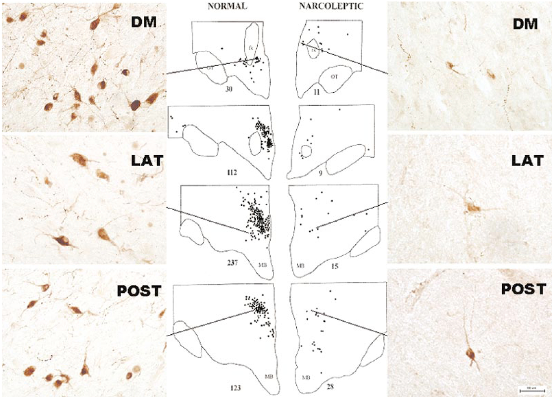
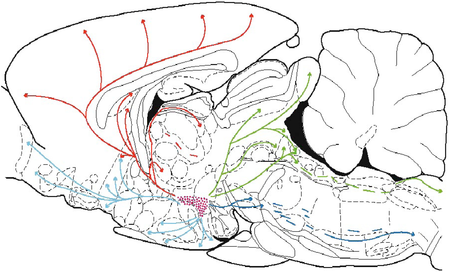
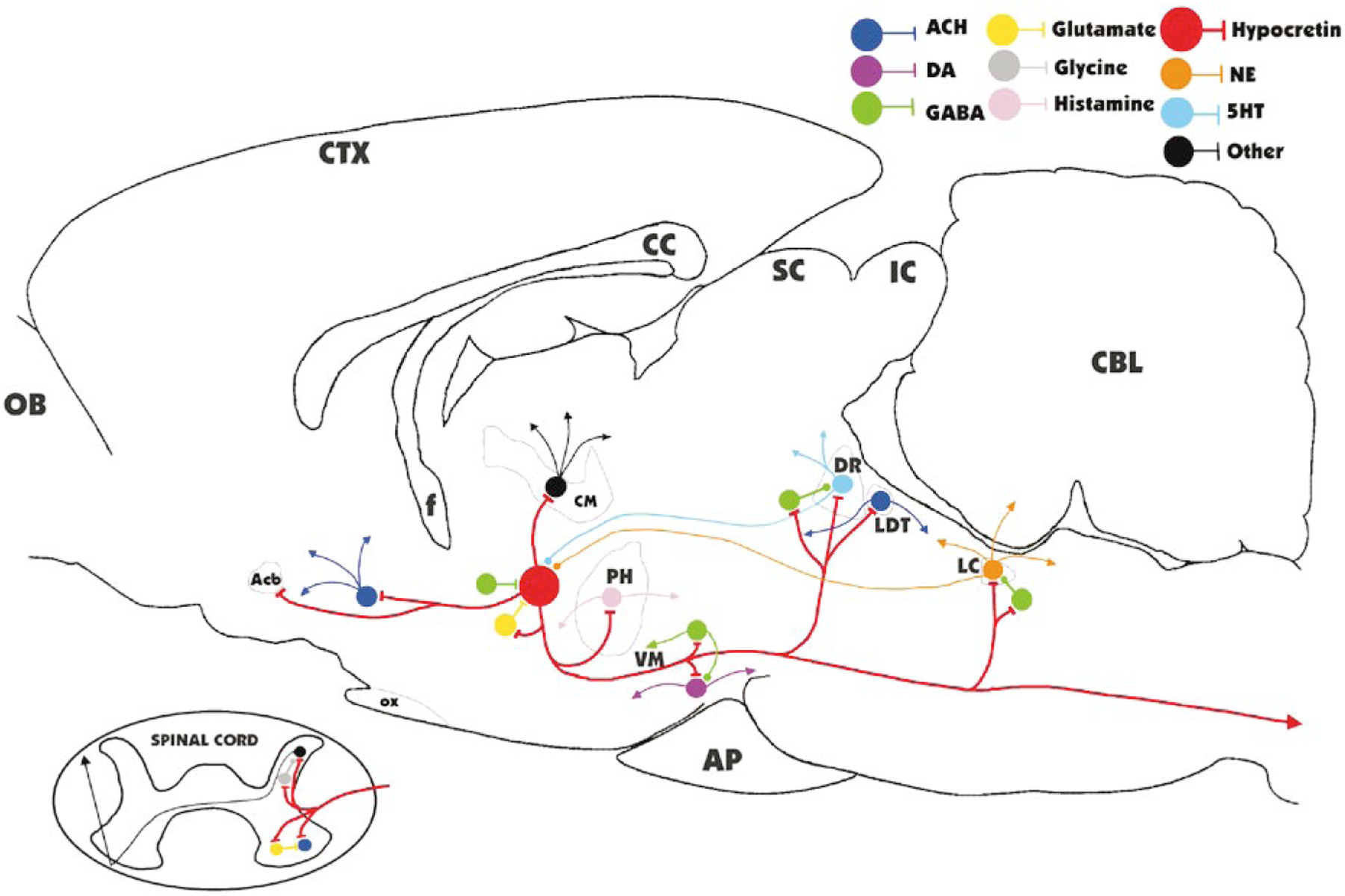
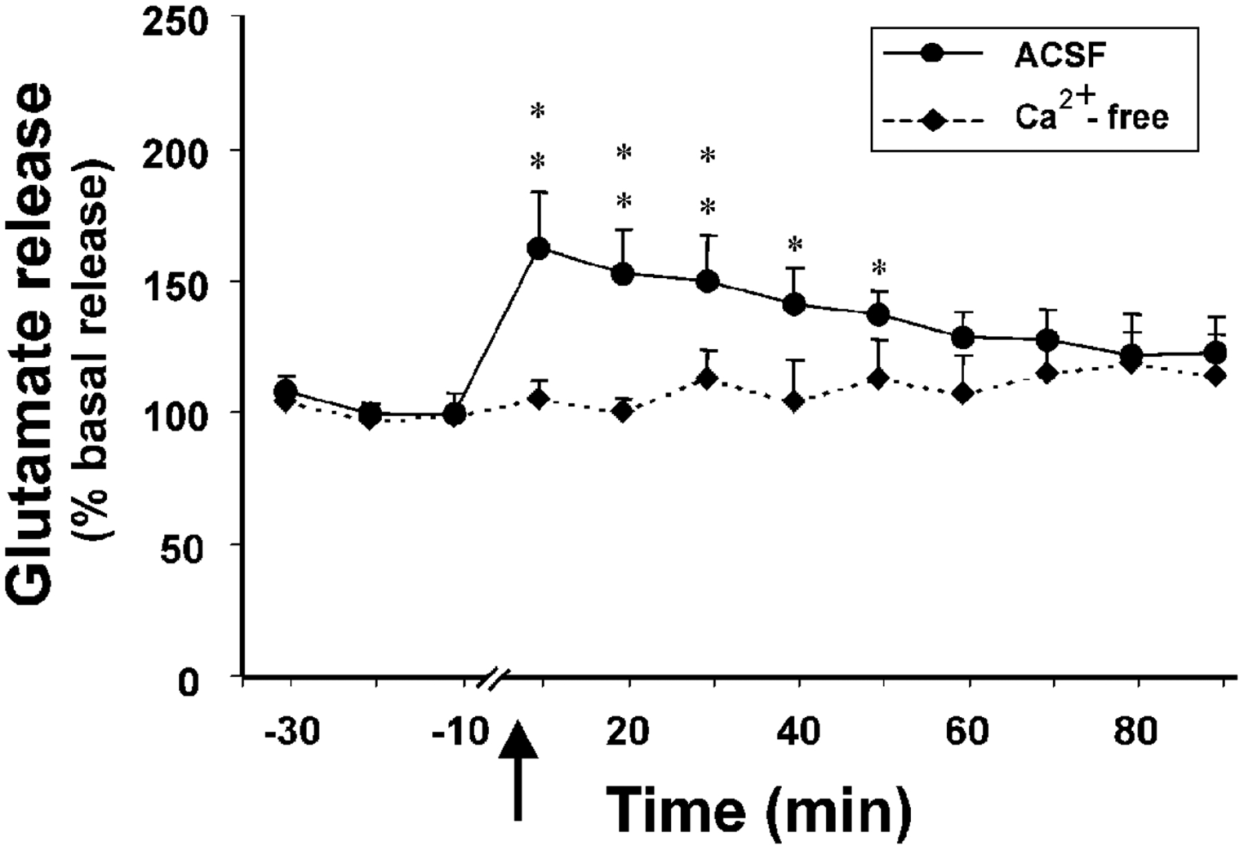
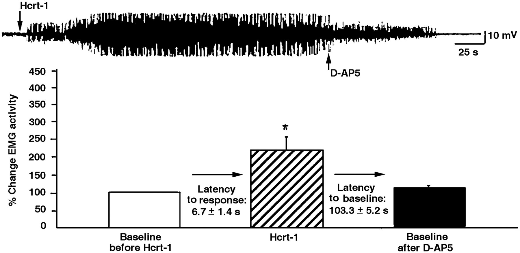
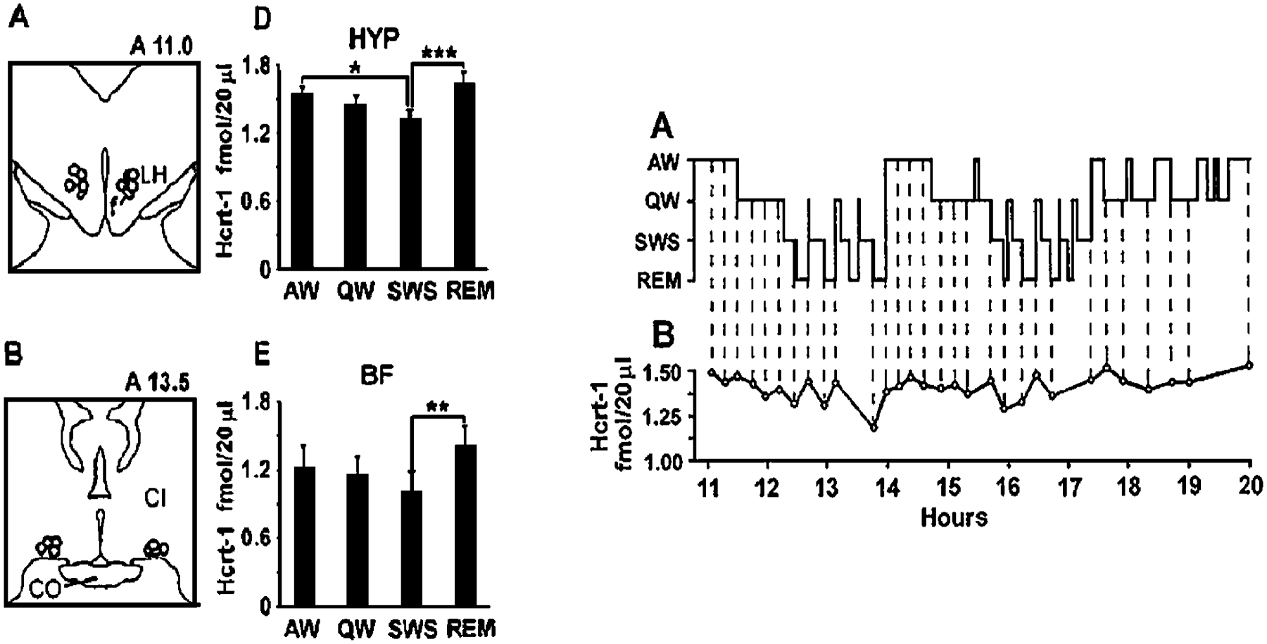
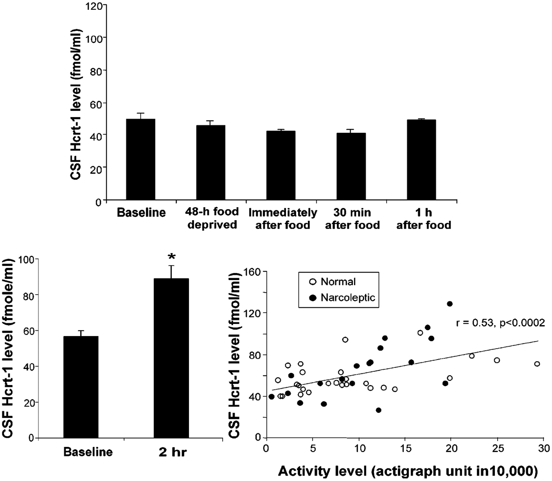
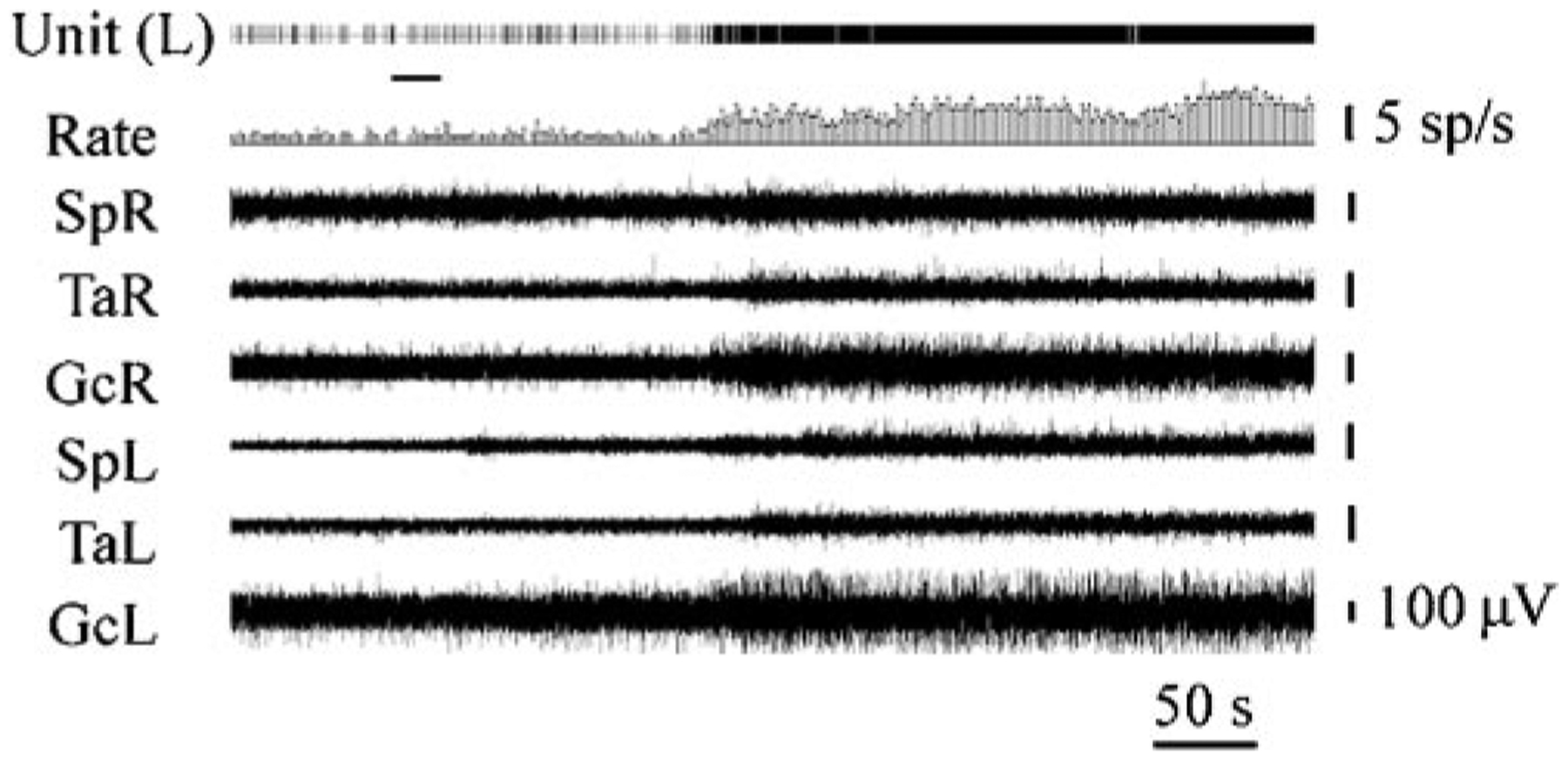
The hypocretins (Hcrts, also known as orexins) are two peptides, both synthesized by a small group of neurons, most of which are in the lateral hypothalamic and perifornical regions of the hypothalamus. The hypothalamic Hcrt system directly and strongly innervates and potently excites noradrenergic, dopaminergic, serotonergic, histaminergic, and cholinergic neurons. Hcrt also has a major role in modulating the release of glutamate and other amino acid transmitters. Behavioral investigations have revealed that Hcrt is released at high levels in active waking and rapid eye movement (REM) sleep and at minimal levels in non-REM sleep. Hcrt release in waking is increased markedly during periods of increased motor activity relative to levels in quiet, alert waking. Evidence for a role for Hcrt in food intake regulation is inconsistent. I hypothesize that Hcrt's major role is to facilitate motor activity tonically and phasically in association with motivated behaviors and to coordinate this facilitation with the activation of attentional and sensory systems. Degeneration of Hcrt neurons or genetic mutations that prevent the normal synthesis of Hcrt or of its receptors causes human and animal narcolepsy. Narcolepsy is characterized by an impaired ability to maintain alertness for long periods and by sudden losses of muscle tone (cataplexy). Administration of Hcrt can reverse symptoms of narcolepsy in animals, may be effective in treating human narcolepsy, and may affect a broad range of motivated behaviors.


Figures

Figure 1
Figure 1
Distribution of Hcrt-labeled cells in normal and narcoleptic humans. (Left) Normal human brain has approximately 70,000 Hcrt cells located in the hypothalamus, both medial and lateral to the fornix. (Right) Narcoleptic human brain has an 85%–95% loss of Hcrt cells. Normal Hcrt cell morphology is visible in surviving Hcrt cells in narcoleptics. DM, dorsomedial; LAT, lateral; POST, posterior hypothalamic nuclei. Calibration 50 μm. (Redrawn from Thannickal et al. 2000)
Figure 2
Figure 2
Locations of Hcrt cells in the hypothalamus in the rat and their ascending and descending projections. Some descending Hcrt axons terminate in the dorsal and ventral horns of the spinal cord. (From Peyron et al. 1998)
Figure 3
Figure 3
Major identified synaptic interactions of Hcrt neurons. Lines terminated by perpendicular lines denote excitation; circular terminations indicate inhibition. ACH, acetylcholine; DA, dopamine; NE, norepinephrine; 5HT, serotonin; OB, olfactory bulb; Acb, nucleus accumbens; f, fornix; OX, optic chiasm; CM, centromedian nucleus of the thalamus; PH, posterior hypothalamus; VM, ventral midbrain; AP, anterior pituitary; SC, superior colliculus; IC, inferior colliculus; DR, dorsal raphe; LDT, laterodorsal tegmental and pedunculopontine; LC, locus coeruleus; CBL, cerebellum.
Figure 4
Figure 4
Effect of intravenous injection of Hcrt on central glutamate release in the amygdala, a Hcrt-innervated region. Intravenous Hcrt injection produces a marked, calcium-dependent increase in glutamate release. No such increase occurs in the cerebellar cortex, a region without substantial Hcrt innervation. This demonstrates that Hcrt effectively crosses the blood-brain barrier and produces a marked release of glutamate in Hcrt-innervated regions. (From John et al. 2003)
Figure 5
Figure 5
Interactions of Hcrt with glutamate after microinjection of Hcrt into motor nuclei. Hcrt microinjections potently excited trigeminal motoneurons, but this effect was completely blocked by the glutamate NMDA receptor antagonist AP-5. Glutamate is a major mediator of Hcrt effects in motor nuclei and in other regions of the brain. (From Peever et al. 2003)
Figure 6
Figure 6
Sleep cycle release of Hcrt. (Left) Hcrt release is maximal in active waking and REM sleep, states with high levels of activity in central motor systems, and is significantly lower in non-REM sleep. Regions that were sampled are indicated in the drawing. CI, internal capsule; HYP, lateral hypothalamus; BF, basal forebrain. (Right) Time course of Hcrt release in successive 5-minute Hcrt samples across the sleep cycle. Maximal values are seen in waking and REM sleep. (From Kiyashchenko et al. 2002)
Figure 7
Figure 7
Behavioral studies of Hcrt release after food deprivation, feeding, and exercise. (Top) Effect of 48-hour food deprivation, feeding, and exercise on Hcrt release in normal dogs. In contrast to the changes in other putative feeding-related peptides, food deprivation does not increase Hcrt levels in cerebrospinal fluid. Feeding after deprivation also does not alter Hcrt levels. (Bottom left) Two-hour period of exercise produces marked increase in Hcrt levels. (Bottom right) Strong positive correlation between motor activity during exercise as measured by actigraph and levels of Hcrt in cerebrospinal fluid. (From Wu et al. 2002b)
Figure 8
Figure 8
Effect of Hcrt microinjection adjacent to the locus coeruleus. This figure shows that Hcrt microinjection into the locus coeruleus raises muscle tone ipsilateral to the injection. The loss of this excitatory effect of Hcrt contributes to cataplexy, the sudden loss of muscle tone experienced by most narcoleptics. Locus coeruleus activity ceases during cataplexy (Wu et al. 1999). SpR, SpL: electromyogram (EMG) of muscle splenius (right and left side); TaR, TaL: EMG of tibialis anterior muscle (right and left side); GcR, GcL: EMG of gastrocnemius muscle (right and left side). (From Kiyashchenko et al. 2001)

References

    1. Abrahamson EE, Moore RY. 2001. The posterior hypothalamic area: chemoarchitecture and afferent connections. Brain Res. 889:1–22 - PubMed
    1. Alam MN, Gong H, Alam T, Jaganath R, McGinty D, Szymusiak R. 2002. Sleep-waking discharge patterns of neurons recorded in the rat perifornical lateral hypothalamic area. J. Physiol 538:619–31 - PMC - PubMed
    1. Aldrich MS. 1998. Diagnostic aspects of narcolepsy. Neurology 50:S2–7 - PubMed
    1. Allen RP, Mignot E, Ripley B, Nishino S, Earley CJ. 2002. Increased CSF hypocretin-1 (orexin-A) in restless legs syndrome. Neurology 59:639–41 - PubMed
    1. Aston-Jones G, Rajkowski J, Kubiak P, Alexinsky T. 1994. Locus coeruleus neurons in monkey are selectively activated by attended cues in a vigilance task. J. Neurosci 14:4467–80 - PMC - PubMed

Publication types