

Particular sensitivity to calcium channel blockers of the fast inward voltage-dependent sodium current involved in the invasive properties of a metastastic breast cancer cell line
- PMID: 14744811
- PMCID: PMC1574233
- DOI: 10.1038/sj.bjp.0705649
Particular sensitivity to calcium channel blockers of the fast inward voltage-dependent sodium current involved in the invasive properties of a metastastic breast cancer cell line
Abstract
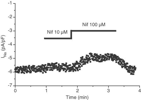
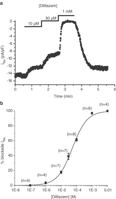
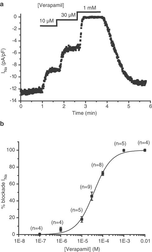
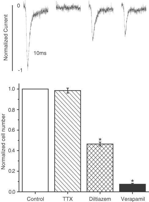
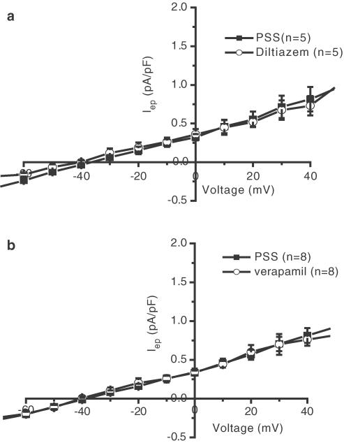
1. A voltage-dependent sodium current has been described in the highly invasive breast cancer cell line MDA-MB-231. Its activity is associated with the invasive properties of the cells. The aim of our study was to test whether this current (I(Na)) is sensitive to three representative calcium channel blockers: verapamil, diltiazem and nifedipine. I(Na) was studied in patch-clamp conditions. 2. I(Na) was sensitive to verapamil (IC(50)=37.6+/-2.5 microM) and diltiazem (53.2+/-3.6 microM), while it was weakly sensitive to nifedipine. 3. The tetrodotoxin (TTX) concentration, which fully blocks I(Na) (30 microM), did not affect cell proliferation. Diltiazem and verapamil, at concentrations that do not fully block I(Na), strongly reduced cell proliferation, suggesting, regarding proliferation, that these molecules act on targets distinct from sodium channels. These targets are probably not other ionic channels, since the current measured at the end of a 500 ms long pulse in the voltage range between -60 and +40 mV was unaffected by verapamil and diltiazem. 4. We conclude that the sodium channel expressed in MDA-MB-231 cells is sensitive to several calcium channel blockers. The present study also underlines the danger of concluding to the possible involvement of membrane channel proteins in any phenomenon on the sole basis of pharmacology, and without an electrophysiological confirmation.
Figures
References
-
- BELPOMME D., GAUTHIER S., PUJADE-LAURAINE E., FACCHINI T., GOUDIER M.J., KRAKOWSKI I., NETTER-PINON G., FRENAY M., GOUSSET C., MARIE F.N., BENMILOUD M., STURTZ F. Verapamil increases the survival of patients with anthracycline-resistant metastatic breast carcinoma. Ann. Oncol. 2000;11:1471–1476. - PubMed
-
- CORNWELL M.M., PASTAN I., GOTTESMAN M.M. Certain calcium channel blockers bind specifically to multidrug-resistant human KB carcinoma membrane vesicles and inhibit drug binding to P-glycoprotein. J. Biol. Chem. 1987;262:2166–2170. - PubMed
-
- FARIAS E.F., AGUIRRE GHISO J.A., LADEDA V., BAL DE KIER JOFFE E. Verapamil inhibits tumor protease production, local invasion and metastasis development in murine carcinoma cells. Int. J. Cancer. 1998;78:727–734. - PubMed
-
- FRASER S.P., DISS J.K.J., MYCIELSKA M.E., COOMBES R.C., DJAMGOZ M.B.A. Voltage-gated sodium channel expression in human breast cancer cells: Possible functional role in metastasis. Breast Cancer Res. Treat. 2002;76 Suppl. 1:S142.
-
- GALPER J.B., CATTERALL W.A. Developmental changes in the sensitivity of embryonic heart cells to tetrodotoxin and D600. Dev. Biol. 1978;65:216–227. - PubMed
MeSH terms
Substances
LinkOut - more resources
Full Text Sources
Medical
Miscellaneous
