. 2004 Feb;45(2):694-701.
doi: 10.1167/iovs.03-0503.

Osmoregulation of taurine transporter function and expression in retinal pigment epithelial, ganglion, and müller cells

Affiliations

Osmoregulation of taurine transporter function and expression in retinal pigment epithelial, ganglion, and müller cells

Amira El-Sherbeny et al. Invest Ophthalmol Vis Sci. 2004 Feb.

Abstract

Purpose: To determine whether taurine transporter (TauT) activity and expression are regulated by hyperosmolarity in RPE, ganglion, and Müller cells.

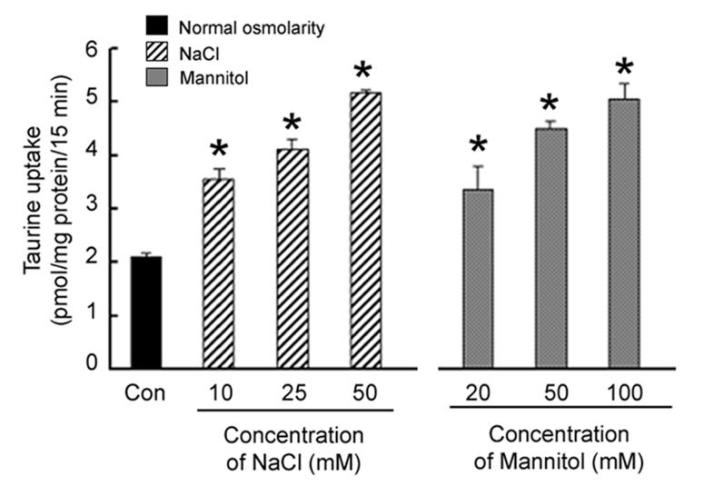
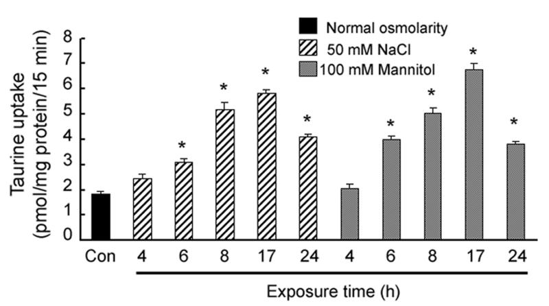
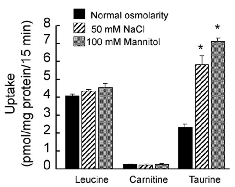
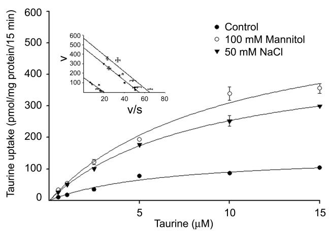
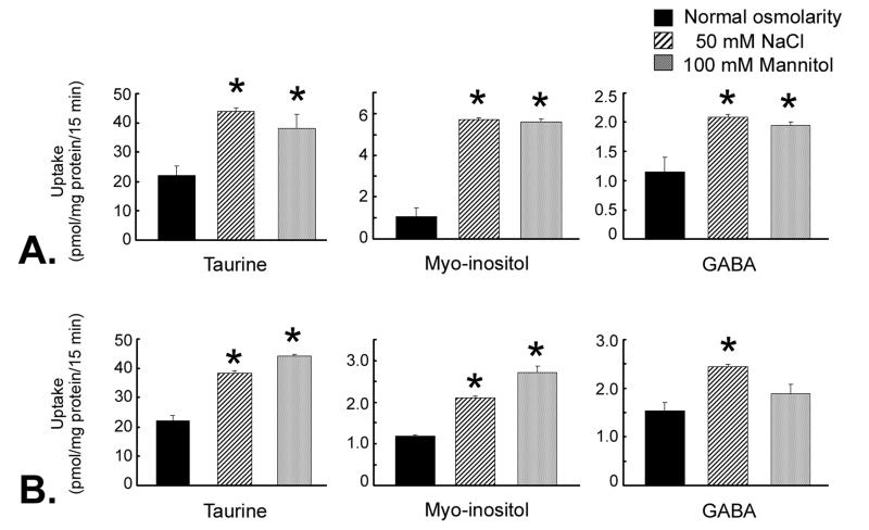
Methods: Uptake of taurine was measured in ARPE-19 cells cultured in DMEM-F12 medium without or with the addition of 50 mM NaCl or 100 mM mannitol. The kinetics of the transport were analyzed. RT-PCR and Northern and Western blot analyses were used to assess TauT mRNA and protein levels. The influence of hyperosmolarity on the uptake of taurine, myo-inositol, and gamma-aminobutyric acid GABA was studied in RPE, RGC-5, and rMC1 cells.

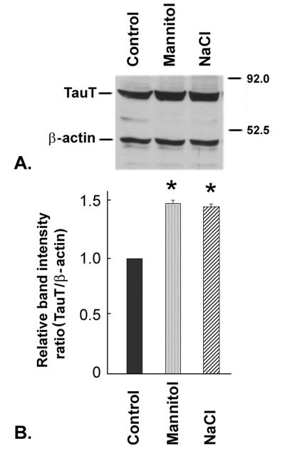
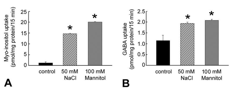
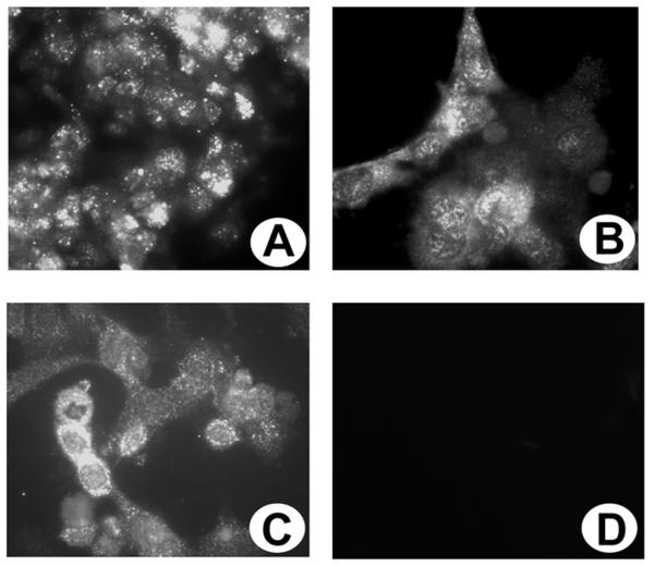
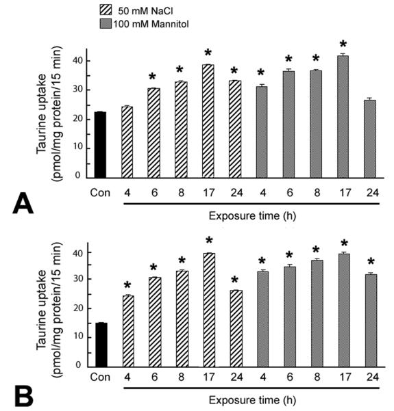
Results: TauT activity was abundant in RPE and was stimulated (3.5-fold) when the cells were exposed to hyperosmolar conditions (DMEM-F12 culture medium plus 50 mM NaCl or 100 mM mannitol). Peak stimulation of taurine uptake occurred after 17 hours of exposure to hyperosmolar medium. Kinetic analysis revealed that the hyperosmolarity-induced stimulation was associated with an increase in V(max) of TauT with no change in K(m). TauT mRNA and protein levels increased in RPE cells exposed to hyperosmolar conditions. Hyperosmolarity also stimulated the uptake of myo-inositol ( approximately 15-fold); GABA uptake was influenced less markedly. Immunofluorescence and functional studies showed that TauT is present in cultured RGC-5 and rMC1 cells. TauT activity was robust in these cells in normal osmolar conditions and increased by approximately twofold in hyperosmolar conditions.

Conclusions: These studies provide the first evidence that hyperosmolarity regulates TauT activity and expression in RPE and that TauT is present in ganglion and Müller cells and is regulated by hypertonicity. The data are relevant to diseases such as diabetes, macular degeneration, and neurodegeneration, in which retinal cell volumes may fluctuate dramatically.


Figures

Fig. 1
Fig. 1. Influence of increasing osmolarity on taurine uptake by RPE cells
Confluent ARPE-19 cells were exposed for 8 h to varying concentrations of NaCl or mannitol added to the normal culture medium and the uptake of taurine (80 nM) was determined. Values represent mean ± SEM for 6 determinations from two independent experiments. *Significantly different from control (p < 0.05).
Fig. 2
Fig. 2. Influence of exposure time to hyperosmotic conditions on taurine uptake in RPE cells
Confluent ARPE-19 cells were exposed for varying lengths of time (0 - 24 h) to either 50 mM NaCl or 100 mM mannitol added to the normal culture medium and the uptake of taurine (80 nM) determined. Values represent mean ± SEM for 6 determinations from two independent experiments. *Significantly different from control (p < 0.05).
Fig. 3
Fig. 3. Specificity of the stimulation of taurine uptake in RPE cells by hyperosmolarity
Confluent ARPE-19 cells were exposed for 17 h to either 50 mM NaCl or 100 mM mannitol added to the normal culture medium and the uptake of leucine (30 nM), carnitine (30 nM) or taurine (80 nM) was determined. Values represent mean ± SEM for 6 determinations from two independent experiments. *Significantly different from cells incubated under normal osmolar conditions (p < 0.05).
Fig. 4
Fig. 4. Kinetic analysis of taurine uptake in RPE cells cultured under normal and hyperosmolar conditions
Confluent ARPE-19 cells were incubated for 17 h in normal culture medium with or without (control) the addition of 100 mM mannitol or 50 mM NaCl. Uptake of taurine was measured over a taurine concentration range of 0.5 – 15 μM. Values are means ± SEM for 6 determinations from 2 independent experiments. Results are presented as plots describing the relationship between taurine concentration and taurine uptake rate. Inset: Eadie-Hofstee plots (v/s vs v, where v is taurine uptake in pmol/mg protein/15 min and s is taurine concentration in μM).
Fig. 5
Fig. 5. Analysis of steady-state levels of TauT mRNA in RPE cells cultured under hyperosmolar conditions
Confluent cells were exposed for 17 h to normal culture medium with or without (control) the addition of 100 mM mannitol or 50 mM NaCl. RNA was isolated from these cells and used for semiquantitative RT-PCR (total RNA) and northern analysis (mRNA). (A) Data from Southern hybridization with 32P-labeled cDNA probes specific for TauT and GAPDH. (B) Data shown in panel A were subjected to phosphorimage analysis and the ratios of the TauT-specific band to the GAPDH-specific band under varying osmolar conditions are shown; the ratio in control cells was taken as 1. (C) Northern blot.
Fig. 6
Fig. 6. Analysis of TauT protein levels in RPE cells cultured under hyperosmolar conditions
Confluent ARPE-19 cells were exposed for 17 h to normal culture medium with or without (control) the addition of 100 mM mannitol or 50 mM NaCl. Protein was isolated and subjected to immunoblotting. (A) Representative blot of proteins from RPE cells cultured under varying osmolar conditions. TauT has a molecular mass of 70 kDa and β-actin (internal control) has a molecular mass of 45 kDa. (B) Quantitation of the ratio of the TauT band to β-actin band. The ratio in control cells was taken as 1.
Fig. 7
Fig. 7. Influence of hyperosmolarity on the uptake of myo-inositol and GABA in RPE cells
Confluent ARPE-19 cells were maintained for 17 h in normal culture medium with or without (control) the addition of 50 mM NaCl, or 100 mM mannitol. Uptake of myo-inositol (100 nM) (A) or GABA (65 nM) (B) was determined in these cells. Values represent mean ± SEM for 6 determinations from two independent experiments. *Significantly different from control. (p < 0.05).
Fig. 8
Fig. 8. Immunolocalization of TauT in cultured human ARPE-19, rat ganglion (RGC-5) and Müller (rMC1) cells
ARPE-19 (A), RGC-5 (B) and rMC1cells (C) were cultured on laminin-coated chamber slides and processed for immunohistochemistry using a primary antibody against TauT followed by an FITC-labeled secondary antibody. (D) ARPE-19 cells incubated with antibody that had been preincubated with the peptide (negative control) showing no positive staining. (Magnifications 600X)
Fig. 9
Fig. 9. Influence of exposure time to hyperosmotic conditions on taurine uptake in RGC-5 and rMC-1 cells
Confluent RGC-5 (A) and rMC1 (B) cells were exposed for varying lengths of time (0 -24 h) to either 50 mM NaCl or 100 mM mannitol added to the normal culture medium (an increase of 100 mOsm in the osmolarity of the medium) and the uptake of taurine (80 nM) determined. Values represent mean ± SEM for 6 determinations from two independent experiments. *Significantly different from control (p < 0.05).
Fig. 10
Fig. 10. Uptake of taurine, myo-inositol and GABA in ganglion cells and Müller cells cultured under hyperosmolar conditions
Confluent RGC-5 cells (A) or rMC1 cells (B) were maintained for 17 h in normal culture medium with no addition (normal osmolarity), 50 mM NaCl, or 100 mM mannitol (an increase of 100 mOsm in the osmolarity of the culture medium). Uptake of taurine (80 nM), myo-inositol (100 nM) or GABA (65 nM) was determined. Values represent mean ± SEM for 6 determinations from two independent experiments. *Significantly different from control (normal osmolarity) (p < 0.05).

References

    1. Pasantes-Morales H, Klethi J, Ledig M, Mandel P. Free amino acids of chicken and rat retina. Brain Res. 1972;41:494–497. - PubMed
    1. Obrosova IG, Minchenko AG, Marinescu V, Fathallah L, Kennedy A, Stockert CM, Frank RN, Stevens MJ. Antioxidants attenuate early up regulation of retinal vascular endothelial growth factor in streptozotocin-diabetic rats. Diabetologia. 2001;44:1102–1110. - PubMed
    1. Hayes KC, Carey RE, Schmidt SY. Retinal degeneration associated with taurine deficiency in the cat. Science. 1975;188:949–951. - PubMed
    1. Pasantes-Morales H, Schousboe A. Volume regulation in astrocytes: a role for taurine as an osmoeffector. J Neurosci Res. 1988;20:503–509. - PubMed
    1. Kimelberg HK, Goderie SK, Higman S, Pang S, Waniewski RA. Swelling-induced release of glutamate, aspartate, and taurine from astrocyte cultures. J Neurosci. 1990;10:1583–1591. - PMC - PubMed

Publication types

MeSH terms