. 2004 Feb 1;18(3):261-77.
doi: 10.1101/gad.1165804. Epub 2004 Jan 26.

Endoplasmic reticulum stress induces p53 cytoplasmic localization and prevents p53-dependent apoptosis by a pathway involving glycogen synthase kinase-3beta

Affiliations

Endoplasmic reticulum stress induces p53 cytoplasmic localization and prevents p53-dependent apoptosis by a pathway involving glycogen synthase kinase-3beta

LiKe Qu et al. Genes Dev. .

Abstract

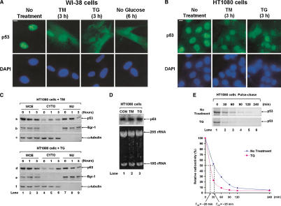
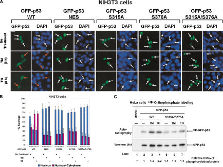
The tumor suppressor p53, a sensor of multiple forms of cellular stress, is regulated by post-translational mechanisms to induce cell-cycle arrest, senescence, or apoptosis. We demonstrate that endoplasmic reticulum (ER) stress inhibits p53-mediated apoptosis. The mechanism of inhibition involves the increased cytoplasmic localization of p53 due to phosphorylation at serine 315 and serine 376, which is mediated by glycogen synthase kinase-3 beta (GSK-3beta). ER stress induces GSK-3beta binding to p53 in the nucleus and enhances the cytoplasmic localization of the tumor suppressor. Inhibition of apoptosis caused by ER stress requires GSK-3beta and does not occur in cells expressing p53 with mutation(s) of serine 315 and/or serine 376 to alanine(s). As a result of the increased cytoplasmic localization, ER stress prevents p53 stabilization and p53-mediated apoptosis upon DNA damage. It is concluded that inactivation of p53 is a protective mechanism utilized by cells to adapt to ER stress.


Figures

Figure 1.
Figure 1.
ER stress alters the subcellular localization of p53. Immunostaining of endogenous p53. WI-38 cells (A) or HT1080 cells (B) were treated with 10 μg/mL of TM, 1 μM of TG, or incubated in glucose-free medium for the indicated time. Endogenous p53 was visualized by immunostaining with anti-p53 monoclonal antibodies (top) as described in Materials and Methods. The exposure time for green fluorescence was identical in all samples (A,B; top). The nucleus was detected by DAPI staining (A,B; bottom). (C) Subcellular fractionation of HT1080 cells. Cells were treated with either 10 μg/mL of TM or 1 μM of TG. At the indicated time points, cytoplasmic and nuclear fractions were prepared as described in Materials and Methods. Whole-cell extracts (WCE; 50 μg of protein), cytoplasmic extracts (CYTO; 40 μg of protein), or nuclear extracts (NU; 8 μg of protein) were subjected to Western blotting with anti-p53 rabbit polyclonal antibody (a,b), anti-Egr-1 rabbit polyclonal antibody (b,e), or anti-α-tubulin mouse monoclonal antibody (c,f). (D) HT1080 cells were treated with either 10 μg/mL of TM or 1 μM of TG for 2 h, total RNA (10 μg) was isolated and subjected to Northern Blotting using [32P]dCTP human p53 cDNA as probe (top). The quality of RNA and equal loading were verified by staining the denaturing gel with ethidium bromide. The position of 28S and 18S rRNA are shown (bottom). (E) HT1080 cells were labeled with [35S]methionine/cysteine for 30 min and chased with radioactive-free medium in the absence or presence of 1 μM of TG for the indicated times. Protein extracts (1 mg) were subjected to immunoprecipitation with anti-p53 rabbit polyclonal antibody followed by SDS-PAGE and autoradiography. The radioactive bands were quantified by PhosphorImager, and the plots of the relative intensity of radioactive p53 bands toward time and the half-life time values are indicated.
Figure 2.
Figure 2.
ER stress prevents p53 stabilization and impairs p53-mediated apoptosis in response to genotoxic stress. (A) WI-38 cells were treated with 1 μM of ADR alone or with either 10 μg/mL of TM or 1 μM of TG for the indicated times. Protein extracts (50 μg) were used for immunoblotting with an anti-p53 rabbit polyclonal antibody. A nonspecific (NS) band on the same blot was used as loading control. (B) WI-38 cells were treated with 5 nM ACD in the absence or presence of either 1 μg/mL of TM or 0.1 μM of TG for the indicated times. Protein extracts (50 μg) were immunoblotted with an anti-p53 rabbit polyclonal antibody. Blot was stripped and reprobed with an anti-actin antibody. (C) HCT116 p53+/+ cells were treated with 375 μM of 5-FU in the absence or presence of either 1 μg/mL of TM or 0.1 μM of TG for the indicated times. The protein levels of p53 were detected by immunoblotting of 50 μg protein extracts with anti-p53 rabbit polyclonal antibody. A nonspecific band on the same blot was used as loading control. (D,E) HCT116 p53 +/+and HCT116 p53-/- cells were pretreated with either 0.3 μg/mL of TM or 0.03 μM of TG for 1 h, followed by treatment with 375 μM of 5-FU for 3 h. The cells were fed with fresh medium and subjected either to FACS analysis 48 h post-treatment (D) or colony formation assay (E) as described in Materials and Methods. The numbers represent the average values of colony formation quantification for each treatment from three independent experiments. Untreated (CON) cells were scored as 100%.
Figure 3.
Figure 3.
Phosphorylation-dependent cytoplasmic localization of p53 in ER-stressed cell. (A) Subcellular localization of GFP-p53. Plasmid DNA (0.5 μg) containing either GFP-p53 WT or each of the indicated GFP-p53 mutants was transiently transfected into NIH-3T3 cells. Twenty-four hours later, cells were left untreated or treated with either 10 μg/mL of TM or 1 μM of TG for 8 h, and then examined for GFP-p53 fluorescence. Cell nuclei were visualized by staining with DAPI. White arrows indicate nuclear localization of GFP-p53 only, whereas orange arrows indicate either cytoplasmic or both cytoplasmic and nuclear localization of GFP-p53. (B) Quantitative analysis of GFP-p53 localization. For each condition, 1000 GFP-p53 positive and live cells were scored. Cells were classified into two groups as follows: the first with predominantly nuclear p53 (blue bars) and the second with p53 both in the nucleus and cytoplasm (red bars). Values are means ± SD from six separate experiments. (C) Induced phosphorylation of GFP-p53 by ER stress in vivo. HeLa cells were transfected with 1 μg of either GFP-p53 WT or GFP-p53 S315A/S376A plasmid DNA. Twenty-four hours later, cells were subjected to [32P]orthophosphate labeling followed by immnunoprecipitation of 500 μg of protein extracts with anti-p53 rabbit polyclonal antibody. Half of the immunoprecipitates were subjected to SDS-PAGE and autoradiography (top; 4 h exposure), the other half to immunoblotting with anti-p53 polyclonal antibody followed by ECL detection for 30 sec (bottom). The relative ratio of p53 phosphorylation was calculated by normalizing the intensity of phosphorylated p53 to the intensity of total p53 protein detected by ECL.
Figure 4.
Figure 4.
Physical and functional interactions between p53 and GSK-3β in ER-stressed cells. (A) ER stress activates GSK-3β. WI-38 cells were treated with 10 μg/mL of TM or 1 μM of TG as indicated. Cell lysates were subjected to Western blotting with anti-phosphoserine 9 antibody of GSK-3β (top, lower bands), which also cross-reacts with phosphoserine 21 of GSK-3α (higher bands). The total levels of GSK-3β on the same blot were detected by immunoblotting (bottom). (B) ER stress promotes GSK-3β nuclear localization and interaction with p53. WI-38 cells untreated or treated with either 10 μg/mL of TM or 1 μM of TG for 2 h were subjected to immunostaining for endogenous p53 (green) or GSK-3β (red). The localization or colocalization (yellow) of both proteins was detected by laser-scanning confocol microscopy. (C) ER stress enhances the interaction between p53 and GSK-3β. WI-38 cells were treated with 10 μg/mL of TM or 1 μM of TG for 45 min. Protein extracts (400 μg) were subjected to immunoprecipitation with 1 μg of a mouse anti-p53 antibody (Ab-6, lanes 2-4). As a control, protein extracts (400 μg) from untreated WI-38 cells were immunoprecipitated with 1 μg of purified mouse IgG. Immunoprecipitates were subjected to immunoblotting with anti-GSK-3β rabbit polyclonal antibody (top) followed by immunoblotting with anti-p53 rabbit polyclonal antibody (second from top). Whole-cell extracts (WCE; 40 μg of protein) were used to detect the amount of either GSK-3β (third from top) or p53 (bottom) in the protein extracts used for p53 immunoprecipitation. (D) GSK-3β phosphorylates the carboxyl terminus of p53 in vitro. HA-tagged GSK-3β (WT or KD) was transiently expressed in HeLa cells. Protein extracts (200 μg) were used for immunoprecipitation with anti-HA rabbit polyclonal antibody followed by in vitro kinase assays with either 1 μg of GST-p53 160-318 (lanes 1,3) or 1 μg of GST-p53 319-393 (lanes 2,4) in the presence of [γ-32P]ATP. Phosphorylated GST-p53 proteins were detected by SDS-PAGE and autoradiography (top), the GST-p53 proteins by Coomassie blue staining (bottom). (E) GSK-3β phosphorylates p53 at serine 376 in vitro. A total of 1 μg of purified GST-p53 319-393 (wild type or S376A mutant) fusion protein was incubated with 30 ng of histidine-tagged GSK-3β and subjected to in vitro kinase assay in the absence or presence of 20 mM LiCl. As a control, 20 mM NaCl in lanes 2 and 4 were used. Phosphorylated GST-p53 was detected by autoradiography (top), the total GST-p53 by Coomassie blue staining (bottom). (F) Phosphorylation of p53 by GSK-3β in vivo. HeLa cells were transfected with 1 μg of either GFP-p53 WT or GFP-p53 S315A/S376A plasmid DNA in the presence of 2 μg of pcDNA/3.1, GSK-3β WT or GSK-3β KD plasmid DNA. Phosphorylation and expression levels of GFP-p53 were detected as described in Fig. 3C.
Figure 5.
Figure 5.
ER stress impairs p53-mediated apoptosis. (A) ER stress inhibits GFP-p53-mediated apoptosis in SAOS-2 cells. Cells were transfected with 0.5 μg of EGFP (negative control), GFP-p53 WT, GFP-p53 S315A, GFP-p53 S376A, or GFP-p53 S315A/S376A plasmid DNA. Four hours post-transfection, ER stress commenced after treatments with 0.4 μg/mL of TM or by growing cells in low-glucose medium. Sixteen hours later, cells were fixed and subjected to fluorescence microscopic observation for GFP. Apoptotic cells are indicated by arrows. (B) Quantification of GFP-p53-induced apoptosis in SAOS-2 cells. GFP-positive cells from A that exhibited distinct nuclear condensation or fragmentation were scored as apoptotic. For each sample, 300 GFP-positive cells were counted. The values in the histograms represent means ± SD from three independent experiments. (C) Ectopic expression of GSK-3β inhibits p53-dependent apoptosis in SAOS-2 cells. SAOS-2 cells were transfected with either 0.5 μg of EGFP plasmid or 0.5 μg of each of the indicated types of GFP-p53 plasmid DNA together with 1 μg of pcDNA3.1, GDK-3β WT, or GSK-3β KD plasmid DNA. Twenty hours later, cells were subjected to fluorescence microscopy for GFP expression and apoptotic phenotype. Quantification of apoptotic Saos-2 cells was done as in B. The values in the histograms represent means ± SD from three independent experiments.
Figure 6.
Figure 6.
ER stress affects p53 localization and p53-mediated apoptosis through GSK-3β. (A) Expression of endogenous p53 in GSK-3β+/+ and GSK-3β-/- MEFs. A total of 50 μg of protein from GSK-3β+/+ and GSK-3β-/- MEFs was subjected to Western blotting with anti-p53 polyclonal antibody or anti-GSK-3β polyclonal antibody. Protein levels were normalized to α-tubulin. (B) GSK-3β mediates ER stress-induced cytoplasmic relocation of p53. GSK-3β-/- MEFs were transfected with 0.05 μg GFP-p53 WT and 0.5 μg of pcDNA/3.1, GSK-3β WT, or GSK-3β KD plasmid DNA. Sixteen hours post-transfection, cells were treated with either 10 μg/mL of TM or 1 μM of TG for 4 h, followed by DAPI staining and fluorescence microscopic observation. (C) Quantification of nuclear to cytoplasmic GFP-p53 in GSK-3β-/- MEFs was done as described previously (Zhang and Xiong 2001; see also Materials and Methods). (D) Inhibition of p53-mediated apoptosis by ER stress in GSK-3β-/- MEFs reconstituted with GSK-3β. GSK-3β-/- MEFs were transfected with 0.2 μg of GFP-p53 WT and 0.5 μg of pcDNA/3.1, GSK-3β WT, or GSK-3β KD plasmid DNA. Four hours post-transfection, cells were treated with 0.4 μg/mL of TM or maintained in low-glucose medium. Sixteen hours later, cells were subjected to fluorescence microscopic observation and quantification of apoptotic cells as in Fig. 5B. The values in the histograms represent means ± SD from three independent experiments. (E) ER stress and genotoxic stress exhibit opposite effects on p53. In cells subjected to various forms of genotoxic stress, p53 is targeted by phosphorylation mainly at serine 15 and serine 20 (serine 18 and 23 for mouse p53) through the activation of ATM and/or ATR- pathway(s). Phosphorylation at these sites promotes p53 nuclear localization and stabilization by disrupting p53/Mdm2 interaction and Mdm2-mediated p53 nucleocytoplasmic export and degradation. On the other hand, ER stress promotes the cytoplasmic localization and degradation of p53 through the activation of GSK-3β, which mediates indirectly or directly the phosphorylation of p53 at serine 315 or serine 376, respectively. In cells subjected to both ER stress and genotoxic stress, p53 stabilization is impaired by the enhanced cytoplasmic relocation of the tumor suppressor caused by ER stress. Although the nuclear-signaling pathways leading to p53 phosphorylation upon DNA damage appear not to be affected by ER stress (as judged by the Chk2 activity levels in Supplementary Fig. 1), decrease in nuclear p53 by ER stress results in diminished activation capacity of the tumor suppressor in response to genotoxic stress.

Comment in

  • p53 and stress in the ER.
    Stavridi ES, Halazonetis TD. Stavridi ES, et al. Genes Dev. 2004 Feb 1;18(3):241-4. doi: 10.1101/gad.1181704. Genes Dev. 2004. PMID: 14871924 No abstract available.

Similar articles

Cited by

References

    1. Aloni-Grinstein R., Zan-Bar, I., Alboum, I., Goldfinger, N., and Rotter, V. 1993. Wild type p53 functions as a control protein in the differentiation pathway of the B-cell lineage. Oncogene 8: 3297-3305. - PubMed
    1. Alt J.R., Cleveland, J.L., Hannink, M., and Diehl, J.A. 2000. Phosphorylation-dependent regulation of cyclin D1 nuclear export and cyclin D1-dependent cellular transformation. Genes & Dev. 14: 3102-3114. - PMC - PubMed
    1. Appella E. and Anderson, C.W. 2001. Post-translational modifications and activation of p53 by genotoxic stresses. Eur. J. Biochem. 268: 2764-2772. - PubMed
    1. Aridor M. and Balch, W.E. 1999. Integration of endoplasmic reticulum signaling in health and disease. Nat. Med. 5: 745-751. - PubMed
    1. Ashcroft M., Taya, Y., and Vousden, K.H. 2000. Stress signals utilize multiple pathways to stabilize p53. Mol. Cell. Biol. 20: 3224-3233. - PMC - PubMed

Publication types

Substances