. 2004 Feb 2;199(3):417-22.
doi: 10.1084/jem.20030997. Epub 2004 Jan 26.

CD1d-dependent activation of NKT cells aggravates atherosclerosis

Affiliations

CD1d-dependent activation of NKT cells aggravates atherosclerosis

Emmanuel Tupin et al. J Exp Med. .

Abstract

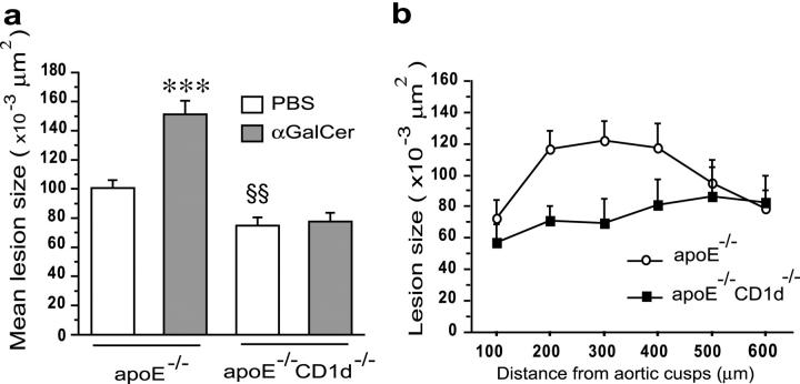
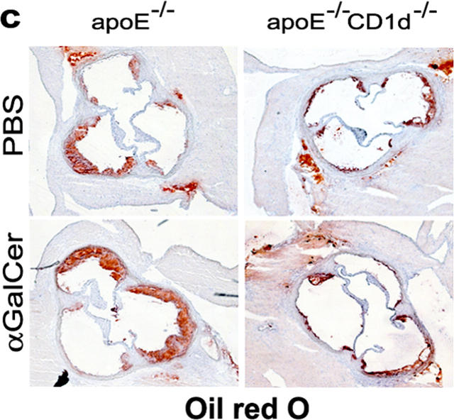
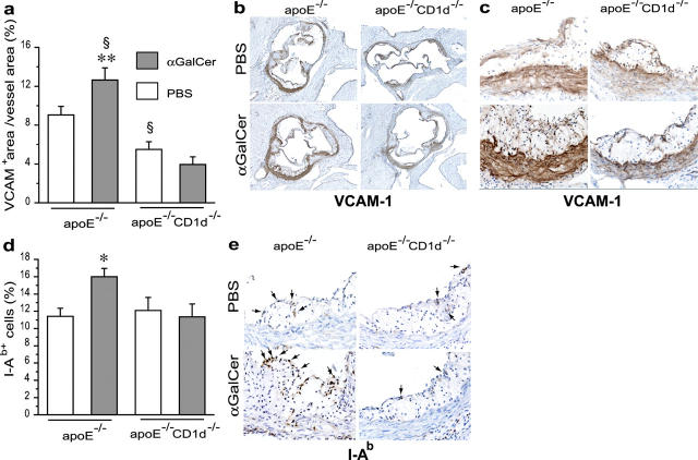
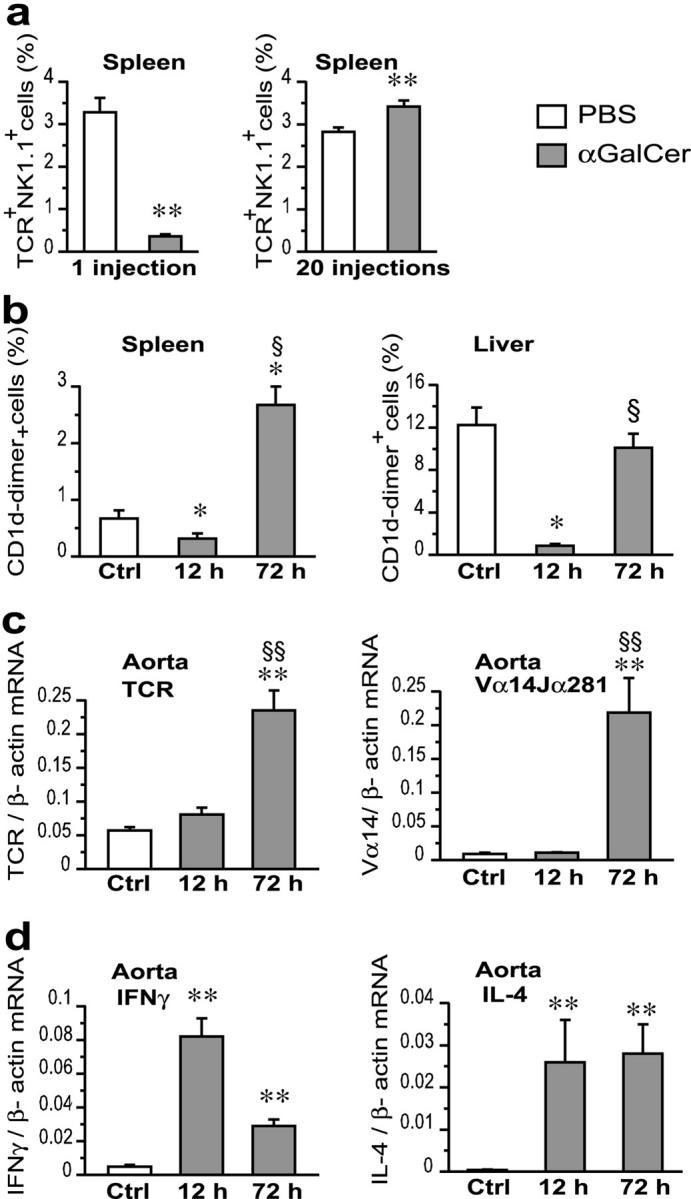
Adaptive and innate immunity have been implicated in the pathogenesis of atherosclerosis. Given their abundance in the lesion, lipids might be targets of the atherosclerosis-associated immune response. Natural killer T (NKT) cells can recognize lipid antigens presented by CD1 molecules. We have explored the role of CD1d-restricted NKT cells in atherosclerosis by using apolipoprotein E-deficient (apoE-/-) mice, a hypercholesterolemic mouse model that develops atherosclerosis. ApoE-/- mice crossed with CD1d-/- (CD1d-/-apoE-/-) mice exhibited a 25% decrease in lesion size compared with apoE-/- mice. Administration of alpha-galactosylceramide, a synthetic glycolipid that activates NKT cells via CD1d, induced a 50% increase in lesion size in apoE-/- mice, whereas it did not affect lesion size in apoE-/-CD1d-/- mice. Treatment was accompanied by an early burst of cytokines (IFNgamma, MCP-1, TNFalpha, IL-2, IL-4, IL-5, and IL-6) followed by sustained increases in IFNgamma and IL-4 transcripts in the spleen and aorta. Early activation of both T and B cells was followed by recruitment of T and NKT cells to the aorta and activation of inflammatory genes. These results show that activation of CD1d-restricted NKT cells exacerbates atherosclerosis.


Figures

Figure 1.
Figure 1.
Effects of CD1d deficiency and αGalCer treatment on atherosclerosis. 5-wk-old female apoE−/− and apoE−/−CD1d−/− mice were injected twice a week for 10 wk with αGalCer or PBS and killed 48 h after the last injection (n = 12 for each group). (a) Mean lesion size in Oil Red O–stained aortic root sections. Mean ± SEM (***P < 0.001 versus apoE−/−-PBS and all apoE−/−CD1d−/−; §§P < 0.01 versus apoE−/−-PBS mice). (b) Lesion size at every 100 μm for the first 600 μm of the aortic root in apoE−/− and apoE−/−CD1d−/− mice. (c) Representative Oil Red O–stained cryosections of aortic roots (magnification ×50).
Figure 1.
Figure 1.
Effects of CD1d deficiency and αGalCer treatment on atherosclerosis. 5-wk-old female apoE−/− and apoE−/−CD1d−/− mice were injected twice a week for 10 wk with αGalCer or PBS and killed 48 h after the last injection (n = 12 for each group). (a) Mean lesion size in Oil Red O–stained aortic root sections. Mean ± SEM (***P < 0.001 versus apoE−/−-PBS and all apoE−/−CD1d−/−; §§P < 0.01 versus apoE−/−-PBS mice). (b) Lesion size at every 100 μm for the first 600 μm of the aortic root in apoE−/− and apoE−/−CD1d−/− mice. (c) Representative Oil Red O–stained cryosections of aortic roots (magnification ×50).
Figure 2.
Figure 2.
Effects of CD1d deficiency and αGalCer treatment on the expression of VCAM-1 and I-Ab in atherosclerotic lesions. Experimental groups were the same as in Fig. 1. (a) VCAM-1 quantitation (VCAM-1+ area/vessel area); (b and c) representative sections of aortic root stained for VCAM-1 by avidin-biotin-immunoperoxidase (brown) (×50 and ×400). (d) I-Ab quantitation (I-Ab+ cells/total hematoxylin+ cells) and (e) representative sections of aortic roots stained for I-Ab by avidin-biotin-immunoperoxidase (brown) (×400). Arrows point at I-Ab+ cells. Mean ± SEM (*P < 0.05 versus apoE−/− treated with PBS and versus all apoE−/−CD1d−/−; **P < 0.01 versus all apoE−/−CD1d−/− mice; §P < 0.05 versus apoE−/−-PBS).
Figure 3.
Figure 3.
Systemic effects of αGalCer in apoE−/− mice. 5-wk-old apoE−/− mice were injected one, three, or five times, twice a week, with αGalCer or PBS. The first injection was i.v. (n = 6 for αGalCer and n = 5 for PBS) and the following ones i.p (n = 5 for αGalCer and n = 3 for PBS). Mice were killed 12 h after the last injection. (a) Serum levels of cytokines after one injection of αGalCer or PBS. In control (PBS) mice, levels of IFNγ, TNFα, and IL-6 were not detectable (nd). (b) Activated spleen T (CD69+TCRαβ+) and B (CD19+B7.2+) cells determined by flow cytometry. (c) IFNγ and IL-4 mRNA in spleens measured by quantitative real-time RT-PCR and normalized to β-actin mRNA after one, three, and five injections. The values for IFNγ are represented in a logarithmic scale. Mean ± SEM (*P < 0.05; **P < 0.01 versus PBS-injected mice).
Figure 4.
Figure 4.
Effect of αGalCer injection on cytokine and TCR transcripts in spleen and aorta of apoE−/− mice. (a) Proportion of spleen cells double positive for TCRβ and NK1.1 in mice injected once and killed at 12 h and in mice injected 20 times, starting at 5 wk of age, and killed 48 h after the last injection. FACS® data expressed as the percentage of forward/side scatter-gated lymphocytes. (b) CD1d-dimer+ NKT cells in spleen and liver of 5-wk-old mice injected once i.v. with PBS (Ctrl) (n = 6) or αGalCer and killed 12 h (n = 5) or 72 h (n = 6) later. FACS® data expressed as the percentage of forward /side scatter-gated lymphocytes. (c and d) Levels of total TCRβ and Vα14Jα281- specific TCR mRNA (c) and of IFNγ and IL-4 mRNA (d) in the aorta of mice treated with PBS or αGalCer as described for panel b. Real-time RT-PCR data are expressed as relative mRNA units normalized to β-actin mRNA. Mean ± SEM (*P < 0.05; **P < 0.01 versus PBS-injected mice; §P < 0.05; §§P < 0.01 versus αGalCer-injected mice, killed at 12 h).

References

    1. Binder, C.J., M.K. Chang, P.X. Shaw, Y.I. Miller, K. Hartvigsen, A. Dewan, and J.L. Witztum. 2002. Innate and acquired immunity in atherogenesis. Nat. Med. 8:1218–1226. - PubMed
    1. Hansson, G.K. 2001. Immune mechanisms in atherosclerosis. Arterioscler. Thromb. Vasc. Biol. 21:1876–1890. - PubMed
    1. Piedrahita, J.A., S.H. Zhang, J.R. Hagaman, P.M. Oliver, and N. Maeda. 1992. Generation of mice carrying a mutant apolipoprotein E gene inactivated by gene targeting in embryonic stem cells. Proc. Natl. Acad. Sci. USA. 89:4471–4475. - PMC - PubMed
    1. Breslow, J.L. 1996. Mouse models of atherosclerosis. Science. 272:685–688. - PubMed
    1. Joyce, S., and L. Van Kaer. 2003. CD1-restricted antigen presentation: an oily matter. Curr. Opin. Immunol. 15:95–104. - PubMed

Publication types