. 2004 Feb 2;164(3):451-9.
doi: 10.1083/jcb.200311112. Epub 2004 Jan 26.

Defective angiogenesis and fatal embryonic hemorrhage in mice lacking core 1-derived O-glycans

Affiliations

Defective angiogenesis and fatal embryonic hemorrhage in mice lacking core 1-derived O-glycans

Lijun Xia et al. J Cell Biol. .

Abstract

The core 1 beta1-3-galactosyltransferase (T-synthase) transfers Gal from UDP-Gal to GalNAcalpha1-Ser/Thr (Tn antigen) to form the core 1 O-glycan Galbeta1-3GalNAcalpha1-Ser/Thr (T antigen). The T antigen is a precursor for extended and branched O-glycans of largely unknown function. We found that wild-type mice expressed the NeuAcalpha2-3Galbeta1-3GalNAcalpha1-Ser/Thr primarily in endothelial, hematopoietic, and epithelial cells during development. Gene-targeted mice lacking T-synthase instead expressed the nonsialylated Tn antigen in these cells and developed brain hemorrhage that was uniformly fatal by embryonic day 14. T-synthase-deficient brains formed a chaotic microvascular network with distorted capillary lumens and defective association of endothelial cells with pericytes and extracellular matrix. These data reveal an unexpected requirement for core 1-derived O-glycans during angiogenesis.


Figures

Figure 1.
Figure 1.
Generation of T-synthase–deficient mice. (A) Schematic of biosynthesis of the four common O-glycan core structures. The cores can be further extended and modified as indicated by additional arrows. The biosynthetic step that T-synthase catalyzes is shown. (B) Wild-type (Wt) T-syn allele showing the site of PCR 1 used for genotyping. (C) Targeted T-syn allele in embryonic stem cells after homologous recombination with the targeting vector. Exons 1 and 2 and a neomycin cassette (Neo) are flanked by three loxP sites. (D) Deletion of loxP-flanked exons 1 and 2 and Neo in embryonic stem cells after in vitro Cre-mediated recombination. The site of PCR 2 used for genotyping is indicated. (E) PCR genotyping of DNA from mouse embryos. PCR 1 amplifies a 430-bp fragment from the Wt allele, and PCR 2 amplifies an 810-bp fragment from the Cre-deleted allele. (F) T-synthase and β1-4-galactosyltransferase (β1-4GalT) activities in E12 tissue extracts. The data represent the mean ± SD of three independent measurements.
Figure 2.
Figure 2.
Targeted disruption of T-syn causes fatal embryonic hemorrhage in mice. (A) Comparison of T-syn +/+ and T-syn −/− embryos at different developmental stages. Blood is visible in the hearts of the older T-syn +/+ embryos. Arrowheads indicate hemorrhage in the brain parenchyma and ventricles, spinal cord, and spinal canal of T-syn −/− embryos. Less blood is detected in T-syn −/− hearts because of anemia. The E14 T-syn −/− embryo is dead and appears pale because blood circulation has ceased. (B) Sagittal section of the E12 T-syn −/− embryo. The red and black boxes indicate hemorrhagic lesions, which are shown at higher magnification in C and D. Bleeding is visible in both the brain parenchyma and ventricles. Arrows indicate erythrocytes, which are nucleated at this stage of development.
Figure 3.
Figure 3.
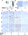
Disruption of T-syn eliminates T antigen expression and exposes the Tn antigen in murine embryos. (A) Blots of E12 tissue extracts probed with PNA, which recognizes nonsialylated core 1 O-glycans, and with HPA, which recognizes the nonsialylated Tn antigen. The extracts were incubated with or without sialidase before electrophoresis and blotting. The asterisk indicates the binding of PNA to the added sialidase. The blots are representative of three experiments. (B–F) Immunohistochemical staining of E12 tissue sections with mAbs to the T antigen, Tn antigen, or sialyl-Tn (sTn) antigen. The sections in D were pretreated with sialidase. Brown reaction product marks sites of antibody binding. Arrows, endothelial cells; arrowheads, epithelial cells; and asterisks, hematopoietic cells. Bars, 50 μm.
Figure 4.
Figure 4.
Expression of α2-3–linked and α2-6–linked sialic acid in T-syn + / + and T-syn / embryos. (A–H) Sections of the indicated tissues were pretreated with or without sialidase and stained with Maackia amurensis hemagglutinin (MAH), which recognizes α2-3–linked sialic acid, or with Sambucus nigra (SNA), which recognizes α2-6–linked sialic acid. Note that MAH binds much less to T-syn −/− embryos than to T-syn +/+ embryos. Pretreatment of the sections with sialidase eliminated binding of MAH and SNA, confirming their specificity. Bars, 50 μm.
Figure 5.
Figure 5.
Plasma-based blood coagulation is equivalent in T-syn + / + and T-syn / embryos. Clotting of individual plasma samples from eight E12 T-syn +/+ embryos and six E12 T-syn −/− embryos (mean ± SD, P > 0.05) was measured by the kinetic activated partial thromboplastin time (A) and the kinetic prothrombin time (B). (C) The activated partial thromboplastin time of human plasma deficient in factor V or factor VIII was measured with or without supplementation with normal human plasma or with pooled plasma from T-syn +/+ or T-syn −/− embryos. The data represent the mean ± SD of three independent experiments.
Figure 6.
Figure 6.
T-syn / embryos develop a chaotic microvascular network. (A–H) Visualization of microvessels in E12 hindbrains using maximal intensity projections of z-stacked confocal images. Endothelial cells were stained with antibodies to CD31 (green); pericytes were stained with antibodies to the proteoglycan NG2 (red); and basement membrane was stained with antibodies to laminin (red). T-syn +/+ embryos form a network of capillaries with uniform diameters and a regular branching pattern (A, C, E, and G), whereas T-syn −/− embryos form capillaries with heterogeneous diameters and excessive, irregular branches (B, D, F, and H). Insets in E and F are thin (5 μm) optical slices of representative vessels enlarged 2.5-fold, which illustrate the abluminal relationship of the NG2-positive pericytes to the CD31-positive endothelial cells. P, perineural vascular plexus; V, ventricle. (I and J) The architecture of large vessels (arrowheads) is similar in T-syn +/+ and T-syn −/− E12 yolk sacs, except that less blood fills the T-syn −/− vessels because of anemia. (K and L) Staining of whole mounts of E12 yolk sacs with antibodies to CD31. More capillary branching and irregular vessel diameters are present in the T-syn −/− yolk sac. Arrowheads indicate narrowed capillaries at branch points, which suggests incomplete vascular pruning. Bars, 50 μm, unless otherwise noted.
Figure 7.
Figure 7.
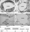
T-syn / embryos have defective capillary structures. (A–D) Electron micrographs of sections from E12 brains. (A) A T-syn +/+ capillary has a uniform lumen and a pericyte that is intimately associated with the endothelial cell. (B–D) T-syn −/− capillaries have distended or distorted lumens with attenuated endothelial cell processes. The upper pericyte in C is in close contact with an endothelial cell, but other pericytes are partially or completely separated from the endothelial cells. Large empty spaces (asterisks) surround the capillaries. Both T-syn +/+ and T-syn −/− capillaries have normal interendothelial cell junctions (arrowheads). Ec, endothelial cell; Pc, pericyte. (E) Sections of capillaries were scored for pericytes closely attached to endothelial cells and for distortions in endothelial cell shape. Bars, 2 μm.

References

    1. Berger, E.G. 1999. Tn-syndrome. Biochim. Biophys. Acta. 1455:255–268. - PubMed
    1. Blixt, O., B.E. Collins, I.M. Van Den Nieuwenhof, P.R. Crocker, and J.C. Paulson. 2003. Sialoside specificity of the siglec family assessed using novel multivalent probes: identification of potent inhibitors of myelin-associated glycoprotein. J. Biol. Chem. 278:31007–31019. - PubMed
    1. Brockhausen, I. 1999. Pathways of O-glycan biosynthesis in cancer cells. Biochim. Biophys. Acta. 1473:67–95. - PubMed
    1. Brockhausen, I., J. Yang, N. Dickinson, S. Ogata, and S.H. Itzkowitz. 1998. Enzymatic basis for sialyl-Tn expression in human colon cancer cells. Glycoconj. J. 15:595–603. - PubMed
    1. Bugge, T.H., Q. Xiao, K.W. Kombrinck, M.J. Flick, K. Holmback, M.J. Danton, M.C. Colbert, D.P. Witte, K. Fujikawa, E.W. Davie, and J.L. Degen. 1996. Fatal embryonic bleeding events in mice lacking tissue factor, the cell-associated initiator of blood coagulation. Proc. Natl. Acad. Sci. USA. 93:6258–6263. - PMC - PubMed

Publication types

MeSH terms