. 2004 Feb 3;101(5):1415-20.
doi: 10.1073/pnas.0307778101. Epub 2004 Jan 26.

Coat protein regulates formation of replication complexes during tobacco mosaic virus infection

Affiliations

Coat protein regulates formation of replication complexes during tobacco mosaic virus infection

S Asurmendi et al. Proc Natl Acad Sci U S A. .

Abstract

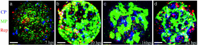
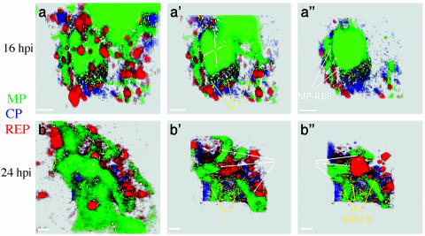
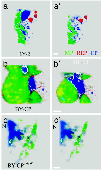
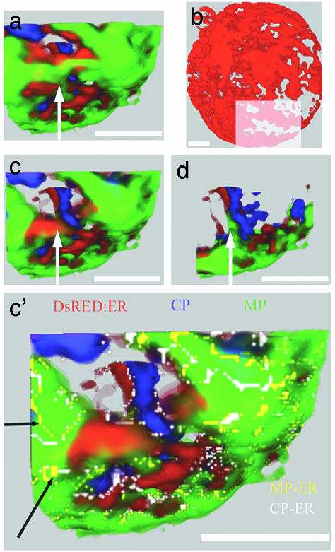
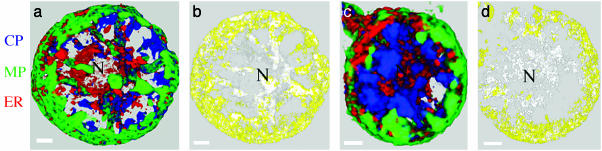
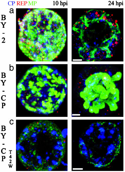
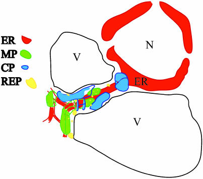
The genome of tobacco mosaic virus (TMV) encodes replicase protein(s), movement protein (MP), and capsid protein (CP). On infection, one or more viral proteins direct the assembly of virus replication complexes (VRCs), in association with host-derived membranes. The impact of CP-mediated resistance on the structures of the replication complexes was examined in nontransgenic and transgenic BY-2 cell lines that produce wild-type CP, mutant CP(T42W), and Ds-Red, which was targeted to endoplasmic reticulum by using immunofluorescence and 3D microscopy. We developed a model of VRCs that shows a clear association of MP with and surrounding the endoplasmic reticulum. Replicase is located within the MP bodies, as well as isolated sites throughout the cell. CP surrounds the VRCs. CP enhances the production of MP and increases the size of the VRC; however, the mutant CP(T42W) reduces the amount of MP and interferes with the formation of VRCs. We propose a regulatory role of the CP in the establishment of the VRC. We suggest that the lack of formation of VRCs restricts the efficiency of virus replication and the formation of virus movement complexes, resulting in restriction of cell-cell spread of infection. This results in higher levels of plant CP-mediated protection provided by CP(T42W).


Figures

Fig. 1.
Fig. 1.
Three-dimensional maximum-intensity projection reconstruction of BY-2 protoplasts infected with TMV through an infection cycle. Protoplasts were collected at 7 hpi (a), 10 hpi (b), 16 hpi (c), and 24 hpi (d), stained with antibodies, and observed by confocal microscopy: images were interpreted by imaris. Immunostaining color code: green, MP; blue, CP; red, replicase. Three-dimensional rotation reconstruction can be observed in Movies 1–4. (Scale bars, 5 μm.)
Fig. 2.
Fig. 2.
Detailed view of VRCs in BY-2 protoplasts infected by TMV. Protoplasts were immunostained and viewed as in Fig. 1, and data were analyzed by focusing on the structure and composition of VRCs. Three cutaway sections of rendered volumes (a, a′, a″, b, b′, and b″) were made through the VRCs along the z axis. Immunostaining color code: green, MP; blue, CP; red, replicase. Colocalization color code: yellow, MP-CP (yellow arrows); white, MP-replicase (white arrows). (a) At 16 hpi. (b) At 24 hpi. (Scale bars, 1 μm.)
Fig. 6.
Fig. 6.
Detailed cutaway view of the VRCs in nontransgenic and transgenic protoplasts 24 hpi with TMV. Shown are two cutaway sections of rendered volumes of cells through VRCs along the z axis. The study demonstrates that infection in BY-CPT42W cells does not result in normal VRCs that contain MP and replicase. (a and a′) Nontransgenic BY-2 cell line. (b and b′) BY-CP cell line. (c and c′) BY-CPT42W cell line. Immunostaining color code: green, MP; blue, CP; red, replicase. Colocalization color code: white, MP-CP. N, nucleus. (Scale bars, 1 μm.)
Fig. 4.
Fig. 4.
ER-DsRed BY-2 protoplasts infected with TMV at 24 hpi after immunostaining to localize CP and MP. Data collected from one region of an infected cell (shown in b) were enlarged to identify the precise manner of colocalization of MP and CP within a single VRC. Color code: green, MP; blue, CP; red, ER-DsRed. Colocalization color code: yellow, MP-ER; white, CP-ER. (b) ER channel only. Shown are a detailed view of the region delineated in b and three cutaway sections of rendered volume (a, c, d, and c′) throughout the VRC along the z axis. (a, b, and c) Fluorophore channels. (c′) Fluorophore and colocalization channels. The black arrows identify the colocalization MP and ER; white arrows indicate the same relative positions in the sectioned volume. Three-dimensional reconstructions are shown in Movies 5 and 6. (Scale bars, 5 μm.)
Fig. 3.
Fig. 3.
Three-dimensional reconstruction of ER-DsRed protoplasts infected with TMV, after immunostaining to identify the location of viral proteins in positional relationship to the ER. Frontal cell cortex was removed in the visualization to observe the interior of the cell. The study shows strong localization of viral proteins with ER and that localization of MP changes during the infection cycles. Immunostaining color code: green, MP; blue, CP; red, ER-DsRed. Colocalization color code: yellow, MP-ER; white, CP-ER. (a) Fluorophore channels, 16 hpi. (b) Colocalization channels, 16 hpi. (c) Fluorophores channels, 24 hpi. (d) Colocalization channels, 24 hpi. N, nucleus. Three-dimensional reconstructions are shown in Movies 5 and 6. (Scale bars, 5 μm.)
Fig. 5.
Fig. 5.
Maximum-intensity projection of TMV-infected protoplasts at 10 and 24 hpi. In the 24-hpi projections, the frontal cell cortex was removed to observe the interior of the cell. The study shows that accumulation of MP is significantly enhanced in BY-CP protoplasts compared with BY-2; the amount of MP in infected BY-CPT42W protoplasts is much reduced compared with the other cell lines. Immunostaining color code: green, MP; blue, CP; red, replicase. (a) BY-2. (b) BY-CP. (c) BY-CPT42W cell line. (Scale bars, 5 μm.)
Fig. 7.
Fig. 7.
Model of VRC structure at late stage of TMV infection. In the middle-to-late stages of infection, VRCs comprising MP and replicase are associated with and surround the cytoplasmic and cortical ER (14). Virus assembly and accumulation is adjacent to VRCs and can accumulate in large aggregates (22) near the nucleus. N, nucleus; V, vacuole; ER, endoplasmic reticulum.

References

    1. Osman, T. A. & Buck, K. W. (1996) J. Virol. 70, 6227–6234. - PMC - PubMed
    1. Noueiry, A. O. & Ahlquist, P. (2003) Annu. Rev. Phytopathol. 41, 77–98. - PubMed
    1. Restrepo-Hartwig, M. A. & Ahlquist, P. (1996) J. Virol. 70, 8908–8916. - PMC - PubMed
    1. Schaad, M. C., Jensen, P. E. & Carrington, J. C. (1997) EMBO J. 16, 4049–4059. - PMC - PubMed
    1. Ivanowski, D. (1903) Z. Pflanzenkr. Pflanzenschutz 13, 1–41.

Publication types

MeSH terms

Substances

LinkOut - more resources