. 2004 Feb 10;101(6):1531-6.
doi: 10.1073/pnas.0308089100. Epub 2004 Jan 26.

The 1.8-A resolution crystal structure of YDR533Cp from Saccharomyces cerevisiae: a member of the DJ-1/ThiJ/PfpI superfamily

Affiliations

The 1.8-A resolution crystal structure of YDR533Cp from Saccharomyces cerevisiae: a member of the DJ-1/ThiJ/PfpI superfamily

Mark A Wilson et al. Proc Natl Acad Sci U S A. .

Abstract

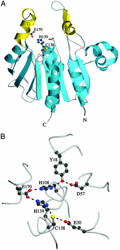
The yeast gene YDR533C encodes a protein belonging to the DJ-1/ThiJ/PfpI superfamily. This family includes the human protein DJ-1, which is mutated in autosomal recessive early-onset Parkinson's disease. The function of DJ-1 and its yeast homologue YDR533Cp is unknown. We report here the crystal structure of YDR533Cp at 1.8-A resolution. The structure indicates that the closest relative to YDR533Cp is the Escherichia coli heat shock protein Hsp31 (YedU), which has both chaperone and protease activity. As expected, the overall fold of the core domain of YDR533Cp is also similar to that of DJ-1 and the bacterial protease PfpI. YDR533Cp contains a possible catalytic triad analogous to that of Hsp31 and an additional domain that is present in Hsp31 but is not seen in DJ-1 and other members of the family. The cysteine in this triad (Cys-138) is oxidized in this crystal structure, similar to modifications seen in the corresponding cysteine in the crystal structure of DJ-1. YDR533Cp appears to be a dimer both in solution and the crystal, but this dimer is formed by a different interface than that found in Hsp31 or other members of the superfamily.


Figures

Fig. 1.
Fig. 1.
A ribbon diagram of the monomer of YDR533Cp. Domain A, common to other members of the DJ-1/ThiJ/PfpI superfamily, is colored blue, and domain P, specific to YDR533Cp and its close relatives, is colored yellow. β-Strands are numbered 1-10, and α-helices are lettered A-J. The diagram was made with molscript (45).
Fig. 2.
Fig. 2.
Ribbon and electrostatic surface representation of the YDR533Cp dimer. (A and B) Views of the YDR533Cp dimer that is generated by crystallographic symmetry. The dimer twofold axis is represented as an arrow and is in the plane of the page in A, and it is perpendicular to the plane of the page and is represented as an ellipse in B. In A and B, monomer A is blue (domain A) and yellow (domain B), and monomer B is green (domain A) and orange (domain B). (C and D) The electrostatic surface for the dimer calculated by grasp in the same orientation as A and B. The saddle-shaped depression in the dimer is highly negatively charged. The figure was made with grasp (46) and molscript (45).
Fig. 3.
Fig. 3.
Two views of the catalytic triad in YDR533Cp. (A) The location of triad residues C138, H139, and E170 on the monomer. (B) The hydrogen-bonding network that encompasses the triad. E170 hydrogen-bonds with both the triad residue H139 and a second histidine, H108. This second histidine is also found in the structurally homologous E. coli chaperone Hsp31. The figure was made with molscript (45).
Fig. 4.
Fig. 4.
A view of the electron density around Cys-138 in the triad. Sigma-A weighted 2 Fo - Fc electron density contoured at 1.0 σ is shown in blue, and sigma-A weighted Fo - Fc electron density contoured at 3.0 σ is shown in green. The Fo - Fc difference electron density around the Sγ of C138 suggests oxidative modification to a mixture of species, including sulfinic acid. E30 is close to the Cys at the nucleophile elbow in several structures of the DJ-1/ThiJ/PfpI superfamily. The figure was made with povscript+ (47).
Fig. 5.
Fig. 5.
Superposition of YDR533Cp, Hsp31, and DJ-1 monomers. (A) YDR533Cp (blue) and Hsp31 (red) are superimposed (Cα rmsd = 2.5 Å), showing the high degree of structural similarity between these two proteins. Most of the structural differences are in the P domains. (B) YDR533Cp (blue) and DJ-1 (yellow) are superimposed in the same orientation as in A (Cα rmsd = 2.1 Å). Despite the better agreement in the core domain A, DJ-1 lacks a P domain, and YDR533Cp and DJ-1 are likely members of different branches of the DJ-1/ThiJ/PfpI superfamily. The figure was made with molscript (45).

References

    1. Gasch, A. P., Spellman, P. T., Kao, C. M., Carmel-Harel, O., Eisen, M. B., Storz, G., Botstein, D. & Brown, P. O. (2000) Mol. Biol. Cell 11, 4241-4257. - PMC - PubMed
    1. Trotter, E. W., Berenfeld, L., Krause, S. A., Petsko, G. A. & Gray, J. V. (2001) Proc. Natl. Acad. Sci. USA 98, 7313-7318. - PMC - PubMed
    1. Beckmann, R. P., Mizzen, L. A. & Welch, W. J. (1990) Science 248, 850-854. - PubMed
    1. Trotter, E. W., Kao, C. M. F., Berenfeld, L., Botstein, D., Petsko, G. A. & Gray, J. V. (2002) J. Biol. Chem. 277, 44817-44825. - PubMed
    1. Ghaemmaghami, S., Huh, W., Bower, K., Howson, R. W., Belle, A., Dephoure, N., O'Shea, E. K. & Weissman, J. S. (2003) Nature 425, 737-741. - PubMed

MeSH terms

Substances

Associated data