. 2004 Feb 10;101(6):1601-6.
doi: 10.1073/pnas.0308212100. Epub 2004 Jan 26.

The Drosophila gene Start1: a putative cholesterol transporter and key regulator of ecdysteroid synthesis

Affiliations

The Drosophila gene Start1: a putative cholesterol transporter and key regulator of ecdysteroid synthesis

Guenther E Roth et al. Proc Natl Acad Sci U S A. .

Abstract

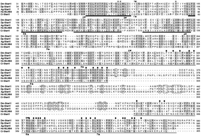
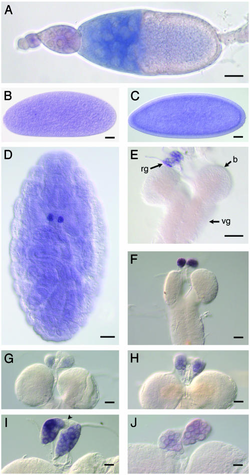
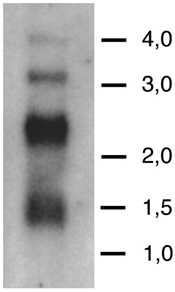
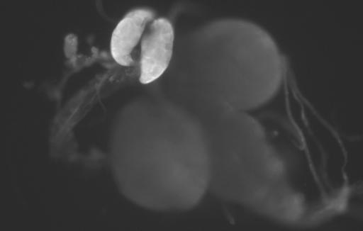
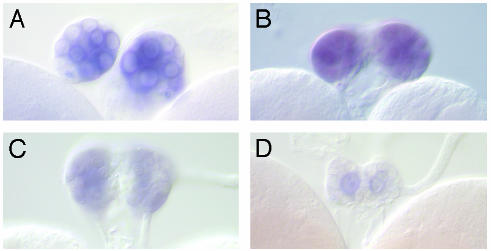
Human metastatic lymph node 64 (MLN64) is a transmembrane protein that shares homology with the cholesterol-binding vertebrate steroid acute regulatory protein (StAR)-related lipid transfer domain (START) and is involved in cholesterol traffic and steroid synthesis. We identified a Drosophila melanogaster gene whose putative protein product shows extensive homology with MLN64 and that we name Start1 (FlyBase CG3522). The putative Start1 protein, derived from Start1 cDNA sequences, contains an additional 122 aa of unknown function within the StAR-related lipid transfer domain. Similar inserts seem to exist in the Start1 homologues of Drosophila pseudoobscura and Anopheles gambiae, but not in the homologous protein of the urochordate Ciona intestinalis. Immunostaining using an insert-specific antibody confirms the presence of the insert in the cytoplasm. Whereas RT-PCR data indicate that Start1 is expressed ubiquitously, RNA in situ hybridizations demonstrate its overexpression in prothoracic gland cells, where ecdysteroids are synthesized from cholesterol. Transcripts of Start1 are detectable in embryonic ring gland progenitor cells and are abundant in prothoracic glands of larvae showing wave-like expression during larval stages. In adults, Start1 is expressed in nurse cells of the ovary. These observations are consistent with the assumption that Start1 plays a key role in the regulation of ecdysteroid synthesis. Vice versa, the expression of Start1 itself seems to depend on ecdysone, as in the ecdysone-deficient mutant ecd-1, Start1 expression is severely reduced.


Figures

Fig. 1.
Fig. 1.
Insect Start1 proteins differ from their chordate homologues by an insert within the START domain. Alignment of Start1 proteins from D. melanogaster (Dm-Start1; AAF47232), D. pseudoobscura (Dp-Start1), A. gambiae (Ag-Start1; EAA03945, see Materials and Methods), C. intestinalis (Ci-Start1), and MLN64 from Homo sapiens (Hs-MLN64; Q14849). Positions with at least one insect residue identical to at least one chordate residue are shown in gray (52%). Identical insert residues are boxed. Putative transmembrane helices for Hs-MLN64 are underlined in black; the START domain for MLN64 (19) is in gray. Triangles 1-6, positions of splice sites conserved between insect-Start1 and Hs-MLN64. Triangles 1′-7′, splice sites common to the chordate proteins. Sites 1 and 6 are common to the five species. ♦, residues playing a role in forming the tunnel of Hs-MLN64 (20), 29% of which are identical and 63% are chemically similar between the species. * indicate the dileucine motif and the tyrosine residue, shown to be necessary for endosomal location of MLN64 (16).
Fig. 2.
Fig. 2.
Start1 mRNA exists in many cell types and contains the insert. RT-PCR on RNA from different larval and adult tissue and from cultured cells using primers spanning six introns. Lane a, total third larva (3L); lane b, 3L brain/ring gland complexes; lane c, 3L imaginal discs; lane d, 3L salivary glands; lane e, adult heads; lane f, ovaries; lane g, testes; lane h, Kc-cells; lane i, genomic DNA control. Approximate positions of bands with the expected size for genomic DNA (1,662 bp), cDNA with insert (1,179 bp), and without (813 bp) are derived from marker lanes. The data are not meant to be quantitative.
Fig. 3.
Fig. 3.
Start1 is expressed in nurse cells of ovaries and the prothoracic cells of the ring gland. Whole-mount RNA in situ hybridization using digoxygenin-labeled Start1 antisense RNA as a probe is shown. (A) Ovariol with egg chambers; nurse cells of stage 10 egg chambers show staining. (B) Praeblastoderm. (C) Blastoderm. (D) Embryo, stage 16; the strongly stained cells are the presumed precursor cells of the PG. (E-J) Brain/ring gland complexes from first larval instar (E) and second larval instar (F). (G-I) third larval instar of different stages: freshly hatched larva (G), feeding larva (H), nonfeeding/crawling larva (I), and white prepupa (J). The difference in expression between the second larva (F) and the young third larva (G) indicates that Start1 transcription is shut off during hatching. Staining conditions were kept constant for all larval stages. No staining was obtained for the sense RNA probes (data not shown). rg, ring gland; b, brain; vg, ventral ganglion. Arrowhead in I indicates unstained corpus allatum cells. (Bar, 40 μm.)
Fig. 4.
Fig. 4.
Northern analysis of Start1. Two micrograms of poly(A) RNA from brain/ring gland complexes was size-fractionated by gel electrophoresis, transferred to a membrane, and hybridized with a 32P-labeled RNA probe complementary to Start1. Exposure time was 14 h. Lines show the position of the high range RNA ladder marker bands (Fermentas; sizes are in kb). The bulk of the signal appears at ≈2.4 kb, as expected for Start1 transcripts, including 100 nt of poly(A) tail. No signal is detected at 2.1 kb, the approximate size for transcripts without the 366-nt ICS. As yet, we have no conclusive explanation for the origin of the other bands.
Fig. 5.
Fig. 5.
Immunofluorescence detection of Start1 protein in the PG cells. As primary antibody we used a polyclonal antibody risen against the insert amino acid sequence. The secondary antibody was labeled with Cy3. There was no noticeable difference in staining with antibodies against the complete START domain, without the transmembrane domains (data not shown). Besides the PG cells, no other tissue showed prominent staining.
Fig. 6.
Fig. 6.
Start1 expression in the PG cells is reduced in the temperature-sensitive ecdysone-deficient mutant ecd-1. Animals were kept at 20°C, the permissive temperature for ecd-1, up to the late second instar. Then one group was allowed to continue development at 20°C, whereas the other group was transferred to 29°C, the restrictive temperature for ecd-1. RNA in situ hybridization was performed on late third instar larvae. WT strain Kochi-R is shown at 20°C(A) and 29°C(B). At this temperature nuclei also show staining. ecd-1 kept at 20°C(C) and 29°C(D) is shown. In ecd-1 at 29°C ring glands are smaller compared to 20°C as observed (35). Only some of the ring glands in the preparation showed staining, and in those only a few cells are stained.

Similar articles

Cited by

References

    1. Becker, H. J. (1962) Chromosoma 13, 341-384.
    1. Thummel, C. S. (2002) Insect Biochem. Mol. Biol. 32, 113-120. - PubMed
    1. Siegmund, T. & Korge, G. (2001) J. Comp. Neurol. 431, 481-491. - PubMed
    1. Gilbert, L. I., Rybczynski, R. & Warren, J. T. (2002) Annu. Rev. Entomol. 47, 883-916. - PubMed
    1. Nagasawa, H., Guo, F., Zhong, X. C., Xia, B. Y., Wang, Z. S., Qui, X. J., Wei, D. Y., Chen, E. I., Wang, J. Z., Suzuki, A., et al. (1980) Sci. Sin. Ser. A 23, 1053-1060. - PubMed

Publication types

MeSH terms

Associated data