. 2004 Feb 10;101(6):1566-71.
doi: 10.1073/pnas.0308319100. Epub 2004 Jan 26.

Estrogen receptor beta inhibits 17beta-estradiol-stimulated proliferation of the breast cancer cell line T47D

Affiliations

Estrogen receptor beta inhibits 17beta-estradiol-stimulated proliferation of the breast cancer cell line T47D

Anders Ström et al. Proc Natl Acad Sci U S A. .

Erratum in

  • Proc Natl Acad Sci U S A. 2006 May 23;103(21):8298

Abstract

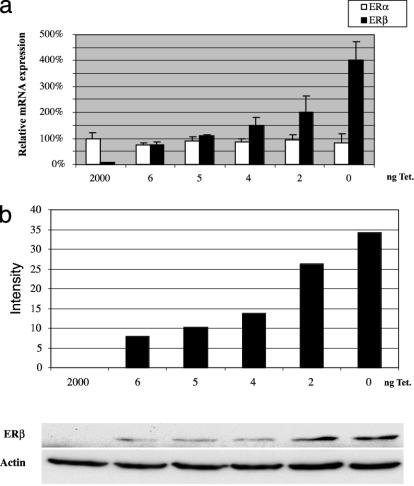
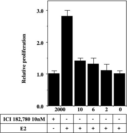
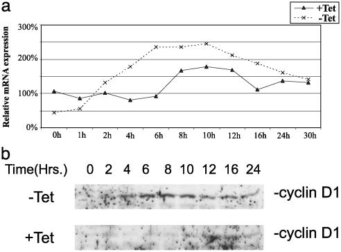
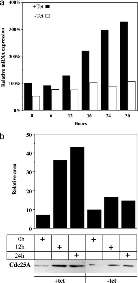
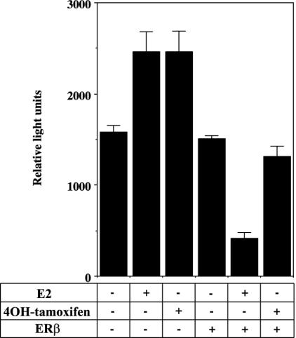
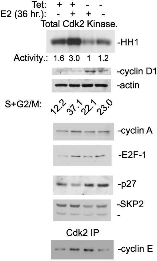
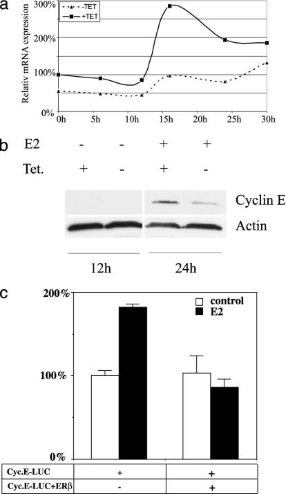
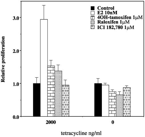
Estrogen receptor (ER) beta counteracts the activity of ERalpha in many systems. In agreement with this, we show in this study that induced expression of ERbeta in the breast cancer cell line T47D reduces 17beta-estradiol-stimulated proliferation when expression of ERbeta mRNA equals that of ERalpha. Induction of ERbeta reduces growth of exponentially proliferating cells with a concomitant decrease in components of the cell cycle associated with proliferation, namely cyclin E, Cdc25A (a key regulator of Cdk2), p45(Skp2) (a key regulator of p27(Kip1) proteolysis), and an increase in the Cdk inhibitor p27(Kip1). We also observed a reduced Cdk2 activity. These findings suggest a possible role for ERbeta in breast cancer and imply that ERbeta-specific ligands may reduce proliferation of ER-positive breast cancer cells through actions on the G(1) phase cell-cycle machinery.


Figures

Fig. 1.
Fig. 1.
Estimation of relative levels of ERα and ERβ using real-time PCR on cDNA prepared from T47D ERβ cells. (a) T47D ERβ cells cultured on six-well plates were treated with different tetracycline concentrations. After 12 h, RNA was prepared, cDNA was synthesized, and real-time PCR was performed by using ERα- and ERβ-specific primers. Each point represents three different treatments. (b) Whole-cell extracts were prepared from 100-mm plates with T47D ERβ cells treated with different concentrations of tetracycline for 12 h. Western blotting was done with 25 μg of protein FLAG antibody to detect tagged ERβ, and β-actin antibody was used as loading control.
Fig. 2.
Fig. 2.
Expression of ERβ inhibits E2-stimulated proliferation. T47D ERβ cells were spread on 24-well plates and synchronized as described. The wells were washed with PBS and medium containing 10 nM E2, 0.5% DCCFBS, and 2,000, 10, 6, 2, or 0 ng of tetracycline/ml. The plates were then placed in an incubator for 5 days, whereupon proliferation assay was performed as described in Materials and Methods.
Fig. 3.
Fig. 3.
ERβ regulates cyclin D1 expression in response to E2 treatment. (a) T47D ERβ cells were spread on six-well plates at a low confluency (40%) and grown as described in Materials and Methods. Tetracycline was removed 12 h before start of treatment with 10 nM E2. Cells was harvested in TRIzol at different time points, and cDNA for real-time PCR was prepared; each point represents an average of two different cDNA preparations. (b) Whole-cell extracts were prepared from synchronized T47D ERβ cells grown on 100-mm plates as described in Materials and Methods. Proteins (50 μg) were separated on SDS/PAGE and electrotransferred to nitrocellulose membrane, and cyclin D1 protein was detected by using antibody directed against cyclin D1 (rabbit polyclonal antibody sc753, Santa Cruz Biotechnology).
Fig. 4.
Fig. 4.
ERβ regulates Cdc25A expression. (a) T47D ERβ cells were spread on six-well plates at a low confluency of 40% and synchronized as described in Materials and Methods. Tetracycline was removed 12 h before start of treatment with 10 nM E2. Cells were harvested in TRIzol at different time points, and cDNA for real-time PCR was prepared. (b) Whole-cell extracts were prepared from synchronized T47D ERβ cells grown on 100-mm plates as described in Materials and Methods. Proteins (50 μg) were separated on SDS/PAGE, electrotransferred to nitrocellulose membrane, and detected with antibody directed against Cdc25A (mouse monoclonal Ab-3, NeoMarkers Fremont, CA). Evaluation of signal strength was done with Bio-Rad chemidoc 1.0 (gel-imaging system).
Fig. 5.
Fig. 5.
Transient transfection of cdc25A promoter into T47D cells. Cdc25A promoter -450 to +126 in pGL3 basic (1 μg) was transfected into normal T47D cells with or without cotransfection with 100 ng of pcDNA3 or Flag 485 ERβ. Transfected cells were treated with 10 nM E2 or 100 nM 4OH-tamoxifen for 24 h before harvest and luciferase assay. Each bar represents an average of measurements from three wells.
Fig. 6.
Fig. 6.
ERβ inhibits Cdk2 activity and prevents reduction of p27Kip1 protein level by reducing expression of p45Skip2. T47D cells with inducible ERβ expression were maintained as an asynchronous proliferating culture. Tetracycline was washed out, and whole-cell extracts were made after 36 h. The proliferative fraction in the culture is reported as S+G2/M.
Fig. 7.
Fig. 7.
ERβ regulates cyclin E expression at mRNA and protein levels. (a) Real-time PCR on cyclin E1 mRNA in T47D cells treated with 10 nM E2 for different times. (b) Western blot using antibody to cyclin E on extracts from T47D cells treated with E2 for 24 h compared to nontreated control. (c) Transient transfection of T47D cells with cyclin E promoter luciferase construct ± transfected pcDNA3 FLAG ERβ.
Fig. 8.
Fig. 8.
ERβ inhibits proliferation using 10 nM E2 or 1 μM solutions of the antiestrogens 4OH-tamoxifen, raloxifene, and ICI182,780. T47D ERβ cells were spread onto 24-well plates and synchronized as described in Materials and Methods. Treatment was started as indicated, the cells were harvested after 5 days, and proliferation assay was performed as described. Each bar represents an average of measurements from six wells.

References

    1. Liu, M. M., Albanese, C., Anderson, C. M., Hilty, K., Webb, P., Uht, R. M., Price, R. H., Jr., Pestell, R. G. & Kushner, P. J. (2002) J. Biol. Chem. 277, 24353-24360. - PubMed
    1. Nelson, H. D., Humphrey, L. L., Nygren, P., Teutsch, S. M. & Allan, J. D. (2002) J. Am. Med. Assoc. 288, 872-881. - PubMed
    1. Lin, C. Q., Singh, J., Murata, K., Itahana, Y., Parrinello, S., Liang, S. H., Gillett, C. E., Campisi, J. & Desprez, P. Y. (2000) Cancer Res. 60, 1332-1340. - PubMed
    1. Strom, A., Arai, N., Leers, J. & Gustafsson, J.-Å. (2000) Oncogene 19, 5951-5953. - PubMed
    1. Dubik, D., Dembinski, T. C. & Shiu, R. P. (1987) Cancer Res. 47, 65117-65121. - PubMed

Publication types

LinkOut - more resources