. 2004 Feb 3;101(5):1141-6.
doi: 10.1073/pnas.0307669100. Epub 2004 Jan 26.

Disruption of Ixodes scapularis anticoagulation by using RNA interference

Affiliations

Disruption of Ixodes scapularis anticoagulation by using RNA interference

Sukanya Narasimhan et al. Proc Natl Acad Sci U S A. .

Abstract

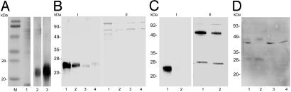
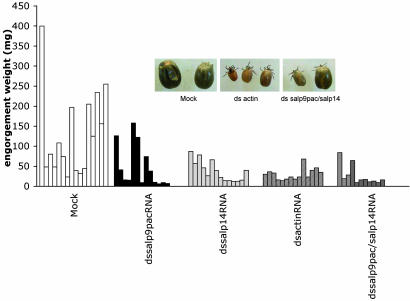
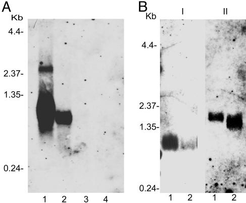
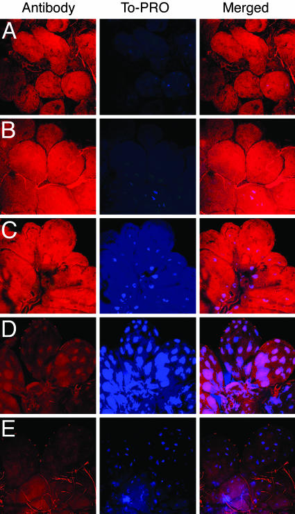
Ixodes scapularis ticks transmit many pathogens, including Borrelia burgdorferi, Anaplasma phagocytophilum, and Babesia microti. Vaccines directed against arthropod proteins injected into the host during tick engorgement could prevent numerous infectious diseases. Salp14, a salivary anticoagulant, poses a key target for such intervention. Salp14 is the prototypic member of a family of potential I. scapularis anticoagulants, expressed and secreted in tick saliva during tick feeding. RNA interference was used to assess the role of Salp14 in tick feeding. Salp14 and its paralogs were silenced, as demonstrated by the reduction of mRNA and protein specific for these antigens. Tick salivary glands lacking Salp14 had reduced anticoagulant activity, as revealed by a 60-80% reduction of anti-factor Xa activity. Silencing the expression of salp14 and its paralogs also reduced the ability of I. scapularis to feed, as demonstrated by a 50-70% decline in the engorgement weights. Because ticks have several anticoagulants, it is likely that the expression of multiple anticoagulants in I. scapularis saliva would have to be ablated simultaneously to abolish tick feeding. These studies demonstrate that RNA interference can silence I. scapularis genes and disrupt their physiologic function in vivo, and they identify vaccine candidates that can alter vector engorgement.
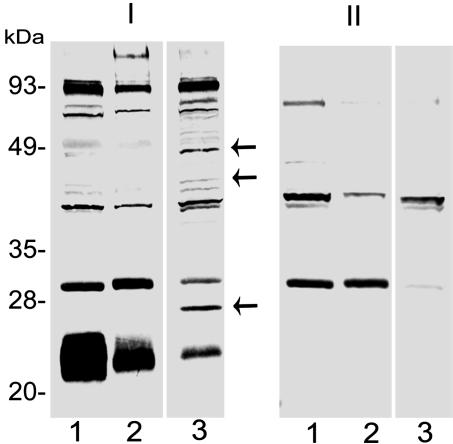

Figures

Fig. 3.
Fig. 3.
Western blot analysis of dsRNA-injected ticks. (A) Untreated adult tick salivary gland extracts (1 μg) separated on an SDS/12% polyacrylamide gel and stained with Coomassie blue (lane 1) or analyzed by immunoblotting with anti-rSalp9pac (lane 2) and anti-rDES-Salp14 (lane 3) antibodies. Lowmolecular-mass protein standards (Bio-Rad) served as markers (lane M). (B) Immunoblotting of protein isolated from the salivary glands of mock-injected (lane 1), actin dsRNA-injected (lane 2), salp14 dsRNA-injected (lane 3), and salp9pac dsRNA-injected (lane 4) ticks probed with anti-rMBP-Salp14 (I) and anti-MBP (II) antibody. (C) Immunoblotting of protein isolated from the salivary glands of mock-injected (lane 1) and salp14/salp9pac dsRNA-injected (lane 2) ticks probed with anti-rMBP-Salp14 (I) and anti-rMBP-Salp25D (II) antibody. (D) Immunoblotting of protein isolated from the salivary glands of mock-injected (lane 1), actin dsRNA-injected (lane 2), salp14 dsRNA-injected (lane 3), and salp9pac dsRNA-injected (lane 4) ticks probed with polyclonal anti-actin antibody.
Fig. 1.
Fig. 1.
Effect of RNAi targeting of Salp14, Salp9pac, and actin on tick feeding. dsRNA complementary to actin, salp14, and salp9pac was injected into the idiosoma of I. scapularis adults, as described in Materials and Methods. The injected ticks (10–15 ticks per experimental group) were allowed to feed, and they were collected after they detached after feeding to repletion. The engorgement weights were recorded and plotted as a series of histograms to assess feeding efficiency. Inset shows that the ticks in the actin dsRNA-injected group appeared pale and were significantly smaller in comparison with other experimental groups.
Fig. 2.
Fig. 2.
Northern blot analysis of dsRNA-injected ticks. Total RNA was isolated from the salivary glands of mock-injected and experimental ticks, and equal amounts were analyzed by Northern blotting, as described in Materials and Methods. (A) Northern blot analysis of RNA isolated from the salivary glands of mock-injected (lane 1), actin dsRNA-injected (lane 2), salp14 dsRNA-injected (lane 3), and salp9pac dsRNA-injected ticks probed with a fluorescein-labeled salp14 DNA fragment (lane 4). (B) Northern blot analysis of RNA isolated from the salivary glands of mock-injected (lane 1) and salp14/salp9pac dsRNA-injected ticks probed with fluorescein-labeled salp14 DNA fragment (I) and fluorescein-labeled salp25D DNA fragment (II).
Fig. 4.
Fig. 4.
Confocal microscopy of dsRNA and mock-injected tick salivary glands. Salivary glands were isolated from mock-injected and salp14/9pac dsRNA-injected ticks, placed on glass slides, and fixed in acetone. Salivary glands were labeled with normal serum, anti-rSalp14 polyclonal antibody, or anti-rSalp25D polyclonal antibody. Binding was visualized by using TRITC-conjugated secondary antibodies, as described in Materials and Methods. Nuclei were counter-stained with To-PRO-3 iodide. Images were collected by using a ×40 objective. Images are shown both in single (Antibody and To-PRO) and merged fluorescent channels. Salivary glands of mock-injected ticks labeled with antibodies to rSalp25D (A) or rSalp14 (C) showed equivalent labeling, as assessed by the TRITC staining of the salivary acini. Salivary glands of salp14/salp9pac dsRNA-injected ticks showed labeling with anti-rSalp25D antibody (B) that was comparable with that observed in salivary glands of mock-injected ticks (A), as judged by TRITC staining. However, salivary glands of salp14/salp9pac dsRNA-injected ticks failed to label with anti-rSalp14 antibody, as evidenced by a significant decrease in the TRITC staining of the salivary glands (D) when compared with mock-injected ticks (C). naïve guinea pig serum showed little or no binding, as visualized by TRITC staining to salivary glands of mock-injected (E) and dsRNA-injected (data not shown) ticks.
Fig. 5.
Fig. 5.
Effect of dsRNA-mediated interference of Salp14 family expression on anticoagulation. (A) aPTT assay. The ability of salivary gland extracts from mockinjected and experimental ticks to alter the coagulation time of human plasma was examined in an aPTT assay, as described in Materials and Methods. Whereas salivary gland extracts of mock-injected ticks prolonged the coagulation time of human plasma by 77 s, salivary gland extracts of experimental ticks prolonged the coagulation time of human plasma by 20 s compared with control. Human plasma incubated with PBS served as a control. Statistical significance was calculated by using Student's t test and is indicated above the mock and experimental groups. (B) Chromogenic assay of factor Xa inhibition. Factor Xa-mediated cleavage of chromogenic substrate (500 pM enzyme/1 mM substrate) was measured in the presence of increasing concentrations of salivary gland extracts (0.5–10 μg) from mock-injected and salp14/salp9pac dsRNA-injected ticks. The ratio of velocities of substrate cleavage in the presence (Vi) and absence (V0) of salivary gland extracts is plotted to show the relative inhibition.
Fig. 6.
Fig. 6.
Disruption of Salp14 family expression and potential secondary effects on the salivary gland proteome. Immunoblotting of protein isolated from the salivary glands of mock-injected (lane 1), actin dsRNA-injected (lane 2), and salp14/salp9pac dsRNA-injected (lane 3) ticks probed with rabbit tick-immune (I) and naïve (II) serum. Arrows indicate proteins that are induced on disruption of Salp14 family expression.

References

    1. Estrada-Pena, A. & Jongejan, F. (1999) Exp. Appl. Acarol. 23, 685–715. - PubMed
    1. Barbour, A. G. & Fish, D. (1993) Science 260, 1610–1616. - PubMed
    1. Telford, S. R., III, Dawson, J. E., Katavolos, P., Warner, C. K., Kolbert, C. P. & Persing, D. H. (1996) Proc. Natl. Acad. Sci. USA 93, 6209–6214. - PMC - PubMed
    1. Mather, T. N., Telford, S. R., III, Moore, S. I. & Spielman, A. (1990) Exp. Parasitol. 70, 55–61. - PubMed
    1. Telford, S. R., III, Armstrong, P. M., Katavolos, P., Foppa, I., Garcia, A. S., Wilson, M. L. & Spielman, A. (1997) Emerg. Infect. Dis. 3, 165–170. - PMC - PubMed

Publication types