

Safety zones for anterior abdominal wall entry during laparoscopy: a CT scan mapping of epigastric vessels
- PMID: 14745325
- PMCID: PMC1356210
- DOI: 10.1097/01.sla.0000109151.53296.07
Safety zones for anterior abdominal wall entry during laparoscopy: a CT scan mapping of epigastric vessels
Abstract
Objective: To determine the efficacy of CT scan in mapping the superior and inferior epigastric vessels, relative to landmarks apparent at laparoscopy.
Summary background data: Trauma to abdominal wall blood vessels occurs in 0.2% to 2% of laparoscopic procedures. Both superficial and deep abdominal wall vessels are at risk. The superficial vessels may be located by transillumination; however, the deep epigastric vessels cannot be effectively located by transillumination and, thus, other techniques should be used to minimize the risk of injury to these vessels.
Methods: Abdominal and pelvic CT images of 100 patients were studied. The location of the superior and inferior epigastric vessels from the midline were determined at five levels, correlated with each other and with the patient age, body mass index, and history of midline laparotomy using Pearson's correlation coefficient and multivariate analysis.
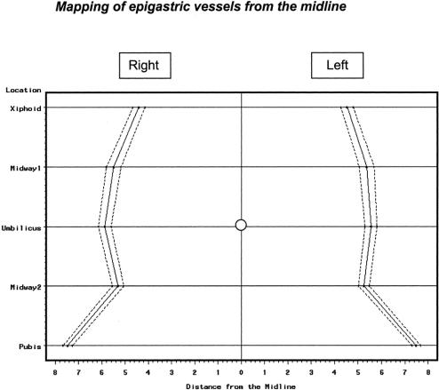
Results: CT scan was successful in mapping the epigastric vessels in 95% of patients. At the xiphoid process level, the superior epigastric vessels (SEA) were 4.41 +/- 0.13 cm from the midline on the right and 4.53 +/- 0.14 cm on the left. Midway between xiphoid and umbilicus, the SEA were 5.50 +/- 0.16 cm on the right of the midline and 5.36 +/- 0.16 cm on the left. At the umbilicus, the epigastric vessels were 5.88 +/- 0.14 cm on the right and 5.55 +/- 0.13 on the left of the midline. Midway between the umbilicus and symphysis pubis, the inferior epigastric (IEA) were 5.32 +/- 0.12 cm on right and 5.25 +/- 0.11 cm on the left. At the symphysis pubis, the IEA were 7.47 +/- 0.10 cm on the right and 7.49 +/- 0.09 cm away from the midline on the left side.
Conclusions: Epigastric vessels are usually located in the area between 4 and 8 cm from the midline. Staying away from this area will determine the safe zone of entry of the anterior abdominal wall.
Figures
References
-
- Zaki H, Penketh R, Newton J. Gynaecological laparoscopy audit: Birmingham experience. Gynecol Endocrinol. 1995;4:251–257.
-
- Aharoni A, Condea A, Leibovitz Z, et al. A comparative study of Foley catheter and suturing to control trocar-induced abdominal wall haemorrhage. Gynecol Endocrinol. 1997;6:31–32.
-
- Vasquez JM. Vascular complications of laparoscopic surgery. J Am Assoc Gynecol Laparosc. 1994;1:163–167. - PubMed
-
- Spitzer M, Golden P, Rehwaldt L, et al. Repair of laparoscopic injury to abdominal wall arteries complicated by cutaneous necrosis. J Am Assoc Gynecol Laparosc. 1996;3:449–452. - PubMed
-
- Hurd WW, Pearl ML, DeLancey JO, et al. Laparoscopic injury of abdominal wall blood vessels: a report of three cases. Obstet Gynecol. 1993;82(4 Pt 2 suppl):673–676. - PubMed
MeSH terms
LinkOut - more resources
Full Text Sources
Medical
