. 2004 Feb;86(2):1002-11.
doi: 10.1016/S0006-3495(04)74175-2.

Response of equatorial x-ray reflections and stiffness to altered sarcomere length and myofilament lattice spacing in relaxed skinned cardiac muscle

Affiliations

Response of equatorial x-ray reflections and stiffness to altered sarcomere length and myofilament lattice spacing in relaxed skinned cardiac muscle

Donald A Martyn et al. Biophys J. 2004 Feb.

Abstract

Low angle x-ray diffraction measurements of myofilament lattice spacing (D(1,0)) and equatorial reflection intensity ratio (I(1,1)/I(1,0)) were made in relaxed skinned cardiac trabeculae from rats. We tested the hypothesis that the degree of weak cross-bridge (Xbr) binding, which has been shown to be obligatory for force generation in skeletal muscle, is modulated by changes in lattice spacing in skinned cardiac muscle. Altered weak Xbr binding was detected both by changes in I(1,1)/I(1,0) and by measurements of chord stiffness (chord K). Both measurements showed that, similar to skeletal muscle, the probability of weak Xbr binding at 170-mM ionic strength was significantly enhanced by lowering temperature to 5 degrees C. The effects of lattice spacing on weak Xbr binding were therefore determined under these conditions. Changes in D(1,0), I(1,1)/I(1,0), and chord K by osmotic compression with dextran T500 were determined at sarcomere lengths (SL) of 2.0 and 2.35 micro m. At each SL increasing [dextran] caused D(1,0) to decrease and both I(1,1)/I(1,0) and chord K to increase, indicating increased weak Xbr binding. The results suggest that in intact cardiac muscle increasing SL and decreasing lattice spacing could lead to increased force by increasing the probability of initial weak Xbr binding.


Figures
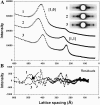
FIGURE 1
FIGURE 1
(A) Representative intensity profiles versus lattice spacing (Å) of the equatorial regions of the low angle x-ray diffraction patterns from a skinned cardiac trabeculae are shown. The dominant integrated peaks attributed to [1,0] and [1,1] reflections on the equator are shown. The profiles were obtained from a single preparation and traces 2 and 3 have been displaced 500 and 1000 units vertically, respectively, for clarity. Profiles were obtained at 25°C and 170-mM μ (trace 1), 5°C and 170-mM μ (trace 2), and 5°C and 50-mM μ (trace 3). Data were analyzed by fitting the intensity scans with the program Peak Fit to determine the positions of the centroids of the peaks and the values of the integrated intensities of each peak. The residuals to the fits of traces 1–3 are shown in B.
FIGURE 2
FIGURE 2
The effects of temperature on lattice spacing (A) and equatorial intensity ratio (B) are illustrated. SL was 2.35 μm and μ was 170 mM. Data (means ± SE) at each temperature was obtained from the same seven preparations.
FIGURE 3
FIGURE 3
Chord stiffness (K) was measured in skinned cardiac preparations under relaxing conditions at 5°C (open symbols) and 25°C (solid symbols) at 2.0 (A) and 2.35 μm (C). Constant amplitude stretches (+0.15% fiber length) were applied at different rates to relaxed (pCa ∼9) skinned trabeculae and the corresponding stiffness (force/stretch amplitude) plotted against the rate of stretch. B and D illustrate the difference in chord K for the corresponding data in A and C, respectively. Data (means ± SE) were obtained from five preparations in A and B and seven preparations in C and D. The dotted lines in B and D are for reference only. For reference, chord K in each preparation is expressed as a fraction of the maximal value of stiffness obtained in a maximal Ca2+-activation (pCa 4.5) contraction at 15°C.
FIGURE 4
FIGURE 4
Lattice spacing (D1,0) was measured at 2.0-μm (•) and 2.35-μm (○) SL in relaxed skinned cardiac trabeculae. Temperature was 5°C. At each SL, lattice spacing was varied independently by osmotic compression with 0–5% dextran T500 in the bathing solutions. Data (means ± SE) were obtained from six preparations. The solid vertical bars in A illustrate the change of lattice spacing between 2.0-μm (top) and 2.35-μm (bottom) SL in intact cardiac trabeculae from rat from Konhilas et al. (2002).
FIGURE 5
FIGURE 5
The effects of myofilament lattice compression with dextran T500 (% w/v) on the intensity ratio (I1,1/I1,0) were determined at 5°C and 170-mM μ. Data (means ± SE) were obtained at 2.0-μm (▴) and 2.35-μm (▵) SL from the same fibers as in Fig. 4. The number next to each symbol is the [dextran] (w/v) used to obtain the corresponding D1,0 value. The values of D1,0 for each [dextran] corresponds to the data in Fig. 4.
FIGURE 6
FIGURE 6
For comparison to corresponding changes in the I1,1/I1,0 equatorial intensity ratio, chord K was measured at 2.0 (A) and 2.35 (C) μm at 5°C and 170-mM μ relaxing solution in the presence (solid symbols) and absence (open symbols) of 5% dextran. The corresponding difference in stiffness measured at each rate of stretch for individual fibers is illustrated in C and D, respectively. Data were obtained from five to seven preparations. The dotted lines in B and D are for reference only.

References

    1. Brenner, B. 1986. The cross-bridge cycle in muscle. Mechanical, biochemical, and structural studies on single skinned rabbit psoas fibers to characterize cross-bridge kinetics in muscle for correlation with the actomyosin-ATPase in solution. Basic Res. Cardiol. 81:1–15. - PubMed
    1. Brenner, B., J. M. Chalovich, L. E. Greene, E. Eisenberg, and M. Schoenberg. 1986. Stiffness of skinned rabbit psoas fibers in MgATP and MgPPi solution. Biophys. J. 50:685–691. - PMC - PubMed
    1. Brenner, B., T. Kraft, G. DasGupta, and E. Reisler. 1996a. Cross-bridge binding to actin and force generation in skinned fibers of the rabbit psoas muscle in the presence of antibody fragments against the N-terminus of actin. Biophys. J. 70:48–56. - PMC - PubMed
    1. Brenner, B., M. Schoenberg, J. M. Chalovich, L. E. Greene, and E. Eisenberg. 1982. Evidence for cross-bridge attachment in relaxed muscle at low ionic strength. Proc. Natl. Acad. Sci. USA. 79:7288–7291. - PMC - PubMed
    1. Brenner, B., S. Xu, J. Chalovich, and L. C. Yu. 1996b. Radial equilibrium lengths of actomyosin cross-bridges in muscle. Biophys. J. 71:2751–2758. - PMC - PubMed

Publication types

MeSH terms

LinkOut - more resources