. 2004 Feb;86(2):1160-8.
doi: 10.1016/S0006-3495(04)74190-9.

Surface functionalized cationic lipid-DNA complexes for gene delivery: PEGylated lamellar complexes exhibit distinct DNA-DNA interaction regimes

Affiliations

Surface functionalized cationic lipid-DNA complexes for gene delivery: PEGylated lamellar complexes exhibit distinct DNA-DNA interaction regimes

Ana Martin-Herranz et al. Biophys J. 2004 Feb.

Abstract

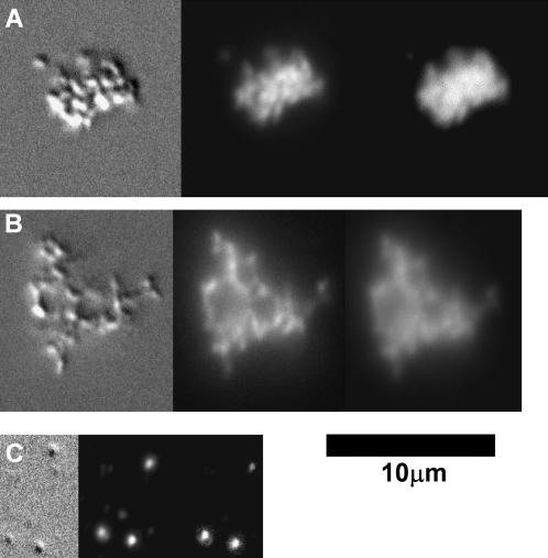
Cationic lipid-DNA (CL-DNA) complexes are abundantly used in nonviral gene therapy clinical applications. Surface functionality is the next step in developing these complexes as competent, target-specific gene carriers. Poly(ethylene glycol) (PEG) is the natural choice to serve as a protective coat or act as a tether for a specific ligand on the surface of these complexes due to its biocompatibility and ability to convey stealth-like properties. Understanding the effect of PEG on the internal structure and surface properties of CL-DNA complexes is essential in developing vectors with more complex derivatives of PEG, such as Arg-Gly-Asp (RGD)-based peptide-PEG-lipids. We report on x-ray diffraction studies to probe the internal structure of CL-DNA complexes consisting of a ternary mixture of cationic lipids, neutral lipids, and PEG-lipids. The PEG-coated complexes are found to exhibit a structure consistent with the lamellar phase. In addition, three distinct DNA interchain interaction regimes were found to exist, due to a), repulsive long-range electrostatic forces; b), short-range repulsive hydration forces; and c), novel polymer-induced depletion attraction forces in two dimensions. Optical microscopy and reporter gene assays further demonstrate the incorporation of the PEG-lipids into the lamellar CL-DNA complexes under biologically relevant conditions, revealing surface modification. Both techniques show that PEG-lipids with a polymer chain of molecular weight 400 do not provide adequate shielding of the PEGylated CL-DNA complexes, whereas PEG-lipids with a polymer chain of molecular weight 2000 confer stealth-like properties. This surface functionalization is a crucial initial step in the development of competent vectors for in vivo systemic gene delivery and suggests that a second type of surface functionality can be added specifically for targeting by the incorporation of peptide-PEG-lipids.


Figures

FIGURE 1
FIGURE 1
Chemical structures and names of the PEG-lipids used in this study.
FIGURE 2
FIGURE 2
Synchrotron SAXS scans for isoelectric PEG-CL-DNA complexes at fixed MDOTAP = 0.33 as a function of increasing concentration of (A) PEG4002+-lipid; (B) PEG20002+-lipid; (C) PEG400-lipid; and (D) PEG2000-lipid. A reference scan of a CL-DNA complex without PEG-lipid is shown at the bottom (shaded plot). The dotted line marks the DNA correlation peak for the reference scan, and the arrows mark the DNA correlation peaks for the others. With the exception of PEG400-lipid, the addition of PEG-lipid leads to a decrease in the DNA spacing compared to the reference scan.
FIGURE 3
FIGURE 3
Interaxial DNA spacing, dDNA, results for CL-DNA complexes incorporating PEG4002+-lipid at various MDOTAP values, obtained from SAXS data. (A) Decrease of the DNA interaxial distance, dDNA, with increasing molar fraction of PEG4002+-lipid at different fixed molar fractions of DOTAP. (B) The experimental dDNA values represented in A are plotted against the membrane charge density (symbols as in A). The solid circles represent the experimental dDNA for the reference system (DOTAP/DOPC without PEG-lipid) and increasing MDOTAP (from left to right, 0.27, 0.33, 0.38, 0.43, 0.53, 0.63, and 0.82). The continuous line represents the predicted dDNA, calculated as described in the text. The data for complexes with added PEG4002+-lipid follow this curve exactly, implying that PEG4002+-lipid behaves as a cationic colipid. The inset shows that no further condensation of DNA occurs upon addition of neutral PEG400-lipid at fixed MDOTAP = 0.33. This verifies the domination of electrostatic interactions.
FIGURE 4
FIGURE 4
Interaxial DNA spacing, dDNA, results for CL-DNA complexes incorporating PEG20002+-lipid at various MDOTAP values, obtained from SAXS data. (A) Decrease of the DNA interaxial distance dDNA with increasing molar fraction of PEG20002+-lipid at different molar fractions of DOTAP. (B) Experimental dDNA data from A are collected and plotted against the membrane charge density (symbols as in A). The continuous line represents the predicted dDNA, calculated as described in the text. The data for complexes with added PEG20002+-lipid show a DNA spacing smaller than the predicted value, implying enhanced DNA condensation. The inset shows similar behavior for the inclusion of neutral PEG2000-lipid and PEG20002+-lipid with increasing concentration of PEG-lipid at fixed MDOTAP = 0.33. This verifies the domination of polymer-induced depletion attraction interactions.
FIGURE 5
FIGURE 5
Schematic of a lamellar CL-DNA complex containing long-chain PEG-lipids. (A) The presence of the polymer is forcing the DNA rods closer together than predicted by electrostatics (depletion attraction force). (B) Cross section of PEG-CL-DNA complex with DNA rich domains (green) and polymer rich domains (blue) between lipid bilayers (gray). (C) Enlarged view of PEG-CL-DNA complex showing internal phase separation as well as the outer shell of polymer chains.
FIGURE 6
FIGURE 6
(A–C) Microscopy images of positive (charge ratio = 2:8) DOTAP/DOPC CL-DNA complexes at MDOTAP = 0.33 with plasmid DNA in the presence of the cell culture medium DMEM, taken in differential interference contrast (left), lipid fluorescence (center), and DNA fluorescence modes. The images show complexes prepared (A) without PEG-lipid, (B) with 10% PEG400-lipid, and (C) with 10% PEG2000-lipid. The aggregation of particles that is seen when no PEG-lipid or 10% PEG400-lipid are added is eliminated by the addition of 10% PEG2000-lipid, indicating steric stabilization by the long-chain polymer.
FIGURE 7
FIGURE 7
Transfection efficiency in fibroblast L-cells as a function of increasing molar fraction of PEG-lipid (MPEG-lipid) added to a DOTAP/DOPC liposome mixture. (A) When initial TE is low, in the low σM regime at MDOTAP = 0.30, addition of PEG400-lipid (open squares) does not effect TE whereas adding the PEG4002+-lipid (open circles) increases TE due to an increase in the overall σM of the DOTAP/DOPC/PEG-lipid system. The PEG2000-lipids (solid squares) and PEG20002+-lipids (solid circles) both decrease TE, due to the presence of the long PEG chains on the CL-DNA surface. (B) In the high σM regime at MDOTAP = 0.80, TE is already at a maximum and addition of PEG4002+-lipid does not aid transfection. Adding PEG20002+-lipid or PEG2000-lipid leads to a decrease in TE by nearly two orders of magnitude with only 6% added PEG-lipid. These findings show that the PEG2000-lipid and PEG20002+-lipid are coating the CL-DNA particles. As a reference, typical TE for naked DNA is on the order of 300,000 RLU/mg protein.

References

    1. Allen, T. M. 1994. Long-circulating (sterically stabilized) liposomes for targeted drug-delivery. Trends Pharmacol. Sci. 15:215–220. - PubMed
    1. Bradley, A. J., D. V. Devine, S. M. Ansell, J. Janzen, and D. E. Brooks. 1998. Inhibition of liposome-induced complement activation by incorporated poly(ethylene glycol) lipids. Arch. Biochem. Biophys. 357:185–194. - PubMed
    1. Bruinsma, R., and J. Mashl. 1998. Long-range electrostatic interaction in DNA cationic lipid complexes. Europhys. Lett. 41:165–170.
    1. Chesnoy, S., and L. Huang. 2000. Structure and function of lipid-DNA complexes for gene delivery. Annu. Rev. Biophys. Biomol. Struct. 29:27–47. - PubMed
    1. Devanand, K., and J. C. Selser. 1991. Asymptotic behavior and long-range interactions in aqueous solutions of poly(ethylene oxide). Macromolecules. 24:5943–5947.

Publication types

LinkOut - more resources