. 2004 Feb;78(4):1918-27.
doi: 10.1128/jvi.78.4.1918-1927.2004.

The K1 protein of Kaposi's sarcoma-associated herpesvirus activates the Akt signaling pathway

Affiliations

The K1 protein of Kaposi's sarcoma-associated herpesvirus activates the Akt signaling pathway

Christine C Tomlinson et al. J Virol. 2004 Feb.

Abstract

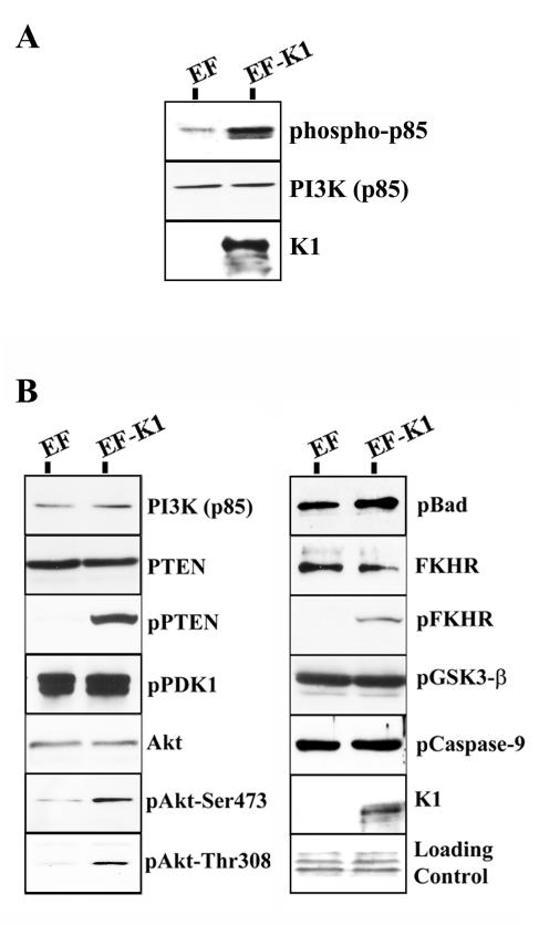
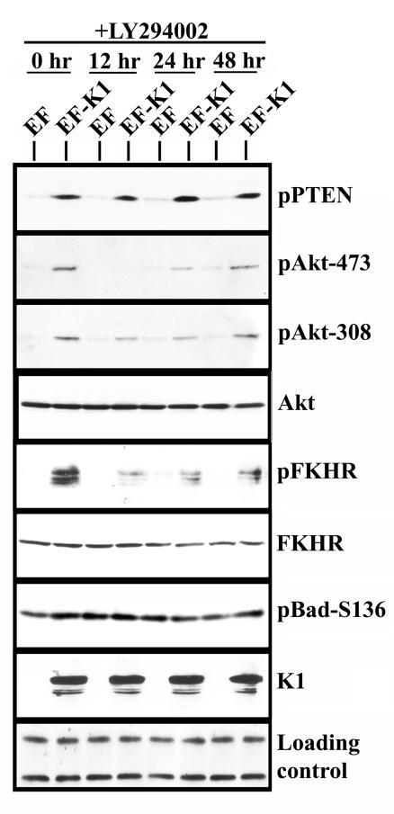
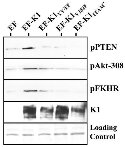
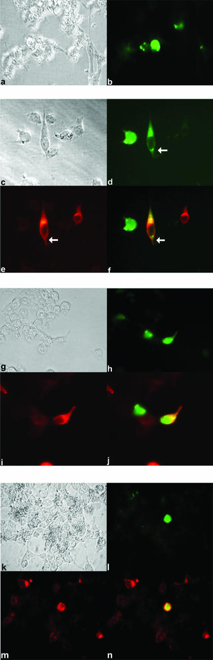
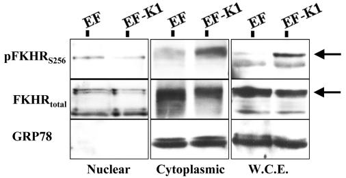
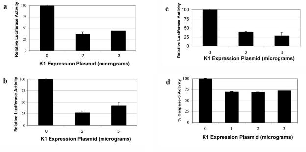
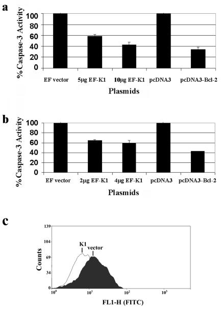
Kaposi's sarcoma-associated herpesvirus (KSHV) has been implicated in Kaposi's sarcoma, as well as in primary effusion lymphoma and multicentric Castleman's disease. The K1 protein of KSHV has been shown to induce cellular transformation and focus formation and to deregulate B-lymphocyte signaling pathways by functionally mimicking the activated B-cell receptor complex. Here we show that expression of K1 in B lymphocytes targets the phosphatidylinositol-3 kinase pathway, leading to the activation of the Akt kinase and the inhibition of the phosphatase PTEN. We also demonstrate that activation of Akt by the K1 protein leads to the phosphorylation and inhibition of members of the forkhead (FKHR) transcription factor family, which are key regulators of cell cycle progression and apoptosis. We demonstrate that K1 can inhibit apoptosis induced by the FKHR proteins and by stimulation of the Fas receptor. Our observations suggest that the K1 viral protein promotes cell survival pathways and may contribute to KSHV pathogenesis by preventing virally infected cells from undergoing apoptosis prematurely.


Figures

FIG. 1.
FIG. 1.
K1 activates the Akt pathway in B cells. (A) BJAB cells were transfected with the EF or EF-K1 expression plasmid as indicated. Cells were harvested, lysed, and subjected to immunoprecipitation with an anti-p85 antibody to pull down PI3K. A Western blot analysis was performed on the immunoprecipitate reactions, using an anti-phospho-Tyr-HRP antibody to detect the phosphorylated p85 subunit of PI3K. (B) BJAB cells were transfected with empty vector (EF) or a K1 expression vector (EF-K1). Equal amounts of proteins were separated by SDS-PAGE, transferred to nitrocellulose, and probed with the indicated antibodies. Ponceau S staining was used to evaluate equivalent loading of the samples. K1 expression was determined by probing with an anti-Flag antibody.
FIG. 2.
FIG. 2.
Activation of Akt by K1 is inhibited by LY294002. EF- or EF-K1-transfected BJAB cells were incubated for 12, 24, or 48 h in the presence of 10 μM LY294002. Lysates were subjected to Western blot analysis and probed with the indicated antibodies.
FIG. 3.
FIG. 3.
Activation of the PI3K/Akt pathway by K1 mutants. BJAB cells were electroporated with the indicated expression plasmids. Cells were harvested as previously described. Equal amounts of proteins were separated by SDS-PAGE, transferred to nitrocellulose, and probed with the indicated antibodies. The individual panels, from top to bottom, represent Western blots probed with an anti-pPTEN, anti-pAKT (T308), and anti-pFKHR antibody, respectively. Ponceau S staining was used to evaluate equivalent loading of the samples. K1 expression was determined by probing with an anti-Flag antibody.
FIG. 4.
FIG. 4.
K1 promotes cytoplasmic localization of FKHR. (a) 293 cells were transfected with empty vector (EF) and FKHR-GFP. Transfected cells were fixed and examined by bright-field microscopy. (b) The same cells as in panel a examined for expression and distribution of FKHR-GFP (green) by immunofluorescence microscopy. Cells expressing FKHR-GFP appear to be undergoing apoptosis based on their rounded morphology. (c) Cells were transfected with EF-K1 and FKHR-GFP expression plasmids. (d) The same cells as in panel c fixed and examined for expression and distribution of FKHR-GFP (green). (e) The same cells as depicted in panels c and d stained for expression of K1 (red) and examined by immunofluorescence microscopy. (f) Merged image of the same cells shown in panels c, d, and e. Yellow represents the colocalization of K1 (red) and FKHR-GFP (green). The white arrow indicates a cell that is coexpressing K1 and FKHR-GFP. The adjacent cell to its left expresses FKHR-GFP, but not K1, and appears to be undergoing apoptosis. (g) Cells were transfected with EF-K1 and the mutant FKHRAAA-GFP expression plasmid. (h) The same cells shown in panel g analyzed for expression and distribution of FKHRAAA-GFP (green). Based on their morphology, these cells appear to be undergoing apoptosis. (i) Cells were examined for expression of K1 (red) by immunofluorescence microscopy. (j) Merged image of the same cells shown in panels g, h, and i. Yellow represents the colocalization of FKHRAAA-GFP (green) and K1 (red). (k) Cells were transfected with FKHR-GFP and the EF-K1ITAM− expression plasmid. Transfected cells were fixed and examined by bright-field microscopy. (l) The same cells shown in panel k analyzed for expression and distribution of FKHR-GFP (green). (m) Cells were examined for expression of the K1ITAM− mutant (red). (n) Merged image of the same cells shown in panels k, l, and m. Yellow represents the colocalization of FKHR-GFP (green) and the K1ITAM− mutant (red).
FIG. 5.
FIG. 5.
Phospho-FKHR is retained in the cytoplasm of K1-expressing B cells. BJAB cells were transfected with EF or EF-K1, and the whole-cell extract (W.C.E.) was fractionated into a nuclear and a cytoplasmic fraction. Equal amounts of cytoplasmic and nuclear fractions from each sample were subjected to Western blot analysis with the indicated antibodies. Left panels, nuclear fraction; middle panels, cytoplasmic fraction; right panels, whole-cell extract. GRP78, a cytoplasmic protein, was used as a marker to assess the purity of the nuclear and cytoplasmic fractions. The arrows point to the FKHR-specific band.
FIG. 6.
FIG. 6.
K1 represses forkhead-regulated promoters and protects cells from FKHR-mediated apoptosis. (a) 293 cells were transfected with 0, 2, or 3 μg of EF-K1 or EF empty vector and 3 μg of a 3XFHRE-luciferase plasmid. A β-Gal construct was also cotransfected to normalize for transfection efficiency. At 48 h posttransfection, cells were lysed and assayed for luciferase expression. Luciferase activity in the absence of K1 was set to 100%, and relative luciferase activities in the presence of different amounts of the K1 expression plasmid were calculated as percentages of this luciferase activity. Error bars represent variations from the means. (b) Cells were transfected similar to the case for panel a, except that anti-Fas antibody was used to stimulate apoptosis with the FHRE-luciferase promoter. Luciferase activity in the absence of K1 and the presence of anti-Fas antibody was set to 100%, and relative luciferase activities in the presence of different amounts of the K1 expression plasmid were calculated as percentages of this luciferase activity. Error bars represent variations from the means. (c) 293 cells were transfected with 0, 2, or 3 μg of EF-K1 or EF empty vector and 3 μg of a 3XIRS-luciferase plasmid. Luciferase activity in the absence of K1 was set to 100%, and relative luciferase activities in the presence of different amounts of the K1 expression plasmid were calculated as percentages of this luciferase activity. Error bars represent variations from the means. (d) Cells were transfected with 0, 1, 2, or 3 μg of EF-K1 or EF empty vector and pCDNA3-FKHR. At 48 h posttransfection, cells were lysed and assayed for caspase-3 activity. Caspase-3 activity in the absence of K1 was set to 100% activity, and relative caspase-3 activities in the presence of different amounts of the K1 expression plasmid were calculated as percentages of this caspase-3 activity. Error bars represent variations from the means.
FIG. 7.
FIG. 7.
K1 protects cells from Fas-mediated apoptosis. (a) KSHV-negative BJAB cells were transfected with 0, 5, or 10 μg of EF-K1 or EF empty vector or pcDNA3 or pcDNA3-Bcl-2 expression plasmid by electroporation. At 48 h posttransfection, cells were stimulated with 1 μg of anti-Fas antibody/ml for 24 h. Cells were lysed and assayed for caspase-3 activity. Caspase-3 activity in the absence of K1 was set to 100%, and relative caspase-3 activities in the presence of different amounts of the K1 expression plasmid were calculated as percentages of this caspase-3 activity. Error bars represent variations from the means. (b) KSHV-positive BCBL-1 cells were transfected with 0, 2, or 4 μg of EF-K1 or EF vector or pcDNA3 or pcDNA3-Bcl-2 expression plasmid by using GenePorter 2 reagent. At 48 h posttransfection, cells were stimulated with 1 μg of anti-Fas antibody/ml for 24 h. Cells were lysed and assayed for caspase-3 activity. Caspase-3 activity in the absence of K1 was set to 100%, and relative caspase-3 activities in the presence of different amounts of the K1 expression plasmid were calculated as percentages of this caspase-3 activity. Error bars represent variations from the means. (c) BJAB cells were transfected with the EF or EF-K1 expression plasmid. Cells were treated with anti-Fas antibody to simulate Fas receptor-dependent apoptosis. Twenty-four hours later, a TUNEL assay was performed. Cells were stained for fragmented DNA by enzymatically labeling the nicked ends with FITC-conjugated dUTP and were assayed by flow cytometry. A total of 14.86% of cells transfected with EF vector alone were FITC or TUNEL positive, while only 7.5% of cells transfected with EF-K1 plasmid were FITC or TUNEL positive.

References

    1. Alam, M. K., S. Davison, N. Siddiqui, J. D. Norton, and J. J. Murphy. 1997. Ectopic expression of Bcl-2, but not Bcl-xL, rescues Ramos B cells from Fas-mediated apoptosis. Eur. J. Immunol. 27:3485-3491. - PubMed
    1. Alessi, D. R., S. R. James, C. P. Downes, A. B. Holmes, P. R. Gaffney, C. B. Reese, and P. Cohen. 1997. Characterization of a 3-phosphoinositide-dependent protein kinase which phosphorylates and activates protein kinase B alpha. Curr. Biol. 7:261-269. - PubMed
    1. Andjelkovic, M., D. R. Alessi, R. Meier, A. Fernandez, N. J. Lamb, M. Frech, P. Cron, P. Cohen, J. M. Lucocq, and B. A. Hemmings. 1997. Role of translocation in the activation and function of protein kinase B. J. Biol. Chem. 272:31515-31524. - PubMed
    1. Bais, C., A. Van Geelen, P. Eroles, A. Mutlu, C. Chiozzini, S. Dias, R. L. Silverstein, S. Rafii, and E. A. Mesri. 2003. Kaposi's sarcoma associated herpesvirus G protein-coupled receptor immortalizes human endothelial cells by activation of the VEGF receptor-2/ KDR. Cancer Cell 3:131-143. - PubMed
    1. Beitz, L. O., D. A. Fruman, T. Kurosaki, L. C. Cantley, and A. M. Scharenberg. 1999. SYK is upstream of phosphoinositide 3-kinase in B cell receptor signaling. J. Biol. Chem. 274:32662-32666. - PubMed

Publication types

MeSH terms