

doi: 10.1128/jvi.78.4.2131-2136.2004.
Distribution of hydrophobic residues is crucial for the fusogenic properties of the Ebola virus GP2 fusion peptide
Affiliations
- PMID: 14747578
- PMCID: PMC369453
- DOI: 10.1128/jvi.78.4.2131-2136.2004
Item in Clipboard
Distribution of hydrophobic residues is crucial for the fusogenic properties of the Ebola virus GP2 fusion peptide
J Virol.
2004 Feb.
Abstract
The lipid-destabilizing properties of the N-terminal domain of the GP2 of Ebola virus were investigated. Our results suggest that the domain of Ebola virus needed for fusion is shorter than that previously reported. The fusogenic properties of this domain are related to its oblique orientation at the lipid/water interface owing to an asymmetric distribution of the hydrophobic residues when helical.
Figures
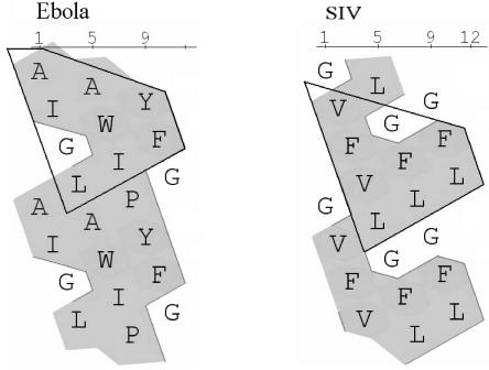
Hydrophobic cluster analysis of the tilted peptide of SIV (residues 1 to 11) and the similar domain in Ebola virus (residues 25 to 35). In this view, the sequence is written on a cylinder that is unrolled and duplicated (11). The hydrophobic amino acids are shaded.
Lowest energy position of the different peptides after IMPALA calculations (α) and representation of the molecular hydrophobicity potential (β). Hydrophobic and hydrophilic envelopes are orange and green, respectively. Mid plane, bilayer center (z = 0); first upper (bottom), lipid acyl chain/polar headgroups interface at 13.5Å from the center; second upper (bottom) plane, lipid/water interface (Z = 18 Å). The N to C orientation was shown. (A) Wild type; (B) PM; (C) TMI; (D) TMII.
Time span of lipid mixing of phosphatidylcholine, phosphatidylethanolamine, sphingomyelin, and cholesterol large unilamellar vesicles (pH 6) induced by wild-type peptide (▪) and SIV peptide (×). The peptide/lipid molar ratio is 0.2 for Ebola virus peptide and 0.02 for the SIV peptide. Peptides, dissolved in TFE, were added to a mixture of R18-labeled and unlabeled large unilamellar vesicles (1:4, wt/wt). The increase in R18 relative fluorescence due to probe dilution is monitored at room temperature. In a control experiment, the same volume of TFE alone (•) is added to the liposome mixture.
Core mixing of phosphatidylcholine, phosphatidylethanolamine, sphingomyelin, and cholesterol large unilamellar vesicles (pH 6) induced by 0.5% Triton X-100 and 10 mM EDTA (▴), TFE alone (▪), wild-type peptide (×; R = 1.2); SIV peptide (•; R = 0.2). R is the peptide/lipid molar ratio. Peptides dissolved in TFE are added to a mixture of calcein-, Co2+-, and EDTA-containing vesicles mixed at a 1:1 weight ratio. The calcein fluorescence is monitored at 520 nm at room temperature as a function of time.
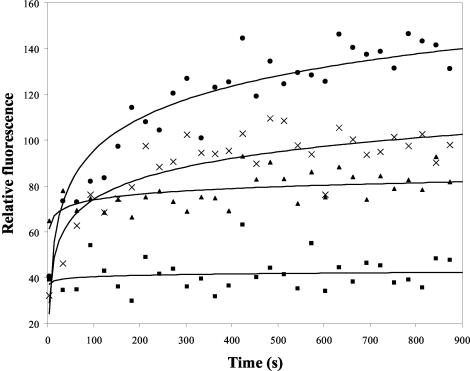
Time course of lipid mixing of phosphatidylcholine, phosphatidylethanolamine, sphingomyelin, and cholesterol large unilamellar vesicles (pH 6) induced by wild-type peptide (•), PM peptide (▪), TMI peptide (×), and TMII peptide (▴). The peptide/lipid molar ratio is 0.2. Peptides dissolved in TFE were added to a mixture of R18-labeled and unlabeled vesicles (1:4 wt/wt). The increase in R18 relative fluorescence due to probe dilution is monitored at room temperature. In a control experiment, the same volume of TFE alone is added to the liposome mixture; this curve has been subtracted from that of the peptides.
References
-
- Atherton, E., C. J. Logan, and R. C. Sheppard. 1981. Peptide synthesis. II. Procedures for solid-phase synthesis by N-fluorenylmethoxycarbonylamino-acids on polyamide supports: synthesis of substance P and of acyl carrier protein 65-74 decapeptide. J. Chem. Soc. Perkin Trans. I 1:538.
-
- Bradshaw, J. P., M. J. Darkes, T. A. Harroun, J. Katsaras, and R. M. Epand. 2000. Oblique membrane insertion of viral fusion peptide probed by neutron diffraction. Biochemistry 39:6581-6585. - PubMed
-
- Brasseur, R. 1991. Differentiation of lipid-associating helices by use of three-dimensional molecular hydrophobicity potential calculations. J. Biol. Chem. 266:16120-16127. - PubMed
-
- Brasseur, R., T. Pillot, L. Lins, J. Vandekerckhove, and M. Rosseneu. 1997. Peptides in membranes: tipping the balance of membrane stability. Trends Biochem. Sci. 22:167-171. - PubMed
-
- Brasseur, R. 2000. Tilted peptides: a motif for membrane destabilization (hypothesis). Mol. Membr. Biol. 17:31-40. - PubMed
Publication types
MeSH terms
Substances
LinkOut - more resources
Full Text Sources
Medical
