. 2004 Feb;24(4):1464-9.
doi: 10.1128/MCB.24.4.1464-1469.2004.

Kinase RIP3 is dispensable for normal NF-kappa Bs, signaling by the B-cell and T-cell receptors, tumor necrosis factor receptor 1, and Toll-like receptors 2 and 4

Affiliations

Kinase RIP3 is dispensable for normal NF-kappa Bs, signaling by the B-cell and T-cell receptors, tumor necrosis factor receptor 1, and Toll-like receptors 2 and 4

Kim Newton et al. Mol Cell Biol. 2004 Feb.

Abstract

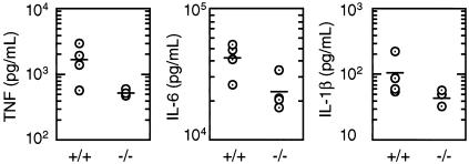
RIP3 is a member of the RIP kinase family. It is expressed in the embryo and in multiple adult tissues, including most hemopoietic cell lineages. Several studies have implicated RIP3 in the regulation of apoptosis and NF-kappa B signaling, but whether RIP3 promotes or attenuates activation of the NF-kappa B family of transcription factors has been controversial. We have generated RIP3-deficient mice by gene targeting and find RIP3 to be dispensable for normal mouse development. RIP3-deficient cells showed normal sensitivity to a variety of apoptotic stimuli and were indistinguishable from wild-type cells in their ability to activate NF-kappa B signaling in response to the following: human tumor necrosis factor (TNF), which selectively engages mouse TNF receptor 1; cross-linking of the B- or T-cell antigen receptors; peptidoglycan, which activates Toll-like receptor 2; and lipopolysaccharide (LPS), which stimulates Toll-like receptor 4. Consistent with these observations, RIP3-deficient mice exhibited normal antibody production after immunization with a T-dependent antigen and normal interleukin-1 beta (IL-1 beta), IL-6, and TNF production after LPS treatment. Thus, we can exclude RIP3 as an essential modulator of NF-kappa B signaling downstream of several receptor systems.


Figures

FIG. 1.
FIG. 1.
Generation of RIP3-deficient mice. (A) Exons 1 to 3 encoding amino acids 1 to 158 of mouse RIP3 were replaced with a PGK-neo selection cassette flanked by loxP sites. (B) Southern blot analysis of genomic DNA from wild-type (+/+), heterozygous (+/−), and homozygous (−/−) rip3 mutant mice. NheI-digested DNAs were hybridized to probe A, which binds sequence that is 5′ of the targeting construct (upper panel). The 4.3- and 6.2-kb DNA fragments correspond to the wild-type and mutant rip3 alleles, respectively. XhoI-digested DNAs were hybridized to probe B, which binds sequence that is 3′ of the targeting construct (lower panel). The 6.5- and 4.3-kb DNA fragments correspond to the wild-type and mutant rip3 alleles, respectively. (C) Western blot analysis of RIP3 protein in embryo fibroblasts (MEFs), purified B and T lymphocytes, and bone marrow-derived macrophages from rip3+/+, rip3+/−, and rip3−/− mice. Blots were probed with a rabbit polyclonal antibody that recognizes amino acids 473 to 486 of mouse RIP3 (upper panel) and then with a mouse monoclonal antibody against β-actin (lower panel).
FIG. 2.
FIG. 2.
Lymphocytes, natural killer cells, and macrophages develop normally in the absence of RIP3. Flow cytometric analysis of cells in the thymus, lymph nodes, bone marrow, and spleen of 8-week-old wild-type (+/+) and RIP3-null (−/−) mice. Numbers indicate the percentage of cells within each quadrant or region. Dot plots are representative of six mice of each genotype.
FIG.3.
FIG.3.
RIP3 is not essential for lymphocyte proliferation or antibody production. (A) Purified spleen B cells from wild-type (+/+; filled circles) and RIP3-deficient (−/−; open circles) mice were stimulated with 20 μg of anti-IgM antibodies (left panel)/ml or with 20 μg of LPS (right panel)/ml. Proliferation was measured by [3H]thymidine uptake for 6 h on the days indicated. Data points represent the mean ± standard deviation of triplicate wells. Results are representative of two independent experiments. (B) Proliferation of purified lymph node T cells stimulated with plate-bound anti-CD3ɛ and anti-CD28 antibodies. Data points represent the mean ± standard deviation for three mice of each genotype. (C) NF-κB DNA binding activity in nuclear extracts prepared from lymph node T cells stimulated with plate-bound anti-CD3ɛ and anti-CD28 antibodies. C1 and C2 are complexes of NF-κB and a DNA probe containing the κB3 binding site from the murine rel promoter. The open arrowhead indicates nonspecific binding to the κB3 probe, and the filled arrowhead indicates free κB3 probe. (D) DNP-specific IgG1 antibodies in the serum of rip3+/+ and rip3−/− mice after immunization with the T-dependent antigen DNP-OVA were quantified by ELISA. Each circle represents one mouse. Horizontal lines indicate the mean antibody level.
FIG. 4.
FIG. 4.
Normal apoptosis and NF-κB signaling in cells lacking RIP3. (A and B) Thymocytes from wild-type (+/+; filled bars) and RIP3-deficient (−/−; open bars) mice were cultured for 3 days in medium alone (A) or were treated for 1 day with 2 ng of phorbol myristate acetate (PMA)/ml, 1 μg of ionomycin (Iono)/ml, 1 μM dexamethasone (Dex), 10 ng of human TNF ± 20 μg of cycloheximide (CHX) per ml, or 100 ng of Flag-tagged human FasL/ml cross-linked with M2 anti-FLAG antibodies (B). Cells were analyzed by flow cytometry after staining with FITC-conjugated annexin V and propidium iodide. The percentage of viable cells not stained by either dye is shown. Data points represent the mean ± standard deviation for three mice of each genotype. (C) Western blot analysis of rip3+/+ and rip3−/− embryo fibroblasts (MEFs) after treatment with 20 ng of human TNF/ml. Blots were probed with antibodies that recognize a phosphorylated form of IκBα (upper panel), all forms of IκBα (middle panel), and β-actin (lower panel). (D) Western blot analysis of bone marrow-derived macrophages from rip3+/+ and rip3−/− mice after treatment with 20 ng of human TNF/ml, 10 μg of peptidoglycan/ml, or 1 μg of LPS/ml. Results are representative of three independent experiments.
FIG. 5.
FIG. 5.
RIP3-deficient mice injected with LPS produce normal amounts of IL-1β, IL-6, and TNF. Wild-type (+/+) and RIP3-null (−/−) female mice aged 9 weeks received 30 μg of LPS/ml by intraperitoneal injection, and levels of IL-1β, IL-6, and TNF in the serum after 2 h were determined by ELISA. Each circle represents one mouse. Horizontal lines indicate the mean cytokine level.

Similar articles

Cited by

References

    1. Devin, A., A. Cook, Y. Lin, Y. Rodriguez, M. Kelliher, and Z. Liu. 2000. The distinct roles of TRAF2 and RIP in IKK activation by TNF-R1: TRAF2 recruits IKK to TNF-R1 while RIP mediates IKK activation. Immunity 12:419-429. - PubMed
    1. Doi, T. S., T. Takahashi, O. Taguchi, T. Azuma, and Y. Obata. 1997. NF-κB relA-deficient lymphocytes: normal development of T cells and B cells, impaired production of IgA and IgG1 and reduced proliferative responses. J. Exp. Med. 185:953-961. - PMC - PubMed
    1. Ghosh, S., M. J. May, and E. B. Kopp. 1998. NF-kappa B and Rel proteins: evolutionarily conserved mediators of immune responses. Annu. Rev. Immunol. 16:225-260. - PubMed
    1. Karin, M. 1999. How NF-kappaB is activated: the role of the IkappaB kinase (IKK) complex. Oncogene 18:6867-6874. - PubMed
    1. Kasof, G. M., J. C. Prosser, D. Liu, M. V. Lorenzi, and B. C. Gomes. 2000. The RIP-like kinase, RIP3, induces apoptosis and NF-kappaB nuclear translocation and localizes to mitochondria. FEBS Lett. 473:285-291. - PubMed

MeSH terms