. 2004 Feb;24(4):1493-504.
doi: 10.1128/MCB.24.4.1493-1504.2004.

Regulation of apoptosis by the Ft1 protein, a new modulator of protein kinase B/Akt

Affiliations

Regulation of apoptosis by the Ft1 protein, a new modulator of protein kinase B/Akt

Ingrid Remy et al. Mol Cell Biol. 2004 Feb.

Abstract

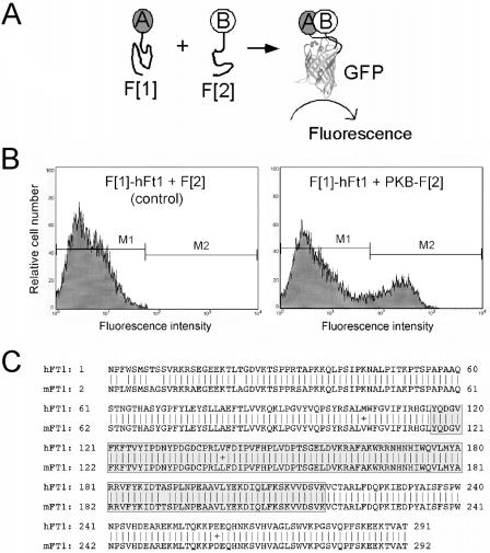
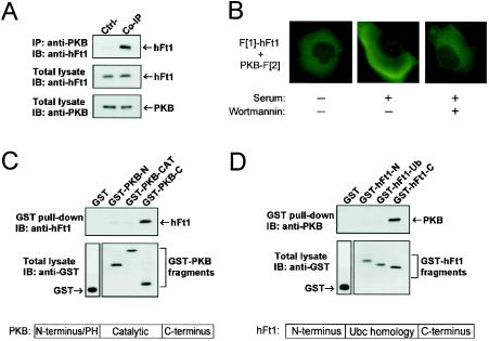
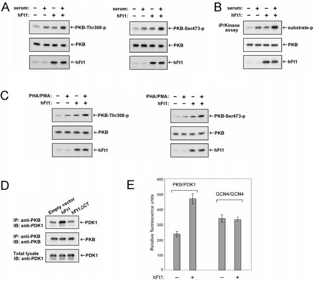
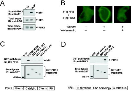
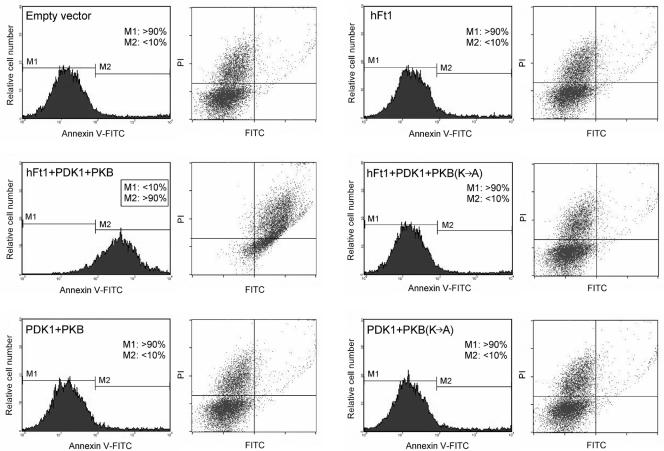
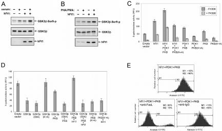
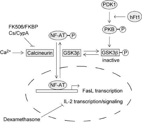
The serine/threonine kinase protein kinase B (PKB)/Akt plays a central role in many cellular processes, including cell growth, glucose metabolism, and apoptosis. However, the identification and validation of novel regulators or effectors is key to future advances in understanding the multiple functions of PKB. Here we report the identification of a novel PKB binding protein, called Ft1, from a cDNA library screen using a green fluorescent protein-based protein-fragment complementation assay. We show that the Ft1 protein interacts directly with PKB, enhancing the phosphorylation of both of its regulatory sites by promoting its interaction with the upstream kinase PDK1. Further, the modulation of PKB activity by Ft1 has a strong effect on the apoptosis susceptibility of T lymphocytes treated with glucocorticoids. We demonstrate that this phenomenon occurs via a PDK1/PKB/GSK3/NF-ATc signaling cascade that controls the production of the proapoptotic hormone Fas ligand. The wide distribution of Ft1 in adult tissues suggests that it could be a general regulator of PKB activity in the control of differentiation, proliferation, and apoptosis in many cell types.


Figures

FIG. 1.
FIG. 1.
Identification of hFt1 as a binding partner of PKB in a GFP PCA-based cDNA library screen. (A) The GFP PCA strategy. The principle of the GFP PCA is that cells simultaneously expressing complementary fragments of GFP (F[1] and F[2]) fused to test proteins (A and B) will produce a fluorescent signal only if the fused proteins physically interact and then bring the complementary fragments of GFP into proximity where it can fold and reassemble into its active form, generating fluorescence. For the GFP PCA-based cDNA library screen, a human brain cDNA library was fused to fragment 1 of GFP (F[1]-cDNA library) and the full-length PKB cDNA was fused to fragment 2 (PKB-F[2]). (B) Interaction of hFt1 with PKB was confirmed by transiently cotransfecting COS-1 cells with the isolated F[1]-cDNA fusion coding for full-length hFt1 (F[1]-hFt1) and the PKB-F[2] fusion, followed by FACS analysis. The physical interaction between PKB and hFt1 induced the folding and reconstitution of GFP from its fragments, generating a fluorescent signal (gate window M2). Cotransfection of cells with the F[1]-hFt1 fusion and free F[2] expression vectors was used as a negative control. (C) Alignment of predicted amino acid sequences of Ft1 from human (accession number NM022476) and mouse (accession number Z67963) origins. The ubiquitin-ligase homology domain (aa 116 to 217) is represented by a gray box.
FIG. 2.
FIG. 2.
Characterization of the interaction between PKB and hFt1. (A) Coimmunoprecipitation of the endogenous interacting partners. Endogenous PKB was immunoprecipitated from HEK293 cells with anti-PKB antibodies, and the immunoprecipitate was run on SDS-PAGE. The presence of hFt1 in the immune complexes was detected by immunoblotting with an anti-hFt1 antibody (upper panel). Expression levels of hFt1 and PKB were detected by immunoblotting total lysates with their corresponding antibodies (middle and lower panels). Immunoprecipitation of a sample with anti-IgG instead of anti-PKB antibodies was used as a control (Ctrl-). (B) Cellular location of the PKB/hFt1 complexes with GFP PCA. COS-1 cells were cotransfected with vectors expressing F[1]-hFt1 or PKB-F[2], serum starved, and untreated or treated with 300 nM wortmannin for 1 h. Afterwards, cells were stimulated for 30 min with 10% serum. Fluorescence microscopy was performed on live cells. (C) hFt1 directly interacts with the C-terminal noncatalytic domain of PKB. Bacterially purified hFt1 was incubated with immobilized GST-PKB fragments (N terminus, catalytic domain, and C terminus). The PKB-bound hFt1 was eluted with glutathione and detected by immunoblotting with anti-hFt1. Levels of the GST fusions were determined by immunoblotting with anti-GST antibodies. (D) PKB directly interacts with the C-terminal domain of hFt1. Bacterially purified PKB was incubated with immobilized GST-hFt1 fragments (N terminus, ubiquitin-ligase homology domain, and C terminus). The hFt1-bound PKB was eluted with glutathione and detected by immunoblotting with anti-PKB antibodies. Levels of the GST fusions were determined by immunoblotting with anti-GST antibodies.
FIG. 3.
FIG. 3.
hFt1 enhances the phosphorylation and activation of PKB and the binding to its upstream kinase PDK1. (A) HEK293T cells were cotransfected with PKB and PDK1 expression vectors, in the presence (+) or absence (−) of hFt1, and stimulated with serum (+) or not stimulated (−). The phosphorylation status of PKB on both of its regulatory sites was analyzed by immunoblotting using the corresponding phospho-specific antibodies (upper panels). Expression levels of PKB and hFt1 were also determined using anti-PKB and anti-hFt1 antibodies (middle and lower panels). (B) In vitro kinase assay to confirm that the hFt1-dependent increase of PKB phosphorylation correlates with an enhancement of its kinase activity. HEK293T cells, transfected and treated as for panel A, were lysed and PKB immunoprecipitated. PKB kinase activity in the immunoprecipitates was measured using a pure substrate (paramyosin fused to a 20-amino-acid GSK-3α/β segment). Phosphorylation of the substrate (substrate-p) was detected by immunoblotting using anti-GSK3α/β-Ser21/9-p antibodies (upper panel). Expression levels of PKB and hFt1 were also determined using anti-PKB and anti-hFt1 antibodies (middle and lower panels). (C) Tag-Jurkat cells were cotransfected with PKB and PDK1 expression vectors, in the presence (+) or absence (−) of hFt1, and stimulated with PHA and PMA (+) or not stimulated (−). The phosphorylation status of PKB on both of its regulatory sites was analyzed as for panel A. (D) hFt1 enhances the formation of PKB/PDK1 complexes. HEK293T cells were cotransfected with PKB and PDK1 expression vectors, in the presence of hFt1 or a truncated form of hFt1 lacking its C-terminal domain (hFt1ΔCT; negative control). PKB was immunoprecipitated using anti-PKB antibodies, and the amount of PDK1 bound to PKB was determined by immunoblotting with anti-PDK1 antibodies (upper panel). The amount of PKB in the immune complexes was detected by immunoblotting with anti-PKB antibodies (middle panel). The expression level of PDK1 was also determined by immunoblotting total lysates with anti-PDK1 antibodies (lower panels). (E) Formation of the PKB/PDK1 complexes was also studied using the GFP PCA in HEK293T cells. PDK1 was fused to the F[1] fragment and PKB was fused to the F[2] fragment of GFP, and the complementary pairs were transiently coexpressed in cells in the presence of hFt1 (without fusion) expression vector (+) or an empty vector (−). The relative amount of reconstituted GFP, a measure of the interaction between the fused protein partners, was detected by fluorometric analysis in intact cells. The constitutive dimerization of the GCN4 leucine zipper fused to the F[1] and F[2] fragments was used as a control. Error bars represent standard errors of the means calculated from three independent samples.
FIG. 4.
FIG. 4.
PDK1 interacts with hFt1 at the plasma membrane in activated cells through its N-terminal noncatalytic domain. (A) Coimmunoprecipitation of endogenous PDK1 and hFt1. Endogenous PDK1 was immunoprecipitated from HEK293 cells with anti-PDK1 antibodies, and the presence of hFt1 in the immune complexes was detected by immunoblotting with an anti-hFt1 antibody (upper panel). Expression levels of hFt1 and PDK1 were detected by immunoblotting total lysates with their corresponding antibodies (middle and lower panels). Immunoprecipitation of a sample with anti-IgG instead of anti-PDK1 antibodies was used as a control (Ctrl-). (B) Cellular location of the PDK1/hFt1 complexes with GFP PCA. COS-1 cells were cotransfected with F[1]-hFt1 and F[2]-PDK1 expression vectors, serum starved, and untreated or treated with 300 nM wortmannin for 1 h. Afterwards, cells were stimulated for 30 min with 10% serum. Fluorescence microscopy was performed on live cells. (C and D) The interaction between hFt1 and PDK1 occurs through the N-terminal domain of PDK1 and the C-terminal domain of hFt1. These studies were carried out as described in the legend for Fig. 2C and D.
FIG. 5.
FIG. 5.
Modulation of PKB activity by hFt1 dramatically enhances the apoptosis susceptibility of Tag-Jurkat cells treated with dexamethasone. Cells were transfected with different combinations of hFt1, PDK1, PKB, or PKB(K→A) expression vectors, as indicated. Cells were treated with 1 μM dexamethasone for 7 to 8 h (in the absence of serum), stained with annexin V-FITC and PI, and analyzed by flow cytometry. Cells that stain positive for annexin V-FITC are undergoing apoptosis (M2; x axis). Cells that stain positive for both annexin V-FITC and PI (upper right square) are either in the late stages of apoptosis or are already dead.
FIG. 6.
FIG. 6.
hFt1 increases production of the proapoptotic hormone Fas ligand via the PKB/GSK3/NF-ATc signaling cascade. (A) hFt1 enhances the PKB-dependent phosphorylation and inactivation of GSK3β. HEK293T cells were cotransfected with GSK3β and PKB expression vectors, in the presence (+) or absence (−) of hFt1, and stimulated with serum (+) or not stimulated (−). The phosphorylation status of GSK3β was analyzed by immunoblotting with the corresponding phospho-specific antibody (upper panel). Expression levels of the proteins were also determined using specific antibodies (middle and lower panels). (B) Tag-Jurkat cells were cotransfected with GSK3β and PKB expression vectors, in the presence (+) or absence (−) of hFt1, and stimulated with PHA and PMA (+) or not stimulated (−). The phosphorylation status of GSK3β was analyzed as for panel A. (C) hFt1 increases NF-AT activity. NF-AT activity was directly quantified in Tag-Jurkat cells with an NF-AT transcription reporter assay. The Tag-Jurkat cell line harbors an integrated β-galactosidase reporter plasmid where three tandem copies of the NF-AT binding site direct transcription of the lacZ gene (15). Cells transfected with the indicated expression vectors were treated with 1 μM dexamethasone, with or without the addition of 300 nM FK506. The conversion of FDG to fluorescein by β-galactosidase was detected by fluorometry. Fluorescence intensity, representing the β-galactosidase activity, is given in relative fluorescence units (y axis). Error bars represent standard errors for the means calculated from three independent samples. (D) Tag-Jurkat cells were transfected with the indicated expression vectors and treated with 1 μM dexamethasone. NF-AT activity was measured as described for panel C. (E) hFt1 increases the production of Fas ligand. Tag-Jurkat cells were transfected with hFt1, PDK1, and PKB expression vectors and treated with 1 μM dexamethasone, with or without the addition of 5 μg of neutralizing anti-human Fas ligand antibody/ml (anti-FasL) or control anti-IgG antibody. Cells were stained with annexin V-FITC and PI and analyzed by flow cytometry.
FIG. 7.
FIG. 7.
Proposed model for the mode of action of hFt1 in T cells. hFt1 enhances the phosphorylation and activation of PKB by promoting its interaction with the upstream kinase PDK1. This hFt1-induced activation of PKB increases the phosphorylation, and thus inactivation, of the NF-ATc kinase GSK3β. In addition to this, an active phosphatase calcineurin (Ca2+ dependent) dephosphorylates NF-ATc. The immunosuppressive drugs FK506 and cyclosporine (Cs) specifically inhibit the activity of calcineurin. The unphosphorylated form of NF-ATc translocates to the nucleus, where it participates in the activation of early immune response genes, including Fas ligand (FasL/CD95L) and IL-2. Treatment of T cells with glucocorticoids downregulates IL-2 expression, leading to an increase of the FasL/IL-2 ratio. This ratio of proliferative versus antiproliferative factors constitutes an important mechanism to preserve the homeostasis of T-cell populations. Consequently, the hFt1-mediated upregulation of Fas ligand expression, via the PDK1/PKB/GSK3/NF-ATc signaling cascade, leads to a Fas ligand-dependent massive cell death in glucocorticoid-treated T cells.

References

    1. Alberola-Ila, J., S. Takaki, J. D. Kerner, and R. M. Perlmutter. 1997. Differential signaling by lymphocyte antigen receptors. Annu. Rev. Immunol. 15:125-154. - PubMed
    1. Alessi, D. R., M. Deak, A. Casamayor, F. B. Caudwell, N. Morrice, D. G. Norman, P. Gaffney, C. B. Reese, C. N. MacDougall, D. Harbison, A. Ashworth, and M. Bownes. 1997. 3-Phosphoinositide-dependent protein kinase-1 (PDK1): structural and functional homology with the Drosophila DSTPK61 kinase. Curr. Biol. 7:776-789. - PubMed
    1. Alessi, D. R., S. R. James, C. P. Downes, A. B. Holmes, P. R. Gaffney, C. B. Reese, and P. Cohen. 1997. Characterization of a 3-phosphoinositide-dependent protein kinase which phosphorylates and activates protein kinase Bα. Curr. Biol. 7:261-269. - PubMed
    1. Beals, C. R., C. M. Sheridan, C. W. Turck, P. Gardner, and G. R. Crabtree. 1997. Nuclear export of NF-ATc enhanced by glycogen synthase kinase-3. Science 275:1930-1934. - PubMed
    1. Belham, C., S. Wu, and J. Avruch. 1999. Intracellular signalling: PDK1—a kinase at the hub of things. Curr. Biol. 9:R93-R96. - PubMed

Publication types

MeSH terms