. 2004 Feb;24(4):1505-15.
doi: 10.1128/MCB.24.4.1505-1515.2004.

Protein kinase B/Akt acts via glycogen synthase kinase 3 to regulate recycling of alpha v beta 3 and alpha 5 beta 1 integrins

Affiliations

Protein kinase B/Akt acts via glycogen synthase kinase 3 to regulate recycling of alpha v beta 3 and alpha 5 beta 1 integrins

Marnie S Roberts et al. Mol Cell Biol. 2004 Feb.

Abstract

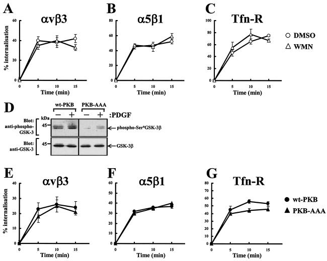
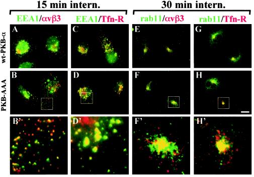
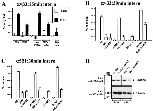
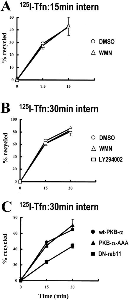
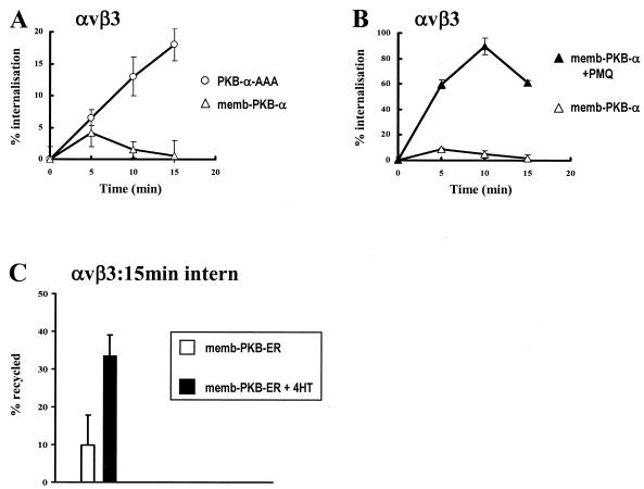
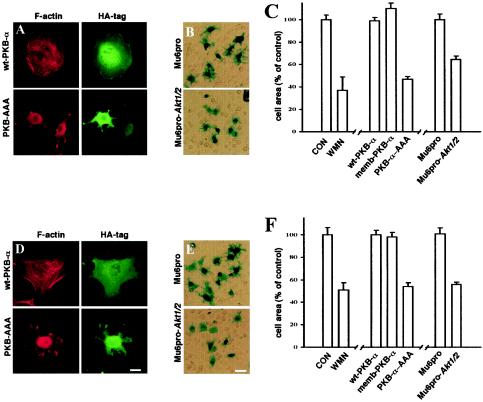
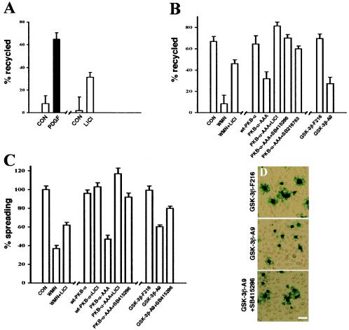
Protein kinase B (PKB)/Akt is known to promote cell migration, and this may contribute to the enhanced invasiveness of malignant cells. To elucidate potential mechanisms by which PKB/Akt promotes the migration phenotype, we have investigated its role in the endosomal transport and recycling of integrins. Whereas the internalization of alpha v beta 3 and alpha 5 beta 1 integrins and their transport to the recycling compartment were independent of PKB/Akt, the return of these integrins (but not internalized transferrin) to the plasma membrane was regulated by phosphatidylinositol 3-kinases and PKB/Akt. The blockade of integrin recycling and cell spreading on integrin ligands effected by inhibition of PKB/Akt was reversed by inhibition of glycogen synthase kinase 3 (GSK-3). Moreover, expression of nonphosphorylatable active GSK-3 beta mutant GSK-3 beta-A9 suppressed recycling of alpha 5 beta 1 and alpha v beta 3 and reduced cell spreading on ligands for these integrins, indicating that PKB/Akt promotes integrin recycling by phosphorylating and inactivating GSK-3. We propose that the ability of PKB/Akt to act via GSK-3 to promote the recycling of matrix receptors represents a key mechanism whereby integrin function and cell migration can be regulated by growth factors.


Figures

FIG. 1.
FIG. 1.
Receptor internalization does not require PKB/Akt. NIH 3T3 fibroblasts were transfected with human αvβ3 (hαvβ3) (A and E), hα5β1 (B and F), or hTfn-R (C and G) together with wt-PKB or PKB-AAA. Cells were serum starved for 30 min at 37°C with the inclusion of 100 nM wortmannin (WMN) or dimethyl sulfoxide (DMSO) for the last 10 min of the starvation period and then surface labeled with 0.2 mg of N-hydroxysuccinimide-S-S-biotin/ml at 4°C for 30 min. Internalization was determined by warming the cells to 37°C for the times indicated in the presence of 0.6 mM primaquine. Biotin was released from proteins remaining at the cell surface by incubation with β-mercaptoethanesulfonic acid (MESNa), and biotinylated proteins were determined by capture ELISA with microtiter wells coated with monoclonal antibodies against hβ3 (A and E) and hα5 (B and F) integrins or human CD71 (C and G). Values are means ± standard errors of the means (n > 4). For analysis of cellular phospho-GSK-3 levels, cells were transfected with wt-PKB or PKB-AAA by using the Amaxa Nucleofector. Cells were then serum starved and incubated in the presence and absence of 10 ng of PDGF-BB/ml for 10 min. Cell lysates were then probed for the presence of GSK-3β phosphorylated at Ser9 and total GSK-3β by Western blotting (D).
FIG. 2.
FIG. 2.
Transport of αvβ3 and Tfn-R to early endosomes and the recycling compartment does not require PKB/Akt. NIH 3T3 fibroblasts were transfected with GFP-EEA1 in combination with human αvβ3 (hαvβ3; A and B) or hTfn-R (C and D) together with wt-PKB (A and C) or PKB-AAA (B and D). Alternatively, cells were transfected with Rab11 in combination with hαvβ3 (E and F) or hTfn-R (G and H) together with wt-PKB (E and G) or PKB-AAA (F and H). Surface receptors were tagged by incubation with a mouse anti-β3 monoclonal antibody (A, B, E, and F) or Texas Red-conjugated Tfn (C, D, G, and H), and the tracer was allowed to internalize for 15 min at 22°C (A to D) or 30 min at 37°C (E to H). Tracer remaining at the cell surface was removed by a low-pH wash at 4°C, and the cells were fixed and permeabilized with detergent. Internalized anti-β3 was visualized with Texas Red-conjugated anti-mouse antibody (A, B, E, and F), and the cells were counterstained with rabbit anti-Rab11, followed by detection with a FITC-conjugated anti-rabbit antibody (E to H). GFP-EEA1 and FITC fluorescence is shown in green, Texas Red fluorescence is shown in red, and yellow indicates colocalization of the two fluorophores. The boxed areas are shown enlarged by a factor of 3.9 in panels B′, D′, F′, and H′. Scale bar, 16 μm.
FIG. 3.
FIG. 3.
The requirement for PI(3)K and PKB/Akt in integrin recycling. NIH 3T3 fibroblasts were transfected with human αvβ3 (hαvβ3; A and B) or hα5β1 (C) in combination with wt-PKB, PKB-AAA, Mu6pro, Mu6pro-akt1/2, N121IRab4 (DN-Rab4), or N124IRab11 (DN-Rab11). Serum-starved cells were surface labeled with 0.2 mg of N-hydroxysuccinimide-S-S-biotin/ml for 30 min at 4°C, and internalization was allowed to proceed for 15 min at 22°C (A) or 30 min at 37°C (B and C). Where appropriate 100 nM wortmannin (WMN) or 60 μM LY294002 was included in the last 10 min of the internalization period. Biotin remaining at the cell surface was removed by exposure to β-mercaptoethanesulfonic acid (MESNa) at 4°C, and internalized integrin was chased back to the cell surface at 37°C for 10 min in the absence (open bars; control) and presence (solid bars) of 10 ng of PDGF-BB/ml for 10 min (A) or for 30 min at 37°C in the absence of PDGF (B and C). Wortmannin and LY294002 were included in the recycling medium where indicated. Cells were then reexposed to MESNa, and biotinylated integrin was determined by capture ELISA using microtiter wells coated with anti-human β3 (A and B) or anti-human α5 (C) integrin monoclonal antibodies. The proportion of integrin recycled to the plasma membrane is expressed as a percentage of the pool of integrin labeled during the internalization period. Values are means ± standard errors of the means (n > 10). To test the ability of the Akt1/Akt2 hairpin RNA to suppress cellular PKB/Akt levels, cells were transfected with Mu6pro or Mu6pro-akt1/2 by using the Amaxa Nucleofector. Cells were lysed for 24 and 48 h following transfection, and the lysates were probed for the presence of PKB/Akt and vinculin (as a loading control) by Western blotting (D).
FIG. 4.
FIG. 4.
125I-Tfn recycles independently of PI(3)K and PKB/Akt. NIH 3T3 fibroblasts were transfected with wt-PKB, PKB-AAA, or S24NRab11 (DN-Rab11) by using the Amaxa Nucleofector. Cells were then serum starved and incubated with 125I-labeled transferrin for 1 h at 4°C. The tracer was allowed to internalize for 15 min at 22°C (A) or 30 min at 37°C (B and C), with 100 nM wortmannin (WMN) or 60 μM LY294002 being included in the incubation for the last 10 min of the internalization time. Tracer remaining at the cell surface was removed, and the internalized 125I-transferrin was allowed to recycle in the absence and presence of wortmannin or LY294002 for the times indicated. The quantity of 125I recycled into the medium is expressed as a percentage of the number of counts incorporated during the internalization period. Values are means ± standard errors of the means (n = 8).
FIG. 5.
FIG. 5.
Active PKB/Akt drives αvβ3 integrin recycling. (A and B) NIH 3T3 fibroblasts were transfected with human αvβ3 (hαvβ3) integrin in combination with constitutively active PKB (memb-PKB) or PKB-AAA. Cells were serum starved and surface labeled with 0.2 mg of N-hydroxysuccinimide (NHS)-S-S-biotin/ml for 30 min at 4°C. Internalization was initiated by warming to 37°C for the times shown in the absence (open symbols) and presence (closed symbols) of 0.6 mM primaquine. Biotin was released from proteins remaining at the cell surface, and biotinylated integrin was determined by capture ELISA using microtiter wells coated with an anti-human β3 integrin monoclonal antibody. (C) Cells were transfected with hαvβ3 in combination with the conditionally active fusion memb-PKB and the hormone-binding domain of the ER (memb-PKB-ER). Serum-starved cells were surface labeled with 0.2 mg of NHS-S-S-biotin/ml for 30 min at 4°C, and internalization was allowed to proceed for 15 min at 22°C. Biotin remaining at the cell surface was removed by exposure to β-mercaptoethanesulfonic acid (MESNa) at 4°C, and internalized integrin was chased back to the cell surface at 37°C for 15 min in the absence (open bars; control) and presence (solid bars) of 300 nM 4-HT. Cells were then reexposed to MESNa, and biotinylated integrin was determined by capture ELISA and expressed as for Fig. 3. Values are means ± standard errors of the means (n = 12).
FIG. 6.
FIG. 6.
PKB/Akt is necessary for cell spreading on vitronectin and fibronectin. NIH 3T3 fibroblasts were transfected with wt-PKB, memb-PKB, PKB-AAA, Mu6pro, or Mu6pro-akt1/2 in combination with a β-galactosidase transfection marker. The cells were then briefly trypsinized and allowed to adhere to either vitronectin (A to C) or fibronectin (D to F) in the presence of 10 ng of PDGF-BB/ml and in the presence and absence of wortmannin (WMN; 100 nM). Attached cells were fixed and stained for β-galactosidase expression and then photographed with a digital camera. In panels C and F, the area of the transfected cells was then determined by delineation of the cell envelope using the National Institutes of Health Image software. Values are means ± standard errors of the means (n > 200 cells). CON, control. Cells expressing HA-tagged PKBs were visualized with a FITC-conjugated anti-HA antibody (green) and counterstained with Texas Red-conjugated phalloidin (red) (A and D). Scale bars, 16 (D) and 80 μm (E).
FIG. 7.
FIG. 7.
Integrin recycling and cell spreading are suppressed by active GSK-3β. (A and B) NIH 3T3 fibroblasts were transfected with human αvβ3 (hαvβ3) in combination with wt-PKB, PKB-AAA, Mu6pro, Mu6pro-akt1/2, GSK-3β-F216, or GSK-3β-A9. Serum-starved cells were surface labeled with 0.2 mg of N-hydroxysuccinimide-S-S-biotin/ml for 30 min at 4°C, and internalization was allowed to proceed for 15 min at 22°C (A) or for 30 min at 37°C (B). Recycling was determined as for Fig. 3; recycling conditions were 10 min at 37°C in the absence (open bars; control) and presence (solid bars) of PDGF-BB (10 ng/ml) (A) or for 30 min at 37°C in the absence of PDGF (B). One hundred nanomolar wortmannin (WMN), 20 mM LiCl, 10 μM SB216763, and 30 μM SB415286 were included during the recycling period where indicated. Values are means ± standard errors of the means (SEM; n > 10). CON, control. (C and D) NIH 3T3 fibroblasts were transfected with wt-PKB, PKB-AAA, Mu6pro, Mu6pro-akt1/2, GSK-3β-F216, or GSK-3β-A9 in combination with a β-galactosidase transfection marker, and cell spreading on vitronectin was determined as for Fig. 6. Cells were spread in the presence of 100 nM wortmannin, 20 mM LiCl, and 30 μM SB415286 as indicated. Values are means ± SEM (n > 80 cells).

References

    1. Bakkeren, D. L., C. M. de Jeu-Jaspars, M. J. Kroos, and H. G. van Eijk. 1987. Release of iron from endosomes is an early step in the transferrin cycle. Int. J. Biochem. 19:179-186. - PubMed
    1. Balendran, A., R. M. Biondi, P. C. Cheung, A. Casamayor, M. Deak, and D. R. Alessi. 2000. A 3-phosphoinositide-dependent protein kinase-1 (PDK1) docking site is required for the phosphorylation of protein kinase Cζ (PKCζ) and PKC-related kinase 2 by PDK1. J. Biol. Chem. 275:20806-20813. - PubMed
    1. Barbieri, M. A., A. D. Kohn, R. A. Roth, and P. D. Stahl. 1998. Protein kinase B/akt and rab5 mediate Ras activation of endocytosis. J. Biol. Chem. 273:19367-19370. - PubMed
    1. Baron, W., S. J. Shattil, and C. ffrench-Constant. 2002. The oligodendrocyte precursor mitogen PDGF stimulates proliferation by activation of αvβ3 integrins. EMBO J. 21:1957-1966. - PMC - PubMed
    1. Byzova, T. V., C. K. Goldman, N. Pampori, K. A. Thomas, A. Bett, S. J. Shattil, and E. F. Plow. 2000. A mechanism for modulation of cellular responses to VEGF: activation of the integrins. Mol. Cell 6:851-860. - PubMed

Publication types