. 2004 Feb;24(4):1560-9.
doi: 10.1128/MCB.24.4.1560-1569.2004.

Mnt loss triggers Myc transcription targets, proliferation, apoptosis, and transformation

Affiliations

Mnt loss triggers Myc transcription targets, proliferation, apoptosis, and transformation

Jonas A Nilsson et al. Mol Cell Biol. 2004 Feb.

Abstract

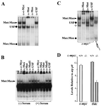
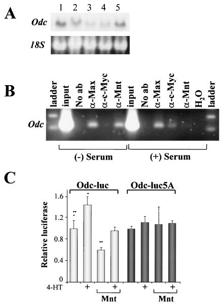
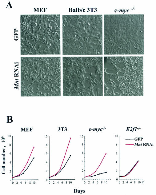
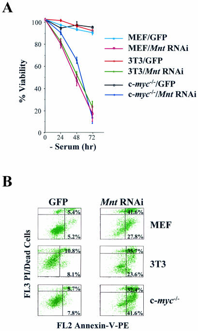
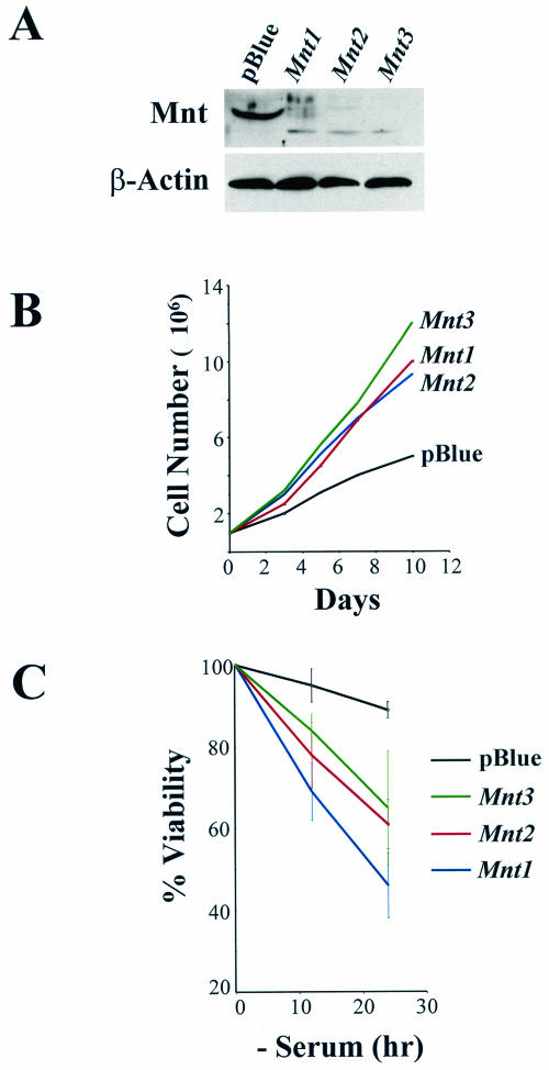
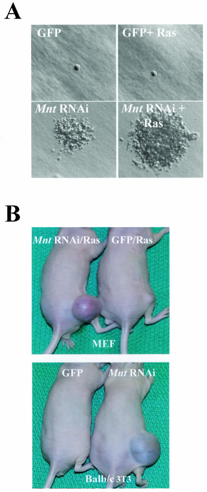
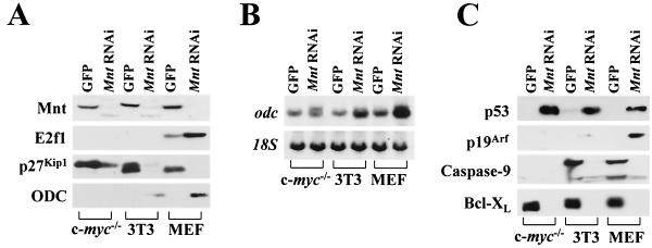
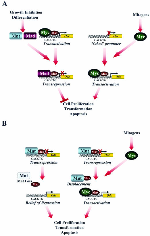
Myc oncoproteins are overexpressed in most cancers and are sufficient to accelerate cell proliferation and provoke transformation. However, in normal cells Myc also triggers apoptosis. All of the effects of Myc require its function as a transcription factor that dimerizes with Max. This complex induces genes containing CACGTG E-boxes, such as Ornithine decarboxylase (Odc), which harbors two of these elements. Here we report that in quiescent cells the Odc E-boxes are occupied by Max and Mnt, a putative Myc antagonist, and that this complex is displaced by Myc-Max complexes in proliferating cells. Knockdown of Mnt expression by stable retroviral RNA interference triggers many targets typical of the "Myc" response and provokes accelerated proliferation and apoptosis. Strikingly, these effects of Mnt knockdown are even manifest in cells lacking c-myc. Moreover, Mnt knockdown is sufficient to transform primary fibroblasts in conjunction with Ras. Therefore, Mnt behaves as a tumor suppressor. These findings support a model where Mnt represses Myc target genes and Myc functions as an oncogene by relieving Mnt-mediated repression.


Figures

FIG. 1.
FIG. 1.
(A and B) Mnt-Max is the predominant complex detected in EMSA using either Odc E-box1 (A) or E-box2 (B and C) probes, using extracts from proliferating FDC-P1 myeloid cells (A) growing (+ Serum) or quiescent (− Serum) BALB/c 3T3 fibroblasts (B). Arrowheads, diamonds, and circles indicate Mnt-Max, USF, and Max-Max complexes, respectively, as established by supershift analyses. Results using E-box probe 1 or 2 were comparable for all cell extracts (data not shown). (C) c-myc loss enhances Mnt-Max DNA binding activity in ES cells. EMSA was performed with extracts from the indicated ES cells. ✧ indicates a complex of unknown origin that is augmented in c-myc−/− ES cells and which contains Max, since it is supershifted by an antiserum against Max. (D). c-myc loss compromises Odc expression in ES cells. RNA was prepared from exponentially growing ES cell cultures, and levels of c-myc and Odc transcripts were determined by real-time PCR. Oligonucleotides and probes for the detection of acidic ribosomal protein P0 mRNA (arp p0) were used as an internal control, since this gene is not regulated by Myc (17).
FIG. 2.
FIG. 2.
Myc activates Odc transcription by relieving Mnt-mediated repression. (A) Odc expression in BALb/c-3T3 cells is serum dependent. Fibroblasts were cultured in the presence (lane 1) or absence of serum for 1, 2, and 3 days (lanes 2 to 4). After 3 days of starvation, fresh medium containing serum was added for 3 h (lane 5). Northern blot analysis was performed using a probe directed against Odc. 18S rRNA served as loading control. (B) Mnt-Max complexes occupy the Odc E-boxes in quiescent BALB/c-3T3 fibroblasts and are displaced by c-Myc-Max complexes in serum-stimulated cells. ChIP assays of serum-starved (2 days) or restimulated (for 3 h) BALB/c-3T3 cells were performed using antibodies directed against Max, c-Myc, or Mnt. The primers used for the PCR encompass the intronic E-boxes of the Odc gene. (C) Mnt represses the Odc promoter, and Mnt repression is relieved by Myc. BALB/c-3T3 cells infected with a Myc-ER-expressing retrovirus (MSCV-Myc-ER-IRES-GFP) were cotransfected with Odc promoter-luciferase constructs (containing intact [Odc-Luc] or mutated [Odc-luc5A] E-boxes) together with a Renilla luciferase control plasmid (pRL-SV40 [Promega]) and, where indicated, a Mnt expression vector (pRc-CMV-HA-Mnt). Transfections were balanced for DNA and promoter content by using the empty pRc vector plasmid. Myc-ER was activated by adding 4-HT for 24 h. Luciferase activity was determined using a luminometer. Results shown are the mean of triplicate assays and are representative of three independent experiments.
FIG. 3.
FIG. 3.
Knockdown of Mnt by stable RNAi accelerates cell proliferation. (A) Morphological changes induced by MntRNAi expression in MEFs, BALB/c-3T3 cells, and c-myc−/− HO.15 fibroblasts are shown. (B) Acceleration of cell proliferation by ablation of Mnt is independent of c-myc but requires E2f1. Growth rates of the indicated MntRNAi-expressing cells were compared to GFP-only expressing fibroblasts.
FIG. 4.
FIG. 4.
Mnt knockdown triggers apoptosis. (A) The indicated cells were shifted to medium containing 0.1% fetal calf serum, and their viability was determined by trypan blue dye exclusion. A representative experiment performed in triplicate is shown. (B) Apoptosis was determined by FACS analysis of annexin V-positive, propidium iodide-stained cells 48 h after serum depletion. The percentages of annexin V-positive cells are given in the quadrants (early apoptotic, lower right panels; late apoptotic, upper right panels). PE, phycoerythrin.
FIG. 5.
FIG. 5.
Phenotypes observed in MEFs on Mnt knockdown are not due to general effects of expressing shRNAi. (A) Immunoblot analyses for Mnt of MEFs infected with control (MSCV-pBlueRNAi-IRES-GFP) or three different MSCV-MntRNAi-IRES-GFP-expressing retroviruses. β-Actin levels were assessed as a control. (B) MEFs expressing any of the three MSCV-MntRNAi-IRES-GFP retroviruses displayed accelerated rates of proliferation compared to MEFs expressing the MSCV-pBlueRNAi-IRES-GFP retrovirus. (C) MEFs expressing any of the three MSCV-MntRNAi-IRES-GFP retroviruses, but not those infected with the retrovirus encoding MSCV-pBlueRNAi-IRES-GFP, undergo rapid apoptosis following serum withdrawal.
FIG. 6.
FIG. 6.
Mnt knockdown provokes transformation in conjunction with activated Ha-Ras. (A) A representative field of soft-agar assays of MEFs expressing GFP, GFP plus Ras, MntRNAi, and MntRNAi plus Ras are shown. MntRNAi-expressing MEFs were capable of growing in an anchorage-independent manner, although colonies were reduced 10- to 20-fold in their numbers and were much smaller than colonies generated by MEFs expressing both Ha-Ras and MntRNAi. (B) MEFs expressing both Ras and MntRNAi are tumorigenic in immunocompromised Nu/Nu mice (n = 5 for each group). BALB/c-3T3 fibroblasts expressing MntRNAi alone were also tumorigenic in this assay (n = 5).
FIG. 7.
FIG. 7.
Mnt knockdown triggers “Myc” transcriptional programs. (A) Immunoblot analyses of the indicated fibroblast strains infected with control (GFP) or MntRNAi-expressing retroviruses. The anti-Mnt and anti-ODC immunoblots demonstrate that the MntRNAi indeed ablates Mnt expression and up-regulates ODC. The absence of ODC and E2f1 signals in the c-myc−/− cells appears to reflect the inability of these antibodies to recognize rat ODC and E2f1. In addition, Mnt loss was associated with a marked down-regulation in levels of p27KipI and in MEFs with an up-regulation of E2f1. (B) Northern blot analysis demonstrates that Odc is up-regulated by MntRNAi in all three fibroblast strains. Evaluation of 18S rRNA levels served as a loading control. (C) The p19Arf-p53 apoptotic pathway and the Bcl-XL apoptotic pathway are triggered by Mnt loss. Knockdown of Mnt was associated with an increase in p53 levels in all fibroblasts and with the induction of Arf in MEFs. The loss of Mnt was also associated with the activation of caspase-9, since larger (inactive) proenzyme form of caspase-9 was lost in MntRNAi-expressing fibroblasts. Again, the absence of caspase-9 protein in the c-myc−/− cells may reflect the inability of this antibody to recognize rat caspase-9. Bcl-XL protein levels were dramatically reduced in all MntRNAi-expressing fibroblast strains.
FIG.8.
FIG.8.
Myc antagonizes Mnt to regulate cell fate. (A) (Left) Conventional models of transcriptional regulation by the Max network. In these models, Mad and Mnt work as antagonists of Myc. (Right) Alternatively, the Myc-Max complex can activate promoters by direct binding to E-boxes not previously occupied by other complexes. (B) In the revised model, Myc functions as an antagonist of Mnt, since Mnt loss triggers most of the“Myc” response, including Myc's transcription targets (e.g., Odc), apoptosis, accelerated proliferation, and transformation. (Right) In this scenario Myc may operate in two steps to induce its targets. First, it may displace Mnt-Max complexes from response elements, thus relieving repression. Second, when bound the Myc-Max complex may transactivate the gene. (Left) The fact that Mnt loss can activate some “Myc” transcription targets even in cells lacking all forms of Myc indicates that Myc binding may not be required to activate some of its targets, which instead are controlled through active repression.

Similar articles

Cited by

References

    1. Askew, D. S., R. A. Ashmun, B. C. Simmons, and J. L. Cleveland. 1991. Constitutive c-myc expression in an IL-3-dependent myeloid cell line suppresses cell cycle arrest and accelerates apoptosis. Oncogene 6:1915-1922. - PubMed
    1. Ayer, D. E., and R. N. Eisenman. 1993. A switch from Myc:Max to Mad:Max heterocomplexes accompanies monocyte/macrophage differentiation. Genes Dev. 7:2110-2119. - PubMed
    1. Ayer, D. E., Q. A. Lawrence, and R. N. Eisenman. 1995. Mad-Max transcriptional repression is mediated by ternary complex formation with mammalian homologs of yeast repressor Sin3. Cell 80:767-776. - PubMed
    1. Baudino, T. A., and J. L. Cleveland. 2001. The Max network gone Mad. Mol. Cell. Biol. 21:691-702. - PMC - PubMed
    1. Baudino, T. A., K. H. Maclean, J. Brennan, E. Parganas, C. Yang, A. Aslanian, J. A. Lees, C. J. Sherr, M. F. Roussel, and J. Cleveland. 2003. Myc-mediated proliferation and lymphomagenesis, but not apoptosis, are compomised by E2f1 loss. Mol. Cell 11:1-20. - PubMed

Publication types

MeSH terms

Substances