. 2004 Feb;24(4):1570-81.
doi: 10.1128/MCB.24.4.1570-1581.2004.

Proapoptotic BH3-only Bcl-2 family member Bik/Blk/Nbk is expressed in hemopoietic and endothelial cells but is redundant for their programmed death

Affiliations

Proapoptotic BH3-only Bcl-2 family member Bik/Blk/Nbk is expressed in hemopoietic and endothelial cells but is redundant for their programmed death

Leigh Coultas et al. Mol Cell Biol. 2004 Feb.

Abstract

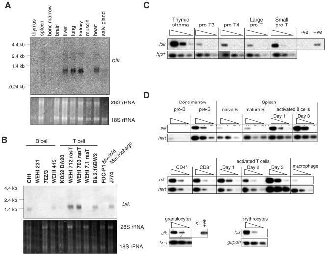
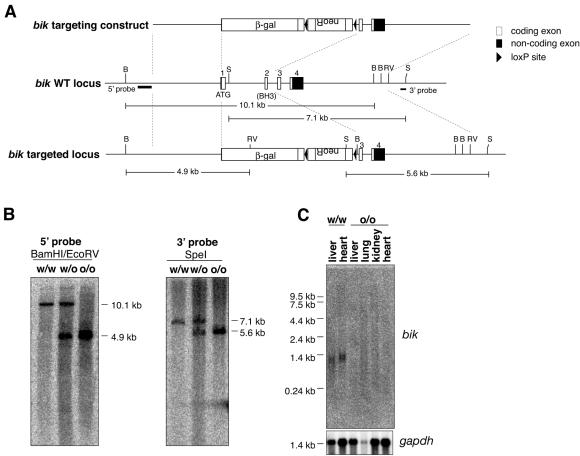
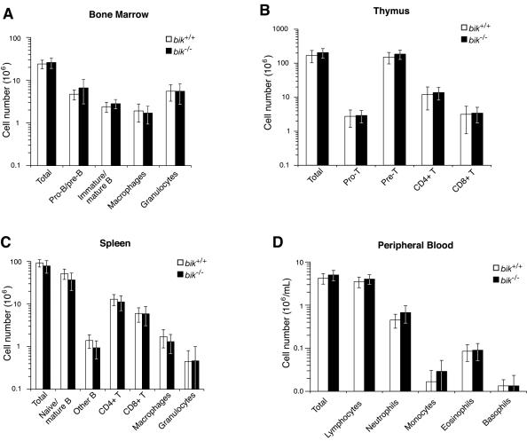
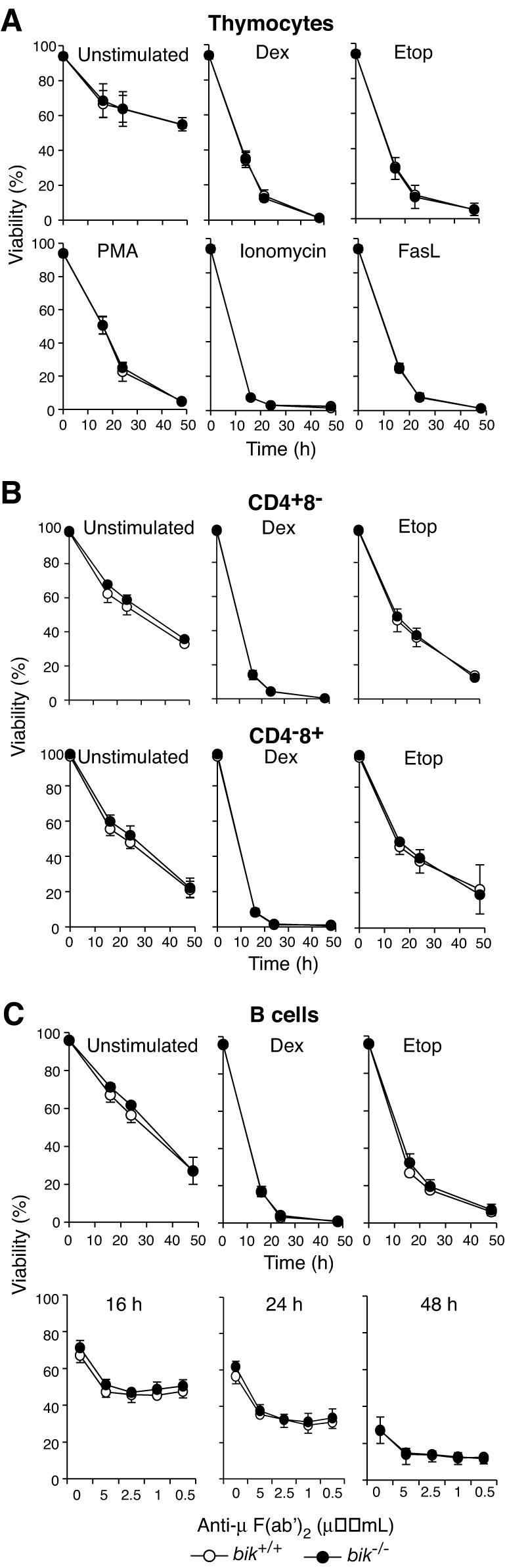
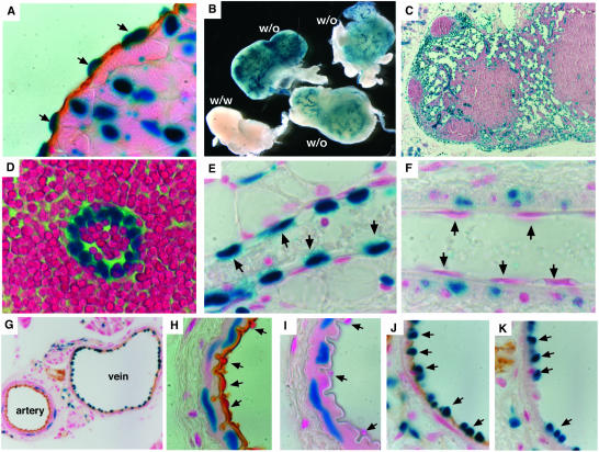
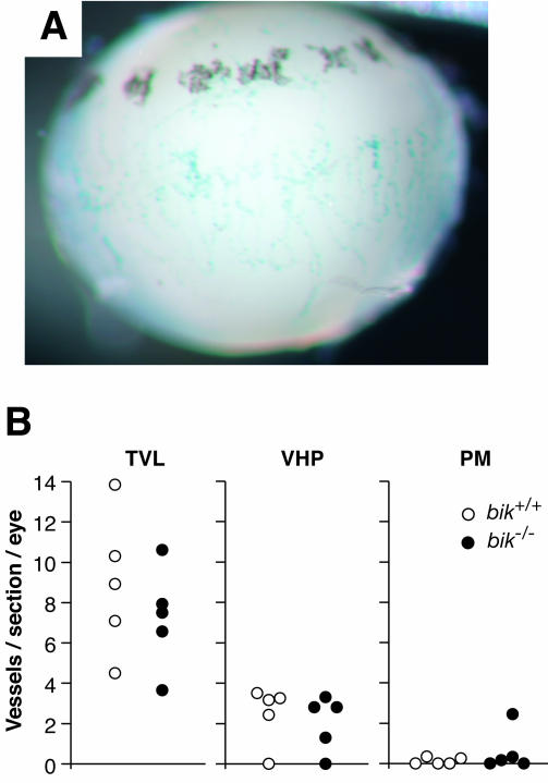
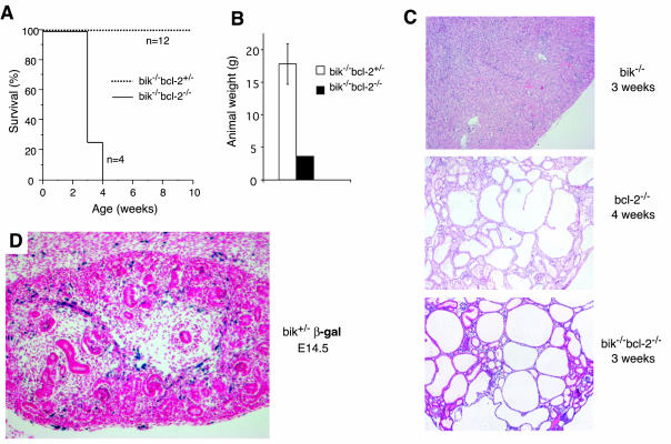
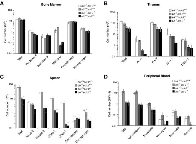
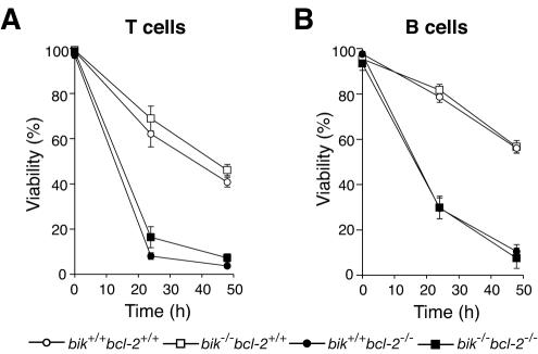
The BH3-only members of the Bcl-2 protein family are essential for initiation of programmed cell death and stress-induced apoptosis. We have determined the expression pattern in mice of the BH3-only protein Bik, also called Blk or Nbk, and examined its physiological function by gene targeting. We found that Bik is expressed widely in the hematopoietic compartment and in endothelial cells of the venous but not arterial lineages. Nevertheless, its loss did not increase the numbers of such cells in mice or protect hematopoietic cells in vitro from apoptosis induced by cytokine withdrawal or diverse other cytotoxic stimuli. Moreover, whereas loss of the BH3-only protein Bim rescued mice lacking the prosurvival protein Bcl-2 from fatal polycystic kidney disease and lymphopenia, loss of Bik did not. These results indicate that any function of Bik in programmed cell death and stress-induced apoptosis must overlap that of other BH3-only proteins.


Figures

FIG. 1.
FIG. 1.
bik expression in adult tissues and hematopoietic cell types. (A) Northern blot analysis of bik mRNA expression on poly(A)+-enriched RNA from indicated tissues of C57BL/6 mice, using a bik cDNA coding region probe. An ethidium bromide stain of 28S and 18S rRNA is included as a loading control. Muscle, skeletal muscle; salv. gland, salivary gland. (B) Northern blot analysis of bik mRNA expression on poly(A)+-enriched RNA from the indicated cell lines. (C) Semiquantitative RT-PCR analysis of bik mRNA expression in FACS-purified thymocyte subsets: pro-T3 (CD25+ CD44), pro-T4 (CD25 CD44), and large (cycling) and small (resting) pre-T (CD4+ CD8+). Analysis was performed on a titration of the cDNA template (neat, 1:5, and 1:25). Each amplicon was confirmed as that for bik by Southern blotting using a bik-specific internal oligonucleotide. Expression of hprt is included as a control for loading. (D) Semiquantitative RT-PCR analysis of bik mRNA expression in FACS-purified hematopoietic cell subsets: pro-B (B220+ CD43+ sIg), pre-B (B220+ CD43 sIg), naive B (B220+ sIgM+ sIgD), mature B (B220+ sIgMlo sIgDhi), CD4+ CD8 T, CD4 CD8+ T, macrophages (Mac-1+ Gr-1), granulocytes (Mac-1+ Gr-1+), or nucleated erythroid progenitors (Ter119+). B cells were activated with IgM cross-linking antibodies in the presence of cytokines. T cells were activated with PMA and ionomycin in the presence of cytokines. Analysis was performed as described in the legend to panel C.
FIG. 2.
FIG. 2.
Targeting of the bik locus. (A) Schematic diagram (to scale) of the bik targeting construct and the wt and targeted bik loci. Dashed lines demarcate regions of the wt locus used in the targeting construct. Locations of 5′ and 3′ external probes for genomic Southern blot analysis and expected fragment sizes from indicated restriction digests are shown. B, BamHI; RV, EcoRV; S, SpeI. (B) Confirmation with 5′ and 3′ genomic probes of the loss of both wt bik alleles in bik−/− mice. (C) Northern blot analysis of poly(A)+ RNA extracted from the indicated tissues of wt and bik knockout mice (littermates). The blot was probed with a cDNA probe containing the entire bik coding region. A gapdh loading control is shown.
FIG. 3.
FIG. 3.
Cell type composition of hematopoietic organs from wt and bik−/− littermates. (A) The total number of bone marrow cells from 5- to 11-week-old wt (n = 6) and bik−/− (n = 8) mice was determined, and the numbers of pro- and pre-B cells (B220+ sIgM sIgD), immature and mature B cells (B220+ sIgM+ sIgD+), macrophages (Mac1+ Grl) and granulocytes (Mac1+ Grl+) was determined by FACS analysis. (B) The total number of thymocytes from 5- to 11-week-old wt (n = 8) and bik−/− (n = 8) animals was determined, and the number of pro-T (CD4 CD8), pre-T (CD4+ CD8+), and mature T (CD4+ CD8 or CD4 CD8+) cells was determined by FACS analysis. (C) The total number of splenocytes from 5- to 11-week-old wt (n = 8) and bik−/− (n = 8) animals was determined, and the number of naive and mature B cells (B220+ sIgM+ sIgD+), other B cells (B220+ sIgM sIgD), mature T cells (CD4+ CD8 or CD4 CD8+), macrophages (Mac1+ Grl), and granulocytes (Mac1+ Grl+) was determined by FACS analysis. (D) Peripheral blood from 30- to 46-week-old wt (n = 6) and bik−/− (n = 9) littermates was examined, and the total number of lymphocytes and granulocytes (neutrophils, monocytes, eosinophils, and basophils) per milliliter was determined.
FIG. 4.
FIG. 4.
bik−/− lymphocyte susceptibility to apoptotic stimuli. (A) Thymocytes from wt (n = 4) and bik−/− (n = 4) littermates were harvested and cultured in the presence of dexamethasone (Dex, 10−7 M), etoposide (Etop, 1 μg/ml), PMA (2 ng/ml), ionomycin (1 μg/ml), or cross-linked FasL (100 ng/ml) or in the absence of exogenous stimuli (unstimulated) for the indicated times. (B) CD4+ or CD8+ T cells were sorted from lymph nodes of wt (n = 4) and bik−/− (n = 4) littermates and cultured in the presence of dexamethasone (10−7 M) or etoposide (1 μg/ml) or in the absence of exogenous stimuli (un-stimulated) for the indicated times. (C) Mature B cells were negatively sorted from lymph nodes of wt (n = 4) and bik−/− (n = 4) littermates and cultured in the presence of dexamethasone (10−7 M), etoposide (1 μg/ml), or anti-IgM cross-linking F(ab')2 fragments at the indicated concentrations or in the absence of exogenous stimuli (unstimulated) for the indicated times.
FIG. 5.
FIG. 5.
β-Galactosidase assay for bik expression. (A) bik expression in endocardium, confirmed by combined staining for β-galactosidase activity (blue) and immunohistochemical staining for CD31 (brown). (B) Whole-mount β-galactosidase staining of lymph nodes from bik+/− mice demonstrating bik expression in lymphoid vasculature. (C and D) The identity of β-galactosidase-positive cells in lymph nodes was determined as endothelium by morphology from thin sections of whole-mount stained nodes. (E and F) Thin sections from whole-mount β-galactosidase-stained mammary gland venule (E) and arteriole (F). Arrows indicate representative endothelial cells. (G) Combination of β-galactosidase staining and immunohistochemical staining for CD31 on frozen sections of blood vessels from the pancreas of bik+/− animals. (H to K) High-power images showing the morphology of the artery (H and I) and vein (J and K) and the distribution of β-galactosidase staining. Anti-B220 antibody staining (I and K) is included as an isotype-matched control for the anti-CD31 antibody (H and J). Arrows indicate representative endothelial cells. In no case was β-galactosidase staining observed in the above-mentioned cell types from wt animals.
FIG. 6.
FIG. 6.
Hyaloid plexus blood vessel numbers in wt and bik−/− mice. (A) β-Galactosidase staining of an eye from a PN7 bik+/− pup indicates bik expression in hyaloid vessels. Shown is a lateral view of the lens with attached β-galactosidase-positive VHP. β-Galactosidase-positive PM is also evident on the anterior surface of the lens (top). No β-galactosidase activity was detected in hyaloid vessels of a control wt littermate. (B) Eyes of PN16 wt (n = 5) and bik−/− (n = 5) mice were sectioned longitudinally and counted for each hyaloid vessel type: TVL, VHP, and pupillary PM. Vessels were counted in 12 nonconsecutive sections taken at the level of the pupil. Each data point represents the average number of vessels observed per section per eye.
FIG. 7.
FIG. 7.
Phenotype of bik−/− bcl-2−/− mice. (A) The survival rate of bik−/− bcl-2−/− mice (n = 4) and their bik−/− bcl-2+/− littermates (n = 12), monitored for a 10-week period and expressed as percent survival over time. (B) Comparison of the weight of bik-2−/− bcl-2−/− and bik−/− bcl-2+/− littermates at PN25. (C) Histological examination of kidneys from bik−/− bcl-2−/− and bcl-2−/− animals at the time of death, showing that they are clearly polycystic by comparison to an age-matched bik−/− control. bik−/− kidneys were indistinguishable from wt. (D) Expression of Bik in the embryonic kidney, assessed by measuring the β-galactosidase activity in a bik+/− E14.5 kidney frozen section.
FIG. 8.
FIG. 8.
Composition of blk−/− bcl-2−/− reconstituted hematopoietic organs. (A) Total number of Ly5.2+ bone marrow cells from 11- to 15-week postreconstitution Ly5.1 recipients reconstituted with Ly5.2 wt (n = 3), bik−/− bcl-2+/+ (n = 5), bik+/+ bcl-2−/− (n = 3), and bik−/− bcl-2−/− (n = 5) fetal liver HSC. Numbers of Ly5.2+ pro- and pre-B cells (B220+ sIgM sIgD), immature B cells (B220+ sIgM+ sIgD), recirculating mature B cells (B220+ sIgMlo sIgDhi), macrophages (Mac1+ Grl), and granulocytes (Mac1+ Grl+) were then determined by FACS. (B) Total number of Ly5.2+ thymocytes and numbers of pro-T cells (CD4 CD8), pre-T cells (CD4+ CD8+), and mature T cells (CD4+ CD8 and CD4 CD8+), determined by FACS. (C) Total number of Ly5.2+ splenocytes and numbers of naive B cells (sIgM+ sIgD), mature B cells (sIgMlo sIgDhi), mature T cells (CD4+ CD8 and CD4 CD8+), macrophages (Mac1+ Grl), and granulocytes (Mac1+ Gr1+), determined by FACS. (D) Peripheral blood from Ly5.1 recipient mice taken 9 to 12 weeks after reconstitution with Ly5.2 wt (n = 7), bik−/− bcl-2+/+ (n = 14), bik+/+ bcl-2−/− (n = 11), and bik−/− bcl-2−/− (n = 15) fetal liver HSC was examined, and the total number of Ly5.2+ lymphocytes and granulocytes (neutrophils, monocytes, eosinophils, and basophils) per milliliter was determined.
FIG. 9.
FIG. 9.
Sensitivity of bik−/− bcl-2−/− lymphocytes to apoptotic stimuli. (A) bik−/− bcl-2−/− T lymphocytes were cultured in the absence of cytokines, and viability was assessed by measuring the PI uptake after 24 and 48 h. Lymphocytes were sorted from lymph nodes of Ly5.1 recipients reconstituted with Ly5.2 wt (n = 5), bik−/− bcl-2+/+ (n = 3), bik+/+ bcl-2−/− (n = 4), and bik−/− bcl-2−/− (n = 5) fetal liver HSC. (B) bik−/− bcl-2−/− B lymphocytes were cultured in the absence of cytokines, and viability assessed as described in the legend to panel A.

References

    1. Adams, J. M. 2003. Ways of dying: multiple pathways to apoptosis. Genes Dev. 17:2481-2495. - PubMed
    1. Amanna, I. J., K. Clise-Dwyer, F. E. Nashold, K. A. Hoag, and C. E. Hayes. 2001. Cutting edge: A/WySnJ transitional B cells overexpress the chromosome 15 proapoptotic Blk gene and succumb to premature apoptosis. J. Immunol. 167:6069-6072. - PubMed
    1. Bouillet, P., S. Cory, L.-C. Zhang, A. Strasser, and J. M. Adams. 2001. Degenerative disorders caused by Bcl-2 deficiency are prevented by loss of its BH3-only antagonist Bim. Dev. Cell 1:645-653. - PubMed
    1. Bouillet, P., D. Metcalf, D. C. S. Huang, D. M. Tarlinton, T. W. H. Kay, F. Köntgen, J. M. Adams, and A. Strasser. 1999. Proapoptotic Bcl-2 relative Bim required for certain apoptotic responses, leukocyte homeostasis, and to preclude autoimmunity. Science 286:1735-1738. - PubMed
    1. Bouillet, P., J. F. Purton, D. I. Godfrey, L.-C. Zhang, L. Coultas, H. Puthalakath, M. Pellegrini, S. Cory, J. M. Adams, and A. Strasser. 2002. BH3-only Bcl-2 family member Bim is required for apoptosis of autoreactive thymocytes. Nature 415:922-926. - PubMed

Publication types

MeSH terms

LinkOut - more resources