. 2004 Feb;24(4):1582-94.
doi: 10.1128/MCB.24.4.1582-1594.2004.

Negative control of the Myc protein by the stress-responsive kinase Pak2

Affiliations

Negative control of the Myc protein by the stress-responsive kinase Pak2

Zhongdong Huang et al. Mol Cell Biol. 2004 Feb.

Abstract

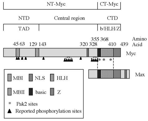
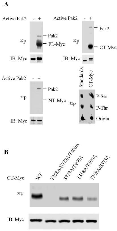
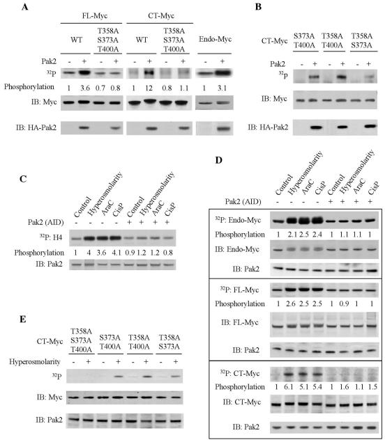
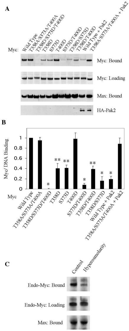
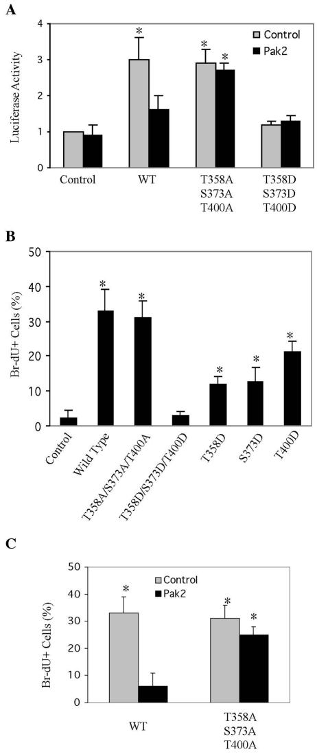
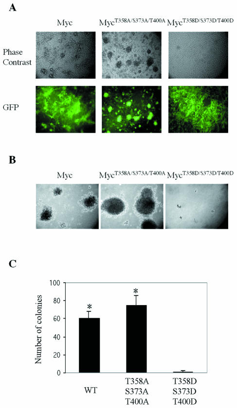
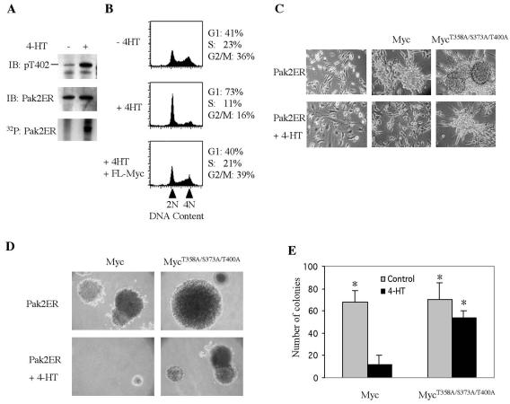
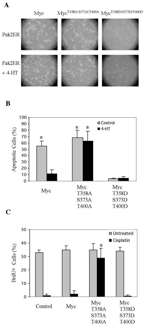
Pak2 is a serine/threonine kinase that participates in the cellular response to stress. Among the potential substrates for Pak2 is the protein Myc, encoded by the proto-oncogene MYC. Here we demonstrate that Pak2 phosphorylates Myc at three sites (T358, S373, and T400) and affects Myc functions both in vitro and in vivo. Phosphorylation at all three residues reduces the binding of Myc to DNA, either by blocking the requisite dimerization with Max (through phosphorylation at S373 and T400) or by interfering directly with binding to DNA (through phosphorylation at T358). Phosphorylation by Pak2 inhibits the ability of Myc to activate transcription, to sustain cellular proliferation, to transform NIH 3T3 cells in culture, and to elicit apoptosis on serum withdrawal. These results indicate that Pak2 is a negative regulator of Myc, suggest that inhibition of Myc plays a role in the cellular response to stress, and raise the possibility that Pak2 may be the product of a tumor suppressor gene.


Figures

FIG. 1.
FIG. 1.
Functional domains of Myc. The amino-terminal domain (NTD) of Myc contains two conserved sequences, Myc Box 1 (MBI) and Myc Box II (MBII), that are required for transcriptional activation activity. The carboxyl-terminal domain (CTD) contains the b/HLH/Z domain, which is responsible for dimerization with Max and binding to E-box DNA. A nuclear localization signal (NLS) is present in the central region of the protein. The arrowheads point to multiple phosphorylation sites reported previously. The Myc fragments used in the following experiments are peptide 1 to 352 (NT-Myc) and peptide 353 to 439 (CT-Myc).
FIG. 2.
FIG. 2.
Pak2 phosphorylates Myc in vitro. Enzymatic reactions were performed as described in Materials and Methods. Reaction products were fractionated by electrophoresis in polyacrylamide gels, followed by immunoblotting (IB) and autoradiography (32P). (A) Phosphorylation of Myc. Recombinant FL-Myc, CT-Myc, and NT-Myc were used as substrates (upper left, upper, right and bottom left panels, respectively). Analysis of phosphorylated amino acids is illustrated on the lower right. (B) Identification of phosphorylated residues. Various alleles of recombinant CT-Myc were used as substrates, as indicated. WT, wild type.
FIG. 3.
FIG. 3.
Pak2 phosphorylates Myc in vivo. (A) Phosphorylation of Myc following overexpression of exogenous Pak2. Molecular clones of Pak2 and various alleles of MYC were transfected into 293T cells. At 24 h later, the cells were labeled with [32P]orthophosphate for 12 h and then harvested for analysis. Immunoprecipitation for endogenous or exogenously expressed Myc was performed and analyzed as described in Materials and Methods. Immunoblots for Myc protein are designated by IB, and autoradiograms for labeled Myc are designated by 32P. WT, wild type. The relative amount of radiolabeled Myc was determined by using ImageQuant (Molecular Dynamics). Immunoblotting was used to ascertain the relative mass of Myc precipitated. (B) Identification of the phosphorylated residues in Myc. In vivo phosphorylation of mutant CT-Myc was performed and analyzed as in panel A. (C) Activation of Pak2 by cellular stress. Exposure of 293T cells to stress and assay for Pak2 activity were performed as described in Materials and Methods. The activity of Pak2 was ascertained from the labeling of the substrate H4 and the relative mass of Pak2 in the reactions by immunoblotting (IB). Pak2-AID was expressed in 293T cells by transfection. AraC, cytosine arabinoside; CisP, cisplatin. (D) Phosphorylation of Myc by Pak2 in response to cellular stress. FL-Myc, CT-Myc, and Pak2-AID were expressed in 293T cells by transfection. The cells were exposed to stress as described in Materials and Methods and analyzed as in panel A. (E) Residues phosphorylated in Myc in response to cellular stress. Various alleles of CT-MYC were transfected into 293T cells. The cells were exposed to hyperosmolarity as described in Materials and Methods and analyzed as in panel A.
FIG. 4.
FIG. 4.
Phosphorylation by Pak2 inhibits the interaction of Myc with Max. (A) Analysis in vitro. Various forms of Myc were produced by translation in vitro. The ability of the Myc substrate to interact with Max after phosphorylation with activated Pak2 was evaluated as described in Materials and Methods. The relative amount of binding was determined by ImageQuant. Aliquots of cell lysates used in the binding assays were subjected directly to SDS-PAGE and immunoblotting for Myc. WT, wild type. (B) Analysis in vivo. The interaction between Myc and Max was evaluated by a two-hybrid assay with 293T cells, as described in Materials and Methods. Interaction was manifested as activation of a luciferase reporter. The expression of the Myc participant in the reaction was determined by immunoblotting. The data shown are the mean and standard deviation of three independent experiments (*, P < 0.005; **, P < 0.03). (C) Activation of Pak2 by stress blocks the interaction of Myc with Max in vivo. The stress conditions were applied to 293T cells as described in the legend to Fig. 3C. After stress treatment, endogenous Myc was immunoprecipitated from cellular extracts. The immunoprecipitates (IP) were examined for Myc and Max by immunoblotting (IB). Where required, the analyses were performed with cells expressing Pak2-AID that had been introduced by transfection. AraC, cytosine arabinoside; CisP, cisplatin.
FIG. 5.
FIG. 5.
Phosphorylation inhibits the binding of Myc to DNA. Pak2 and various alleles of Myc were expressed by transfection into 293T cells. Analysis of cellular extracts for the binding of Myc and endogenous Max to E-box oligonucleotide was performed as described in Materials and Methods. (A) Coprecipitation of Myc and Max with biotinylated E-box oligonucleotide. Precipitated Myc and Max were detected by immunoblotting. Aliquots of cell lysates used in the binding assays were subjected to SDS-PAGE and immunoblotting for Myc. (B) Quantification of results. The histogram illustrates the mean and standard deviation of three independent experiments, quantified with ImageQuant (*, P < 0.005; **, P < 0.02). (C) Coprecipitation of endogenous Myc and Max with E-box DNA was detected by immunoblotting as in panel A.
FIG. 6.
FIG. 6.
Phosphorylation by Pak2 reduces activation of transcription and stimulation of cellular proliferation by Myc. (A) Transactivation assay. Pak2 and various alleles of Myc were expressed by transfection into 293T cells, with a reporter plasmid as described previously (38). At 24 h later, the cells were harvested and assayed for luciferase activity as described in Materials and Methods. The histogram shows the mean and standard deviation of three independent experiments (*, P < 0.03). WT, wild type. (B) Effect on proliferation by mutations of Myc that mimic phosphorylation by Pak2. To evaluate the impact on cellular proliferation, NIH 3T3 cells expressing various mutant alleles of Myc were propagated to confluence, held for 48 h, and then labeled with BrdU for 30 min and analyzed as described in Materials and Methods. The mean and standard deviation of the percentage of BrdU-positive cells are shown (*, P < 0.04). (C) Effect of Pak2 on Myc-induced proliferation. NIH 3T3 cells expressing Pak2 and various alleles of Myc were treated and analyzed as in panel B. The mean and standard deviation of the percentage of BrdU-positive cells are shown (*, P < 0.01).
FIG. 7.
FIG. 7.
Mutants that mimic phosphorylation by Pak2 reduce cellular transformation by Myc. Retroviral vectors were used to express wild-type and mutant alleles of Myc in NIH 3T3 cells. EGFP was coexpressed with Myc by using the pMIG retroviral vector as described in Materials and Methods. (A) Morphological transformation. Photomicroscopy was performed at a magnification of ×200. (B) Anchorage-independent growth. Assays with soft agar were performed as described in Materials and Methods. Photomicroscopy was performed at a magnification of ×40. (C) Quantification of anchorage-independent growth. Colonies were counted in 10 random fields for each specimen. The histograms represent the mean of three experiments, with error bars indicating standard deviation (*, P < 0.01). WT, wild type.
FIG. 8.
FIG. 8.
Phosphorylation by Pak2 reduces cellular transformation by Myc. Retroviral vectors were used to express the Pak2ER construct and various alleles of Myc in NIH 3T3 cells. The enzymatic activity of the Pak2ER protein was elicited by exposure of the cells to 4-HT, as described in Materials and Methods. (A) Induction of enzymatic activity by 4-HT. Cells were incubated with 4-HT for 2 h, then labeled with [32P]orthophosphate for 12 h, and harvested for analysis by immunoprecipitation of the Pak2ER protein. The precipitates were analyzed by immunoblotting (IB) for phosphorylation of T402 in Pak2 and for the total mass of Pak2ER protein. Labeling of the protein by 32P was detected by autoradiography. (B) Induction of Pak2 activity arrests cells in G1. Cells were exposed to 4-HT for 2 days and then analyzed by fluorescence-activated cell sorting after labeling with propidium iodide, gating on Myc-positive cells with GFP as the indicative parameter. (C) Induction of Pak2 activity reduces morphological transformation by Myc. Cells were exposed to 4-HT for 6 days and then subjected to photomicroscopy at a magnification of ×200. (D) Induction of Pak2 reduces anchorage-independent growth elicited by Myc. Colonies in soft agar were photographed at a magnification of ×40 after 21 days in either the absence or presence of 4-HT. (E) Quantification of anchorage-independent growth. Colonies were counted in 10 random fields. The histograms represent the mean from three experiments, with error bars indicating standard deviation (*, P < 0.02).
FIG. 9.
FIG. 9.
Activation of Pak2 reduces apoptosis induced by Myc. (A) NIH 3T3 cells expressing Pak2ER were infected with a retroviral vector expressing various alleles of MYC. At 24 h after infection, cells were treated for 12 h with 4-HT. They were then kept in serum-free medium with 4-HT for 4 days. Induction of Pak2 activity reduced apoptosis induced by Myc. Photomicroscopy was performed at a magnification of ×40. (B) Quantification of apoptotic cells in panel A. Apoptotic cells were labeled by 7-aminoactinomycin D staining and analyzed by flow cytometry. The histograms represent the mean of three experiments, with error bars indicating standard deviation (*, P < 0.01). (C) A Myc mutant that cannot be phosphorylated by Pak2 drives cell cycle progression under stress. NIH 3T3 cells expressing various alleles of Myc were treated for 24 h with cisplatin, labeled with BrdU for 30 min, and analyzed as described in Materials and Methods. The histograms represent averages from three experiments, with error bars indicating standard deviation (*, P < 0.01).

References

    1. Alarcon-Vargas, D., W. P. Tansey, and Z. Ronai. 2002. Regulation of c-myc stability by selective stress conditions and by MEKK1 requires aa 127-189 of c-myc. Oncogene 21:4384-4391. - PubMed
    1. Alexandrow, M. G., M. Kawabata, M. Aakre, and H. L. Moses. 1995. Overexpression of the c-Myc oncoprotein blocks the growth-inhibitory response but is required for the mitogenic effects of transforming growth factor beta 1. Proc. Natl. Acad. Sci. USA 92:3239-3243. - PMC - PubMed
    1. Alitalo, K., G. Ramsay, J. M. Bishop, S. O. Pfeifer, W. W. Colby, and A. D. Levinson. 1983. Identification of nuclear proteins encoded by viral and cellular myc oncogenes. Nature 306:274-277. - PubMed
    1. Amati, B., K. Alevizopoulos, and J. Vlach. 1998. Myc and the cell cycle. Front. Biosci. 3:D250-D268. - PubMed
    1. Axelson, H., M. Henriksson, Y. Wang, K. P. Magnusson, and G. Klein. 1995. The amino-terminal phosphorylation sites of C-MYC are frequently mutated in Burkitt's lymphoma lines but not in mouse plasmacytomas and rat immunocytomas. Eur. J. Cancer 31A:2099-2104. - PubMed

Publication types

MeSH terms