. 2004 Feb;24(4):1595-607.
doi: 10.1128/MCB.24.4.1595-1607.2004.

Functional interaction between poly(ADP-Ribose) polymerase 2 (PARP-2) and TRF2: PARP activity negatively regulates TRF2

Affiliations

Functional interaction between poly(ADP-Ribose) polymerase 2 (PARP-2) and TRF2: PARP activity negatively regulates TRF2

Françoise Dantzer et al. Mol Cell Biol. 2004 Feb.

Abstract

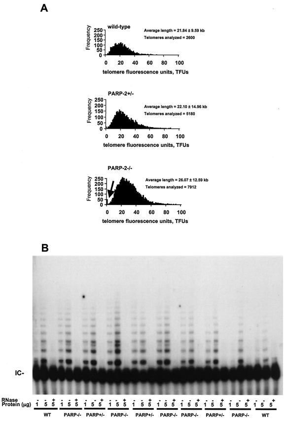
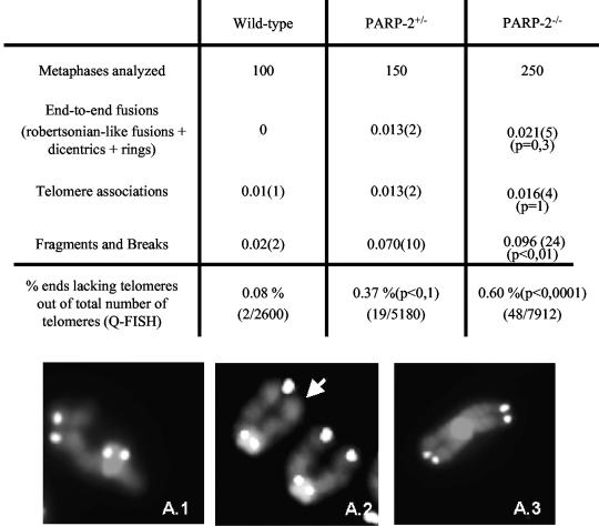
The DNA damage-dependent poly(ADP-ribose) polymerase-2 (PARP-2) is, together with PARP-1, an active player of the base excision repair process, thus defining its key role in genome surveillance and protection. Telomeres are specialized DNA-protein structures that protect chromosome ends from being recognized and processed as DNA strand breaks. In mammals, telomere protection depends on the T(2)AG(3) repeat binding protein TRF2, which has been shown to remodel telomeres into large duplex loops (t-loops). In this work we show that PARP-2 physically binds to TRF2 with high affinity. The association of both proteins requires the N-terminal domain of PARP-2 and the myb domain of TRF2. Both partners colocalize at promyelocytic leukemia bodies in immortalized telomerase-negative cells. In addition, our data show that PARP activity regulates the DNA binding activity of TRF2 via both a covalent heteromodification of the dimerization domain of TRF2 and a noncovalent binding of poly(ADP-ribose) to the myb domain of TRF2. PARP-2(-/-) primary cells show normal telomere length as well as normal telomerase activity compared to wild-type cells but display a spontaneously increased frequency of chromosome and chromatid breaks and of ends lacking detectable T(2)AG(3) repeats. Altogether, these results suggest a functional role of PARP-2 activity in the maintenance of telomere integrity.


Figures

FIG. 1.
FIG. 1.
PARP-2 interacts with TRF2. (A) Lysates from Cos1 cells expressing Myc-hTRF2 fusion protein (lanes 1 to 5) together with either GST (lane 1) or GST-mPARP-2 fusion protein (lanes 2 to 5) were analyzed by GST pull down under increasing stringency conditions of washing buffers as indicated, followed by Western blotting using, successively, anti-Myc (top) and anti-GST (bottom) antibodies. (B) Conditions of interaction between PARP-2 and TRF2. Interaction of GST (lane 1) or GST-mPARP-2 fusion protein (lanes 2 to 5) with Myc-hTRF2 fusion protein (lanes 1 to 5) in Cos1 cells either untreated (lane 2) or treated with 4 mM N-nitroso-N-methylurea (lane 3), 2 mM 3-AB (lane 4), or 10 μg of ethidium bromide/ml (lane 5). Proteins were analyzed by GST pull down and Western blotting using, successively, anti-Myc, anti-GST, and antipolymer antibodies as indicated.
FIG. 2.
FIG. 2.
The N-terminal domain of PARP-2 interacts with the myb domain of TRF2. (A) Upper panel: schematic representation of mPARP-2. DBD, DNA binding domain. Lower panel: GST (lane 1), GST-tagged mPARP-2 (lane 2), and GST-tagged deletion mutants of mPARP-2 (lanes 3 to 5) were expressed in Cos1 cells together with myc-hTRF2 fusion protein (lanes 1 to 5). Interacting proteins were analyzed by GST pull down and Western blotting with anti-Myc and, subsequently, anti-GST antibodies. (B) Upper panel: schematic representation of hTRF2. NLS, nuclear localization signal. Lower panel: lysates from Cos1 cells expressing either GFP (lanes 1, 3, and 5) or GFP-tagged mPARP-2 (lanes 2, 4, and 6 to 9) were mixed with lysates from Cos1 cells expressing either GST (lanes 3 and 4), GST-hTRF2 (lanes 5 and 6), or GST-tagged deletion mutants of hTRF2 (lanes 7 to 9). Proteins were analyzed by GST pull down and Western blotting with, respectively, anti-GFP and anti-GST antibodies.
FIG. 3.
FIG. 3.
Colocalization of PARP-2 and TRF2 in U2OS cells. U2OS cells were transfected with GFP-mPARP-2 proteins (green), fixed 13 h postrelease of a double thymidine block, and stained red with Alexa 568-labeled rabbit anti-PML antibody (a to c) or Alexa 594-labeled goat anti-TRF2 antibody (d to i). To detect PARP activity, cells were treated with 5 mM H2O2 for 10 min before fixation and costained with Alexa 594-labeled anti-TRF2 (red) and fluorescein isothiocyanate-conjugated antipolymer antibodies (green) (g to i).
FIG. 4.
FIG. 4.
The central domain of hTRF2 is poly(ADP-ribosyl)ated. GST, GST-hXRCC1141-572, GST-hTRF2, and GST-tagged deletion mutants of hTRF2 were expressed in Cos1 cells, extracted by GST pull down, and incubated with or without mPARP-2 as indicated in activity buffer containing [α-32P]NAD+ and DNase I-activated DNA. Where indicated, 3-AB was added throughout the experiment. (Right panel) Autoradiography. (Left panel) Subsequently, fusion proteins were analyzed by Western blotting with anti-GST antibody.
FIG. 5.
FIG. 5.
PARP-2 activity negatively regulates TRF2 DNA binding activity. (A) Schematic representation of the telomeric probe dsT4S1. The telomeric repeats are shown in bold. (B) Increasing amounts of either purified mPARP-2 (lanes 1 to 6) or purified hTRF2 (lanes 7 to 12) were incubated with the radiolabeled telomeric probe under binding conditions for 30 min at 20°C. Complexes were analyzed by electrophoresis at 4°C through a nondenaturing 1% agarose gel and phosphorimaging of the dried gel. The positions of free DNA and covalent complexes (mPARP-2-DNA and hTRF2-DNA) are indicated. (C) Purified mPARP-2 was incubated with the radiolabeled telomeric probe for various times as indicated. The complexes were analyzed on a nondenaturing 1% agarose gel as described for panel B. The positions of free DNA and covalent complexes (mPARP-2-DNA) are indicated. (D) Purified hTRF2 (100 nM) was preincubated with the radiolabeled telomeric probe for 10 min under binding conditions at 20°C, and purified mPARP-2 (100 nM) was subsequently added for 10 min (lanes 4 to 6), followed by the addition of the indicated antibodies (lanes 5 and 6). In lanes 1 to 3, the radiolabeled telomeric substrate was incubated with, respectively, no protein, hTRF2 (100 nM), and mPARP-2 (100 nM) for 30 min under binding conditions at 20°C. In lanes 7 to 10, purified hTRF2 (lanes 7 and 9) or purified mPARP-2 (lanes 8 and 10) was preincubated with the radiolabeled probe before the addition of the indicated antibodies. The binding products were analyzed on a nondenaturing 1% agarose gel as described for panel B. The positions of free DNA, covalent complexes (mPARP-2-hTRF2-DNA, hTRF2-DNA, and mPARP-2-DNA), and supershifts are indicated. (E) Purified hTRF2 (100 nM) was preincubated with the radiolabeled telomeric substrate for 10 min under binding conditions as described for panel D, and purified mPARP-2 (100 nM) was subsequently added for 10 min (lanes 4 to 10) followed by the addition of NAD+ for various times as indicated in the absence (lanes 4 to 9) or in the presence of 3-AB (lane 10). In lanes 1 to 3, the radiolabeled telomeric substrate was incubated with, respectively, no protein, mPARP-2 (100 nM), or hTRF2 (100 nM) for 30 min under binding conditions. The binding products were analyzed on a nondenaturing 1% agarose gel as described for panel B. The positions of free DNA, covalent complexes, and supershifts are indicated. Note that the migration of the mPARP-2-hTRF2-DNA complex was only slightly retarded compared to that of the hTRF2-DNA complex.
FIG. 6.
FIG. 6.
Poly(ADP-ribose) inhibits the DNA binding activity of hTRF2. (A) Purified hTRF2 (30 nM) was incubated with the radiolabeled telomeric probe and increasing amounts of poly(ADP-ribose) as indicated for 30 min at 20°C under binding conditions as described in the legend for Fig. 5. The binding products were analyzed by autoradiography after nondenaturing 1% agarose gel electrophoresis. The positions of free DNA and covalent complexes (hTRF2-DNA) are indicated. (B) Poly(ADP-ribose) binds to purified recombinant hTRF2. Upper panel: 1 μg of purified recombinant hTRF2, hXRCC1, or mPARP-2 was spotted onto nitrocellulose and incubated with [32P]poly(ADP-ribose) as described in Materials and Methods. Lower panel: the amount of protein loaded was controlled by Coomassie staining. (C) Poly(ADP-ribose) binds to the myb domain of hTRF2. GST (lane 1), GST-hXRCC1141-572 (lane 2), GST-hTRF2 (lane 3), and GST-tagged deletion mutants of hTRF2 (lanes 4 to 6) were expressed in Cos1 cells, extracted by GST pull down, separated on SDS-12% PAGE, blotted onto nitrocellulose membrane, renatured, and incubated with [32P]poly(ADP-ribose) as described for panel B. Lane 7, hTRF2 (1 μg). Upper panel: autoradiography. Lower panel: subsequently, fusion proteins were analyzed by Western blotting with anti-GST.
FIG. 7.
FIG. 7.
PARP-2 deficiency does not alter telomere length nor telomerase activity. (A) Telomere length analysis by Q-FISH. Histograms show telomere length frequencies (i.e., the number of telomeres of a given length, indicated on the x axis) in primary MEFs from littermate wild-type, PARP-2+/−, and PARP-2−/− mice. The histograms indicate a similar distribution of telomere length frequencies in the different genotypes. One telomere-forming unit (TFU) corresponds to 1 kb of TTAGGG repeats. The increase in the number of signal-free ends (telomeres with undetectable TTAGGG signal) in PARP-2+/− and PARP-2−/− MEFs is indicated with an arrow. (B) Telomerase activity. S-100 extracts were prepared from primary MEFs of the indicated genotypes. Extracts were pretreated or not with RNase. The protein concentration used (in micrograms) is indicated. The internal control (IC) for PCR efficiency is indicated.
FIG. 8.
FIG. 8.
Chromosomal instability in wild-type, PARP-2+/−, and PARP-2−/− primary MEFs. (Upper panel) Frequency of the indicated chromosomal aberrations per metaphase in the indicated number of metaphases in wild-type, PARP-2+/−, and PARP-2−/− primary MEFs. In parentheses is the number of aberrations in the total number of metaphases. The percentage of undetectable telomeric signals out of the total number of telomeres analyzed is also indicated. (Lower panel) Representative images of a chromosome break (A1), a signal-free end (as indicated by an arrow in A2), and an end-to-end Robertsonian-like fusion (A3).

References

    1. Ame, J. C., V. Rolli, V. Schreiber, C. Niedergang, F. Apiou, P. Decker, S. Muller, T. Hoger, J. Menissier-de Murcia, and G. de Murcia. 1999. PARP-2, a novel mammalian DNA damage-dependent poly(ADP-ribose) polymerase. J. Biol. Chem. 274:17860-17868. - PubMed
    1. Ancelin, K., M. Brunori, S. Bauwens, C. E. Koering, C. Brun, M. Ricoul, J. P. Pommier, L. Sabatier, and E. Gilson. 2002. Targeting assay to study the cis functions of human telomeric proteins: evidence for inhibition of telomerase by TRF1 and for activation of telomere degradation by TRF2. Mol. Cell. Biol. 22:3474-3487. - PMC - PubMed
    1. Augustin, A., C. Spenlehauer, H. Dumond, J. Menissier-De Murcia, M. Piel, A. C. Schmit, F. Apiou, J. L. Vonesch, M. Kock, M. Bornens, and G. De Murcia. 2003. PARP-3 localizes preferentially to the daughter centriole and interferes with the G1/S cell cycle progression. J. Cell Sci. 116:1551-1562. - PubMed
    1. Bailey, S. M., M. N. Cornforth, A. Kurimasa, D. J. Chen, and E. H. Goodwin. 2001. Strand-specific postreplicative processing of mammalian telomeres. Science 293:2462-2465. - PubMed
    1. Bianchi, A., and T. de Lange. 1999. Ku binds telomeric DNA in vitro. J. Biol. Chem. 274:21223-21227. - PubMed

Publication types

MeSH terms