. 2004 Feb;24(4):1614-27.
doi: 10.1128/MCB.24.4.1614-1627.2004.

Cdc6 chromatin affinity is unaffected by serine-54 phosphorylation, S-phase progression, and overexpression of cyclin A

Affiliations

Cdc6 chromatin affinity is unaffected by serine-54 phosphorylation, S-phase progression, and overexpression of cyclin A

Mark G Alexandrow et al. Mol Cell Biol. 2004 Feb.

Abstract

Ectopically expressed Cdc6 is translocated from the nucleus during S phase in a cyclin A-Cdk2-dependent process, suggesting that reinitiation of DNA replication is prevented by removal of phosphorylated Cdc6 from chromatin after origin firing. However, whether endogenous Cdc6 translocates during S phase remains controversial. To resolve the questions regarding regulation of endogenous Cdc6, we cloned the cDNA encoding the Chinese hamster Cdc6 homolog and specifically focused on analyzing the localizations and chromatin affinities of endogenous and exogenous proteins during S phase and following overexpression of cyclin A. In agreement with other reports, ectopically expressed Cdc6 translocates from the nucleus during S phase and in response to overexpressed cyclin A. In contrast, using a combination of biochemical and immunohistochemical assays, we show convincingly that endogenous Cdc6 remains nuclear and chromatin bound throughout the entire S period, while Mcm5 loses chromatin affinity during S phase. Overexpression of cyclin A is unable to alter the nuclear localization of Cdc6. Furthermore, using a phosphospecific antibody we show that phosphoserine-54 Cdc6 maintains a high affinity for chromatin during the S period. Considering recent in vitro studies, these data are consistent with a proposed model in which Cdc6 is serine-54 phosphorylated during S phase and functions as a chromatin-bound signal that prevents reformation of prereplication complexes.


Figures
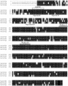
FIG. 1.
FIG. 1.
Alignment of the amino acid sequences of Chinese hamster Cdc6, human Cdc6, and murine Cdc6. The alignment was performed using Clustal W algorithms, followed by shading with the program Boxshade, both available at the Internet location http://restools.sdsc.edu. Residues identical between two or among three species are indicated by dark shading, and similar residues are indicated by light shading. The immunogenic peptide to which the Molecular Probes anti-Cdc6 monoclonal antibody (antibody 4) was made is indicated above the amino acid sequence, as are the Walker A and B boxes (43). Conserved serines that are putative substrates of cyclin-dependent kinases in vitro and in vivo are indicated below the amino acid sequence (see text for references). GenBank accession numbers are as follows: human Cdc6, NM_001254; murine Cdc6, NM_011799; Chinese hamster Cdc6, AY491989.
FIG. 2.
FIG. 2.
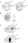
Characterization of antibodies. (A) (Left) Western analysis of TCEs from asynchronous HeLa, 3T3, and CHO cells or from CHO cells transfected with pRcLac-CgCdc6, probed with polyclonal anti-Cdc6 (antibody 1). Arrowheads indicate Cdc6 bands. (Right) Western analysis of the same samples probed with monoclonal anti-Cdc6 (antibody 2). (B) (Left) TCEs from asynchronous HeLa, 3T3, and CHO cells analyzed by immunoblotting using mouse monoclonal anti-Cdc6 (antibody 3). (Right) Histidine-tagged CgCdc6 purified from Escherichia coli was analyzed by immunoblotting using monoclonal anti-Cdc6 (antibody 3). Note the degradation product below the predominant full-length Cdc6 band. (C) Asynchronous HeLa and CHO TCEs, HeLa and CHO nuclear fractions (Nucl; isolated as described for log-phase P3 pellet in Fig. 3), bacterially expressed GST-tagged CgCdc6, and log-phase CHO cells transfected with pRcLac-CgCdc6 were used to analyze the specificity of the anti-Cdc6-phosphoserine-54 polyclonal antibody. (Top) Cells probed with phosphoserine-54-specific anti-Cdc6 antibody. (Bottom) Cells probed with nonphosphospecific anti-Cdc6 polyclonal antibody 1. Arrowheads in the lower panel indicate two Cdc6-specific bands in this experiment, while a third is visible upon longer exposure (data not shown). Numbers at left of panels indicate molecular masses in kilodaltons.
FIG. 3.
FIG. 3.
Chromatin association and detergent sensitivity of Cdc6 and Cdc654P during G0, G1, and S phases. CHO cells were synchronized in G0 by isoleucine deprivation, followed by release into G1 and S phases. TCEs, cytosolic-nucleosolic S1 fractions, and detergent-resistant P3 chromatin fractions were isolated at the times indicated above. Asynchronous CHO cells (Log) were also collected as a reference sample. Western blotting with anti-Cdc6 polyclonal antibody 1 (Cdc6), anti-Cdc654P polyclonal antibody (Cdc6-P), and anti-cyclin A polyclonal antiserum was performed. Probing with anti-lamin A/C and antitubulin monoclonal antibodies confirmed that the fractionation procedure was effective. BrdU analysis using immunohistochemistry was performed to verify that cells had been adequately synchronized (Fig. 6).
FIG. 4.
FIG. 4.
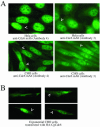
Immunohistochemical analysis of endogenous Cdc6 in CHO and HeLa cells and transfected CgCdc6 in CHO cells. (A) Asynchronous HeLa or CHO cells were analyzed by immunohistochemistry with two independently derived monoclonal antibodies to mammalian Cdc6 (antibodies 3 and 4). The secondary antibody was anti-mouse antibody conjugated with FITC. Arrowheads indicate discrete cytoplasmic focal staining with either antibody. At least 300 nuclei were observed; representative photographs are shown. (B) Asynchronous CHO cells were transfected with the pc2HA-CgCdc6 vector and fixed and analyzed by immunohistochemistry with anti-HA monoclonal antibody followed by anti-mouse antibody conjugated with FITC. Arrowheads indicate diffuse and focal cytoplasmic staining for exogenous HA-CgCdc6. More than 50 transfected cells were observed, and representative cells were photographed.
FIG. 5.
FIG. 5.
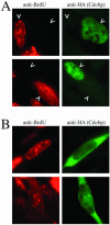
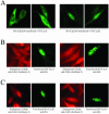
Transfected CgCdc6 is cytoplasmic in S-phase cells. Asynchronous CHO cells were transfected with pc2HA-CgCdc6 and analyzed for exogenous protein expression and BrdU incorporation by immunohistochemistry. BrdU was added to cells for 30 min prior to fixation. HA-CgCdc6 was detected with anti-HA monoclonal antibody conjugated with FITC (green), and BrdU was detected with anti-BrdU monoclonal antibody conjugated with Alexa-594 fluorophore (red). More than 100 transfected cells were analyzed, and representative photographs are shown. (A) Cells expressing Cdc6 in the nucleus do not simultaneously show BrdU labeling and are therefore non-S-phase cells. Arrowheads indicate nuclei with a clear reciprocal relationship between anti-BrdU and anti-HA (Cdc6p) staining. (B) Cells in S phase are labeled with BrdU and consistently show ectopic Cdc6 expression in the cytoplasm.
FIG. 6.
FIG. 6.
BrdU incorporation in synchronized CHO cells and staging of BrdU patterns. CHO cells were synchronized in G0 by isoleucine deprivation (Iso− sample) and were released into G1 and S phase in complete medium for the times indicated above (see Materials and Methods). Cells were pulsed with BrdU for 30 min prior to fixing. Asynchronous cells were also labeled with BrdU (log-phase sample). BrdU labeling was visualized using anti-BrdU monoclonal antibody followed by secondary anti-mouse antibody conjugated with FITC. (Top) Multiple fields were observed for each time point, and representative fields are shown at ×20 magnification. Log-phase cells generally showed 30 to 50% BrdU incorporation. At the peak periods (12 and 15 h), 90 to 95% of cells were labeled. (Bottom) Magnifications of ×100 showing details of nuclei at different stages of S phase. The 9-h point showed primarily stage 1 BrdU patterns. The 12-h point showed a few stage 1, many stage 2, and some stage 3 BrdU patterns. The 15-h point showed primarily stage 3 and 4 BrdU patterns. The 18-h point showed mostly stage 4 and 5 BrdU patterns. At least 100 nuclei were analyzed at each time point, and representative photographs are shown.
FIG. 7.
FIG. 7.
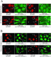
Endogenous Cdc6 remains nuclear and chromatin bound throughout the entire S phase. Asynchronous HeLa and CHO cells were labeled for 30 min with BrdU, fixed immediately (A) or following Triton X-100 treatment (B), and analyzed by immunohistochemistry for BrdU incorporation and endogenous Cdc6 localization. Cdc6-labeled nuclei and BrdU-labeled nuclei are shown in horizontal pairs. Cdc6 was visualized with anti-Cdc6 monoclonal antibodies 3 and 4, and both Cdc6 monoclonal antibodies were detected with anti-mouse secondary antibody conjugated with FITC (green). Following Cdc6 probing, cells and Cdc6 primary and secondary antibodies were fixed again, and then BrdU was detected with anti-BrdU monoclonal antibody conjugated with Alexa-594 (red). Labeled arrowheads point to S-phase nuclei staged according to the results of Fig. 6. Unlabeled arrowheads indicate non-S-phase cells. For each pair of pictures, at least 100 nuclei were analyzed, and representative pictures are shown.
FIG. 8.
FIG. 8.
Pretreatment with Triton X-100 effectively removes Mcm5 from S-phase nuclei. Asynchronous HeLa cells were labeled for 30 min with BrdU, fixed immediately (A) or following Triton X-100 treatment (B), and analyzed by immunohistochemistry for BrdU incorporation and Mcm5 localization. Mcm5 was detected with polyclonal anti-Mcm5 followed by secondary anti-rabbit antibody conjugated with Texas Red (red). BrdU was detected with monoclonal anti-BrdU followed by secondary anti-mouse antibody conjugated with FITC (green). Arrowheads point to non-S-phase cells or S-phase cells staged according to the results in Fig. 6. At least 100 nuclei were examined, and representative photographs are shown.
FIG. 9.
FIG. 9.
Overexpression of cyclin A causes exogenous, transfected CgCdc6 to translocate to the cytoplasm but has no effect on endogenous CgCdc6. (A) Asynchronous CHO cells were transfected with the HA-tagged CgCdc6 expression vector and a fivefold molar excess of either untagged pcDNA3-HsCyclin A (left panels) or untagged pcDNA3-HsCyclin E (right panels). Cells expressing HA-CgCdc6 were detected with monoclonal anti-HA followed by secondary anti-mouse antibody conjugated with FITC. (B and C) Asynchronous CHO cells were transfected with pc2HA-HsCyclin A (B) or pc3HA-HsCyclin E (C) and analyzed for endogenous Cdc6 in HA-cyclin-expressing cells. Cdc6 was detected with monoclonal anti-Cdc6 antibody 4 (panel pairs on left) or monoclonal anti-Cdc6 antibody 3 (panel pairs on right), followed by secondary anti-mouse antibody conjugated with Texas Red (red images). Cells and Cdc6 primary and secondary antibodies were fixed again, and HA-cyclin A (B) or HA-cyclin E (C) was detected with monoclonal anti-HA conjugated with FITC (green images). In all experiments, ∼50 HA-expressing cells were analyzed, and representative photographs are shown.

Similar articles

Cited by

References

    1. Alcasabas, A. A., A. J. Osborn, J. Bachant, F. Hu, P. J. Werler, K. Bousset, K. Furuya, J. F. Diffley, A. M. Carr, and S. J. Elledge. 2001. Mrc1 transduces signals of DNA replication stress to activate Rad53. Nat. Cell Biol. 3:958-965. - PubMed
    1. Bell, S. P., and A. Dutta. 2002. DNA replication in eukaryotic cells. Annu. Rev. Biochem. 71:333-374. - PubMed
    1. Bell, S. P., and B. Stillman. 1992. ATP-dependent recognition of eukaryotic origins of DNA replication by a multiprotein complex. Nature 357:128-134. - PubMed
    1. Brush, G. S., and T. J. Kelly. 2000. Phosphorylation of the replication protein A large subunit in the Saccharomyces cerevisiae checkpoint response. Nucleic Acids Res. 28:3725-3732. - PMC - PubMed
    1. Bueno, A., and P. Russell. 1992. Dual functions of CDC6: a yeast protein required for DNA replication also inhibits nuclear division. EMBO J. 11:2167-2176. - PMC - PubMed

Publication types

MeSH terms

Associated data

LinkOut - more resources