. 2004 Feb;24(4):1667-79.
doi: 10.1128/MCB.24.4.1667-1679.2004.

Placental failure and impaired vasculogenesis result in embryonic lethality for neuropathy target esterase-deficient mice

Affiliations

Placental failure and impaired vasculogenesis result in embryonic lethality for neuropathy target esterase-deficient mice

Markus Moser et al. Mol Cell Biol. 2004 Feb.

Abstract

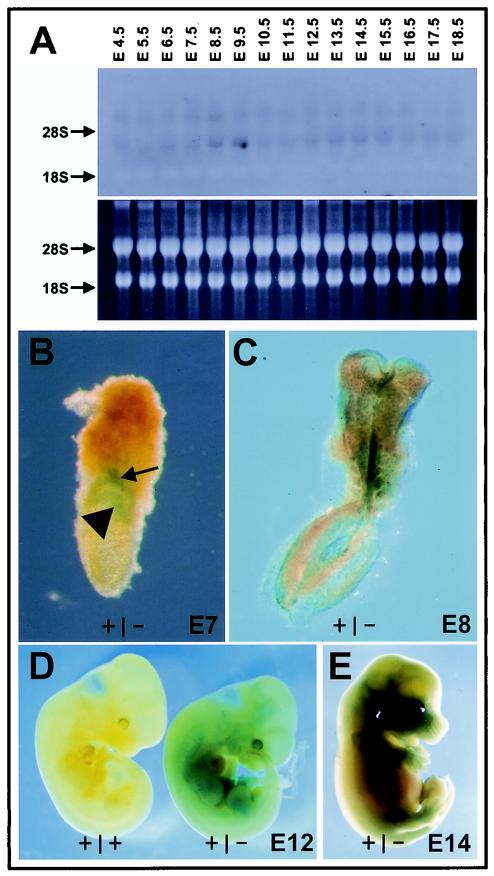
Age-dependent neurodegeneration resulting from widespread apoptosis of neurons and glia characterize the Drosophila Swiss Cheese (SWS) mutant. Neuropathy target esterase (NTE), the vertebrate homologue of SWS, reacts with organophosphates which initiate a syndrome of axonal degeneration. NTE is expressed in neurons and a variety of nonneuronal cell types in adults and fetal mice. To investigate the physiological functions of NTE, we inactivated its gene by targeted mutagenesis in embryonic stem cells. Heterozygous NTE(+/-) mice displayed a 50% reduction in NTE activity but underwent normal organ development. Complete inactivation of the NTE gene resulted in embryonic lethality, which became evident after gastrulation at embryonic day 9 postcoitum (E9). As early as E7.5, mutant embryos revealed growth retardation which did not reflect impaired cell proliferation but rather resulted from failed placental development; as a consequence, massive apoptosis within the developing embryo preceded its resorption. Histological analysis indicated that NTE is essential for the formation of the labyrinth layer and survival and differentiation of secondary giant cells. Additionally, impairment of vasculogenesis in the yolk sacs and embryos of null mutant conceptuses suggested that NTE is also required for normal blood vessel development.


Figures

FIG. 1.
FIG. 1.
NTE targeting vector and genotyping of mutant mice. (A) Schematic representation of the partial NTE gene locus and the targeting construct. Boxes, exons and the probe used for Southern hybridization of the neomycin-resistant stem cell clones, which recognizes the 5.5-kb wild-type (wt) and 2-kb mutant EcoRI fragments; arrowheads, primers used for screening ES cell DNA by genomic PCR. ko, knockout. (B) Homologous recombination resulted in generation of a 1.8-kb fragment (asterisk). (C) PCR analysis of yolk sac DNA from embryos derived from heterozygous matings. The positions of primer pairs for amplification of the 190-bp wild-type and 380-bp mutant PCR fragments are shown in panel A. (D) Western blots of cell lysates from in vitro-cultured wild-type, heterozygous, and NTE-null embryos were probed with rabbit anti-mouse NTE serum. Equal protein loading is shown by antitubulin staining.
FIG. 2.
FIG. 2.
NTE mutant embryos reveal growth retardation. (A to D) Embryos at E7. NTE mutants (C and D) are already slightly growth retarded at E7 compared to wild-type (A) and heterozygous (B) embryos from the same litter. Nevertheless, mutant embryos develop with embryonic (arrow) and extraembryonic (arrowhead) ectoderms. (E and F) At E8 growth retardation of the NTE knockout embryos (F) becomes more evident in comparison with a wild-type embryo (E). The allantois (arrows) and amnion (arrowheads) of the wild-type embryo are shown. (G) At E9 the developmental retardation becomes more obvious. (H) LacZ staining of a wild-type and NTE-heterozygous embryos at E9. LacZ is expressed throughout the heterozygous embryo, including head structures, neural tissue, somites, and tail bud, whereas no staining is present in the wild-type embryo. Panels A to D have the same magnification, as do panels E and F.
FIG. 3.
FIG. 3.
Histological analyses of wild-type and NTE mutant embryos at E7.5 to E9.5. (A to C) Sagittal sections through E7.5 wild-type (A) and NTE mutant (B and C) embryos prove the formation of three germ layers, the allantois, and a thinner chorion in NTE-null embryos. (D to F) Chorioallantoic fusion occurs at E8 in wild-type embryos (D), whereas in NTE-null embryos fusion is delayed (E) and occurs at E8.5 (F; arrow). (G and H) Strong growth retardation at E9.5 can be seen by comparing histological sections of wild-type (G) and mutant (H) embryos. (I) Chorioallantoic fusion of E9.5 NTE-deficient mice. Note nucleated red blood cells in vessels of the allantois, indicating a functional connection with the chorion (arrow). al, allantois; am, amnion; ch, chorion; cp, chorionic plate; ec, ectoderm; en, endoderm; me, mesoderm. Panels A to C have the same magnification, as do panels D to F and panels G and H.
FIG. 4.
FIG. 4.
Expression of NTE during embryonic development and in adult tissues. (A) Northern blot from total embryos between E4.5 and E18.5 reveals two transcripts of about 4.4 and 9 kb at all different time points examined. (B to E) LacZ staining of heterozygous embryos from E7 (B), E8 (C), E12 (D), and E14 (E) shows NTE expression in the extraembryonic ectoderm of the chorion (arrowhead) and ectoplacental cone (arrow) at E7; expression is visible in almost all tissues of the embryo at later stages. A wild-type embryo at E12 (D) lacks any LacZ staining and served as a control for staining specificity.
FIG. 5.
FIG. 5.
Cell proliferation and cell death in NTE mutant embryos. BrdU labeling experiments reveal no labeled cells in NTE-deficient embryos (B and C), whereas wild-type embryos reveal strong proliferation throughout the embryo (A). However, Ki67 staining indicates that cell proliferation in knockout mice (E and F) is indistinguishable from that in normal embryos (D). (G to J) TUNEL staining of E9.5 (G to I) and E10.5 (J) mouse embryos of wild-type (G) and NTE-deficient mice (H to J) indicates massive cell death already at E9.5 in various regions of the embryo in comparison to only a few positive cells in the wild-type embryo (G). The same magnification was used for all panels.
FIG. 6.
FIG. 6.
NTE-deficient cells survive in vitro. Isolated cells from E8 NTE-null embryos survive several days in culture and differentiate into a number of different cell types (A and B), including cells (C) with long (arrow) and branched (arrowhead) neurite-like processes.
FIG. 7.
FIG. 7.
Defects in placental development in NTE mutants. Histology of the placenta in NTE mutant conceptuses. (A to D) Histological sections of wild-type and NTE mutant placentas at E7.5. (C and D) In situ hybridizations with the giant cell marker Pl-1. Arrows, giant cells. Note that the compact cells of the ectoplacental cone are almost absent in NTE mutants and that the chorion is much thinner than it is in wild-type extraembryonic tissue. At E8.0 the chorionic plate is curved in NTE mutant placentas (F), compared to the flat chorionic plate of wild-type placentas (E) (bar and curve). (G and H) Higher-magnification version of panels E and F showing the smaller ectoplacental cone (epc) and the thinner chorionic plate (cp) of mutant placentas. (I to L) At E8.5 defects in placental development become more evident. The sizes of the ectoplacental cone and the chorion are strongly reduced, and fewer giant cells (L; arrows) are detectable in mutants. In addition, the nuclei and cells of the ectoplacental cone seem to be bigger in NTE-deficient placentas. Panels K and L are higher-magnification versions of panels I and J, respectively). (M to P) Placentas at developmental stages E9.5 and E10.5. Locations of the allantois (al), labyrinth (la), spongiotrophoblast layer (sp), giant cells (gi), and maternal deciduae (de) are indicated. Big arrows (N and P), giant cells in mutant placentas; small arrows (N), folds within the chorion, where primitive embryonic vessels start to form. At E10.5 the mutant placenta is totally collapsed. Only a few giant cells (arrow) and a thin layer of chorion and spongiotrophoblast cells remain (P). Compared pictures were taken at the same magnification. Dotted lines mark the interface between trophoblast giant cells and decidua.
FIG. 8.
FIG. 8.
Trophoblast marker analysis of NTE mutants at E8.0, E8.5, and E9.5. Serial sections of wild-type (wt) and mutant (knockout [ko]) conceptuses were probed with antisense riboprobes for Pl-1, Mash2, and 4311. Pl-1 is expressed in trophoblast giant cells; Mash2 is expressed in the chorionic plate at E8.0, the ectoplacental cone at E8.5, and the labyrinth and spongiotrophoblast cells at E9.5; and 4311 is expressed in the spongiotrophoblast layer of E9.5 placentas. Note that at E9.5 almost no giant cells are labeled and no labyrinth has formed in NTE mutants, whereas spongiotrophoblast cells are present in NTE-deficient placentas. Strikingly these cells are negative for Mash2. All pictures were taken with the same magnification. Dotted lines indicate the size and shape of the ectoplacental cone. H&E, hematoxylin and eosin.
FIG. 9.
FIG. 9.
Apoptosis and cell proliferation in NTE-deficient placentas. There is significantly higher apoptotic cell death in the placentas of NTE mutants. (A to D) TUNEL staining reveals apoptosis already at E8.0 within the ectoplacental cone (arrows) and giant cells (arrowhead) of NTE mutants, whereas only a few apoptotic cells are stained at the interface between the ectoplacental cones and deciduae of wild-type placentas. There are more apoptotic cells in the ectoplacental cone of NTE-null placentas. Proliferation is not impaired in the placentas of NTE mutants but is mainly restricted to the chorionic plate at E8.0 (F) and to the chorionic stem cells at E9.5 (H). Even at E10.5, the remaining chorionic stem cells still proliferate (J). Proliferating cells were detected by anti-Ki67 immunohistochemistry. Dotted lines indicate the border between ectoplacental cone and giant cells.
FIG. 10.
FIG. 10.
Vascular defects in yolk sac and within the embryos of NTE mutants. (A to D) Vascularization of the yolk sac initially occurs normally. Blood islands are lined by endothelial cells (ec), the mesoderm (m), and the visceral endoderm (en). (E and F) Vascularization collapses at E9.5, when vasculogenesis occurs in wild-type yolk sacs. (G) Sagittal sections through an E9.5 NTE mutant embryo reveal severe hemorrhages in the head (arrow) or extremely dilated dorsal aortae (*). (H) In other embryos the paired dorsal aortae seemed to be less affected (*) but other blood vessels are dilated (arrow).

Similar articles

Cited by

References

    1. Anson-Cartwright, L., K. Dawson, S. J. Fisher, R. A. Lazzarini, and J. C. Cross. 2000. The glial cells missing-1 protein is essential for branching morphogenesis in the chorioallantoic placenta. Nat. Genet. 25:311-314. - PubMed
    1. Carmeliet, P., V. Ferreira, G. Breier, S. Pollefeyt, L. Kieckens, M. Gertsenstein, M. Fahrig, A. Vandenhoeck, K. Harpal, C. Eberhardt, C. Declercq, J. Pawling, L. Moons, D. Collen, W. Risau, and A. Nagy. 1996. Abnormal blood vessel development and lethality in embryos lacking a single VEGF allele. Nature 380:435-439. - PubMed
    1. Colosi, P., F. Talamantes, and D. I. Linzer. 1987. Molecular cloning and expression of mouse placental lactogen I complementary deoxyribonucleic acid. Mol. Endocrinol. 1:767-776. - PubMed
    1. Cross, J. C., M. L. Flannery, M. A. Blanar, E. Steingrimsson, N. A. Jenkins, N. G. Copeland, W. J. Rutter, and Z. Werb. 1995. Hxt encodes a basic helix-loop-helix transcription factor that regulates trophoblast cell development. Development 121:2513-2523. - PubMed
    1. Ehrich, M., and B. S. Jortner. 2001. Organophosphorus-induced delayed neuropathy, p. 987-1012. In R. Krieger (ed.), Handbook of pesticide toxicology, 2nd ed. Academic Press, San Diego, Calif.

Publication types

MeSH terms