. 2004 Feb;24(4):1799-808.
doi: 10.1128/MCB.24.4.1799-1808.2004.

Enhancer I predominance in hepatitis B virus gene expression

Affiliations

Enhancer I predominance in hepatitis B virus gene expression

Gilad Doitsh et al. Mol Cell Biol. 2004 Feb.

Abstract

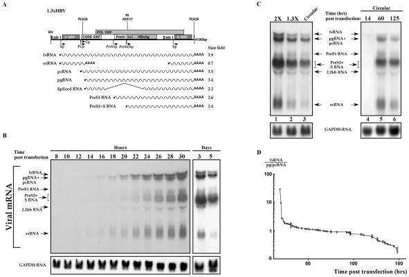
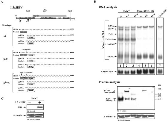
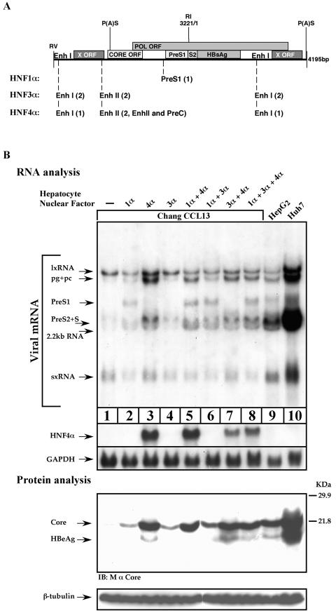
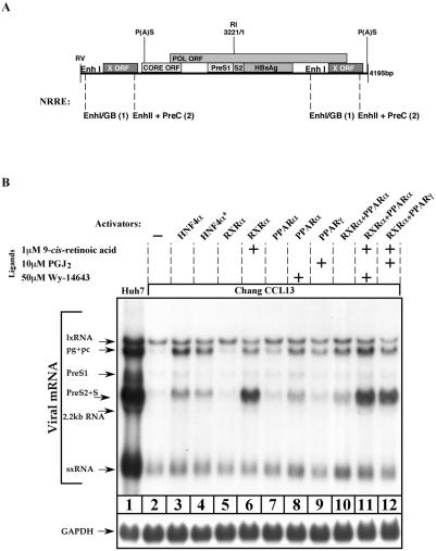
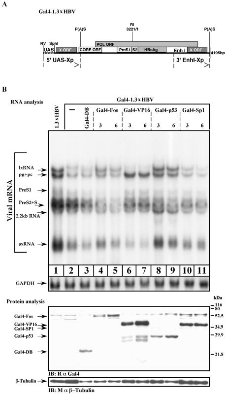
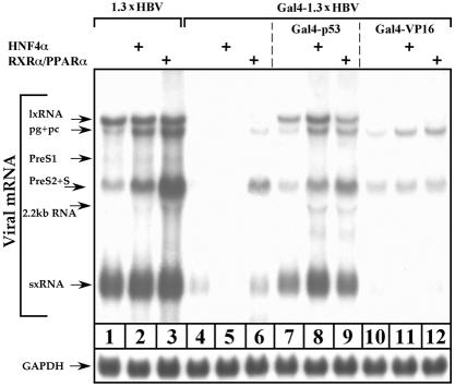
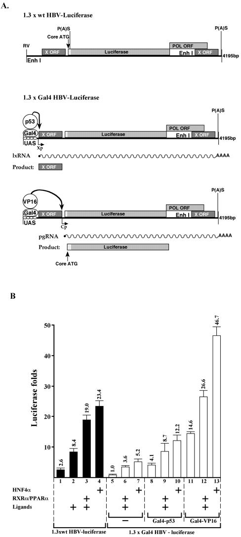
Previous studies of human hepatitis B virus (HBV) transcription revealed the requirement of two enhancer elements. Enhancer I (EnhI) is located upstream of the X promoter and is targeted by multiple activators, including basic leucine zipper proteins, and enhancer II (EnhII) is located upstream to the PreCore promoter and is targeted mainly by nuclear receptors (NRs). The mode of interplay between these enhancers and their unique contributions in regulating HBV transcription remained obscure. By using time course analysis we revealed that the HBV transcripts are categorized into early and late groups. Chang (CCL-13) cells are impaired in expression of the late transcripts. This could be corrected by overexpressing EnhII activators, such as hepatocyte nuclear factor 4 alpha, the retinoid X receptor alpha, and the peroxisome proliferator-activated receptor alpha, suggesting that in Chang cells EnhI but not EnhII is active. Replacing the 5'-end EnhI sequence with a synthetic Gal4 response (UAS) DNA fragment ceased the production of the early transcripts. Under this condition NR overexpression poorly activated EnhII. However, activation of the UAS by Gal4-p53 restored both the expression of the early transcripts and the EnhII response to NRs. Thus, a functional EnhI is required for activation of EnhII. We found a major difference between Gal4-p53 and Gal4-VP16 behavior. Gal4-p53 activated the early transcripts, while Gal4-VP16 inhibited the early transcripts but activated the late transcripts. These findings indicate that the composition of the EnhI binding proteins may play a role in early to late switching. Our data provides strong evidence for the role of EnhI in regulating global and temporal HBV gene expression.


Figures

FIG. 1.
FIG. 1.
Temporal HBV gene expression. (A) Schematic illustration of the 1.3xHBV DNA construct used in this study and the expected mRNA species. The different HBV ORFs and promoters (arrows) are shown. P(A)S indicates the position of the polyadenylation signal. The EcoRV (RV) and EcoRI (RI) unique sites are indicated. (B) Huh7 cells were transfected with plasmids containing 1.3 copies of HBV DNA and harvested at the indicated time points (hours/days) posttransfection. RNA was extracted, separated on a formaldehyde-agarose gel, and analyzed by using a 32P-X gene DNA probe. A GAPDH probe was used to quantify RNA in each lane. Arrows indicate the position of the known viral transcripts. The 2.2-kb transcript is an HBV spliced RNA (data not shown). Note that lx- and sxRNA are the first to be visible (16 h posttransfection) and are the first to disappear at later time points. (C) The pattern of HBV transcription is not template dependent. The transcription pattern obtained by three different HBV DNA configurations is shown in lanes 1 to 3. At lane 1 two tandem copies of complete HBV genome ligated at the unique EcoRI site was used (2X). For lane 2 the construct shown in panel A was used. For lane 3 HBV DNA was linearized at the EcoRI site and was self ligated as described previously (12) to obtain a circular intact HBV genome. The circular HBV DNA template was used in a time course experiment (lanes 4 to 6). (D) Time-dependent ratio between the lxRNA and the pg/pcRNA level. The levels of lxRNA and the pg/pcRNA were measured by phosphorimager at different time points and were plotted in a semilogarithmic scale versus time (hours). The data summarize 14 different experiments, each with about 10 different time points (n = 153).
FIG. 2.
FIG. 2.
Chang cells are defective in late gene expression. (A) Schematic drawing of HBV transcripts and proteins produced by the three HBV constructs. The wild-type (wt) 1.3xHBV construct programs the synthesis of the X, Core, and HBeAg proteins by the lxRNA, pgRNA, and pcRNA, respectively. The X-C construct, harboring a fused X-Core gene, directs the synthesis of a 40-kDa X-Core fused product by the lxRNA. The X-C mutant directs the synthesis of Core protein by the pcRNA but is incapable of producing the HBeAg by the pcRNA, since the HBeAg gene resides upstream of the frameshift mutation site (12). The Xko-C mutant harbors an additional stop mutation at position 27 of the 5′-end X gene and is therefore incapable of producing both the X-Core fused product and the HBeAg. (B) Huh7 and Chang cells were transfected with either the wt 1.3xHBV DNA (lanes 1 and 4), the X-C mutant (lanes 2 and 5), or the Xko-C mutant (lanes 3 and 6). Lane 7 shows the RNA pattern obtained upon transfecting Chang cells with a circular HBV DNA. RNA was analyzed as described in the legend to Fig. 1. For Northern blotting hybridization the X-gene DNA probe was used. For protein analysis, monoclonal mouse anti-Core (mAb 22) and a monoclonal mouse anti-β-tubulin antibody were used. (C) The Huh7-transfected cells with wt HBV DNA were analyzed for pX production by using anti-pX-specific monoclonal antibody generated in our laboratory. IB, immunoblot.
FIG. 3.
FIG. 3.
Overexpression of HNF4α is sufficient to restore HBV gene expression. (A) A schematic drawing depicting the different hepatocyte nuclear factor-responsive elements situated along the 1.3xHBV DNA. Each element is depicted by its position within the HBV DNA, and the number of binding sites is shown in parenthesis. (B) Chang cells were transfected with wild-type HBV DNA alone or together with 200 ng of different combinations of plasmids expressing HNF1α, HNF3α, or HNF4α proteins. As a positive control highly differentiated HepG2 and Huh7 cells were transfected only with HBV DNA. Cells were harvested 40 h posttransfection for total RNA and protein analysis. The same blotting membrane was subsequently used for hybridization with the X-gene DNA, HNF4α DNA, and GAPDH DNA probes. For protein analysis monoclonal mouse anti-Core or anti-β-tubulin antibody was used. IB, immunoblot
FIG. 4.
FIG. 4.
Induction of the HBV late transcripts by overexpression of NRs. (A) Schematic presentation of the different NRREs situated along the 1.3xHBV DNA. Each element is depicted by its position within the HBV DNA, and the number of binding sites is indicated in parenthesis. (B) Chang cells were transfected with wild-type HBV DNA alone or together with 200 ng of different combinations of plasmids that express HNF4α, RXRα, PPARα, and PPARγ proteins. As a positive control, highly differentiated Huh7 cells were transfected only with HBV DNA. Cells (except control cells of line 4*) were plated in medium supplemented with 8% charcoal-treated fetal bovine serum from 24 h prior to transfection up to harvesting. After removal of transfected excess DNA, 9-cis-retinoic acid, prostaglandin J2 (PGJ2), and Wy-14643 were added to the cell media at the amounts indicated in the final concentrations. These are the ligands of RXRα, PPARγ, and PPARα, respectively. All cells were harvested 40 h posttransfection for total RNA analysis. For Northern blotting hybridization the X-gene DNA probe was used. GAPDH probe was used to quantify RNA in each lane.
FIG. 5.
FIG. 5.
EnhI regulates HBV transcription. (A) Schematic illustration of the Gal4-1.3xHBV construct. The 5′ copy of EnhI has been replaced by four repeats of a synthetic Gal4-responsive element (UAS). EcoRV and SphI sites are the unique restriction sites flanking the UAS region. Arrows indicate the redundant termini. (B) Huh7 cells were transfected with Gal4-1.3xHBV DNA alone or together with 3 or 6 μg of Gal4-Fos, Gal4-VP16, Gal4-p53, and Gal4-Sp1 expression plasmids as indicated. Cells were harvested 40 h posttransfection for total RNA and protein analysis. For Northern blotting hybridization, the X-gene DNA probe was used. GAPDH probe was used to quantify RNA in each lane. For protein analysis, polyclonal rabbit anti-Gal4 (RαGal4) or anti-β-tubulin antibodies were used. The position of the different Gal4 chimera is shown. IB, immunoblot.
FIG. 6.
FIG. 6.
EnhI predominates HBV gene expression; analysis by Northern blotting. Chang cells were transfected with a wild-type 1.3xHBV DNA or were cotransfected with a mutant Gal4-1.3xHBV DNA together with 3 μg of Gal4-p53 or Gal4-VP16 expression plasmids as indicated. In addition, cells were cotransfected with 200 ng of HNF4α or 150 ng of RXRα and PPARα expression plasmids. Cells were plated in medium supplemented with 8% charcoal-treated fetal bovine serum from 24 h prior to transfection until harvesting. After removal of transfected excess DNA, 1 μM 9-cis-retinoic acid and 50 μM Wy-14643 were added to the RXRα-PPARα-transfected cells. Cells were harvested 40 h posttransfection for total RNA analysis as performed above.
FIG. 7.
FIG. 7.
EnhI predominates HBV gene expression; analysis by HBV-based reporter plasmids. (A) The structure of the constructed HBV-based reporter plasmids is shown. Also indicated are the predicted modes of activity of the different Gal4-chimera activators and the resultant transcripts and proteins. (B) Chang cells were transfected with the plasmids indicated in panel A, and level of Luciferase activity was measured. The reporter plasmids were cotransfected with the indicated plasmids and were treated with ligands (1 μM 9-cis-retinoic acid and 50 μM Wy-14643).

References

    1. Antonucci, T. K., and W. J. Rutter. 1989. Hepatitis B virus (HBV) promoters are regulated by the HBV enhancer in a tissue-specific manner. J. Virol. 63:579-583. - PMC - PubMed
    1. Batterson, W., and B. Roizman. 1983. Characterization of the herpes simplex virion-associated factor responsible for the induction of alpha genes. J. Virol. 46:371-377. - PMC - PubMed
    1. Ben-Levy, R., O. Faktor, I. Berger, and Y. Shaul. 1989. Cellular factors that interact with the hepatitis B virus enhancer. Mol. Cell. Biol. 9:1804-1809. - PMC - PubMed
    1. Bock, C. T., S. Kubicka, M. P. Manns, and C. Trautwein. 1999. Two control elements in the hepatitis B virus S-promoter are important for full promoter activity mediated by CCAAT-binding factor. Hepatology 29:1236-1247. - PubMed
    1. Cattaneo, R., H. Will, N. Hernandez, and H. Schaller. 1983. Signals regulating hepatitis B surface antigen transcription. Nature 305:336-338. - PubMed

Publication types

MeSH terms