. 2004 Jan 28;24(4):773-80.
doi: 10.1523/JNEUROSCI.0949-03.2004.

Prefrontal cortical-ventral striatal interactions involved in affective modulation of attentional performance: implications for corticostriatal circuit function

Affiliations

Prefrontal cortical-ventral striatal interactions involved in affective modulation of attentional performance: implications for corticostriatal circuit function

Anastasia Christakou et al. J Neurosci. .

Abstract

Anatomically segregated systems linking the frontal cortex and the striatum are involved in various aspects of cognitive, affective, and motor processing. In this study, we examined the effects of combined unilateral lesions of the medial prefrontal cortex (mPFC) and the core subregion of the nucleus accumbens (AcbC) in opposite hemispheres (disconnection) on a continuous performance, visual attention test [five-choice serial reaction-time task (5CSRTT)]. The disconnection lesion produced a set of specific changes in performance of the 5CSRTT, resembling changes that followed bilateral AcbC lesions while, in addition, comprising a subset of the behavioral changes after bilateral mPFC lesions previously reported using the same task. Specifically, both mPFC/AcbC disconnection and bilateral AcbC lesions markedly affected aspects of response control related to affective feedback, as indexed by perseverative responding in the 5CSRTT. These effects were comparable, although not identical, to those in animals with either bilateral AcbC or mPFC/AcbC disconnection lesions. The mPFC/AcbC disconnection resulted in a behavioral profile largely distinct from that produced by disconnection of a similar circuit described previously, between the mPFC and the dorsomedial striatum, which were shown to form a functional network underlying aspects of visual attention and attention to action. This distinction provides an insight into the functional specialization of corticostriatal circuits in similar behavioral contexts.


Figures

Figure 1.
Figure 1.
Representative lesion photomicrographs. A, Photomicrograph of left unilateral mPFC lesion. B, Same lesion as in A but at a higher magnification, with arrowheads indicating recognizable “landmarks” for reference. C, Photomicrograph of left AcbC lesion, with dotted line circumscribing the affected area. D, Left sham-operated control AcbC. Scale bars, 100 μm. aca, Anterior commissure; AcbS, nucleus accumbens shell; Cg1, anterior cingulate cortex; LV, lateral ventricle; M2, motor cortex.
Figure 2.
Figure 2.
Schematic representation of lesion extent. Schematic representation of the extent of mPFC lesions (A) and AcbC lesions (B); the extent of the largest (clear) and smallest (black) lesions is depicted. For details on lesion extent, see Results. The schematics were prepared based on the electronic version of Paxinos and Watson (1998).
Figure 3.
Figure 3.
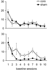
Bilateral AcbC lesions: impulsive responding. The average number of premature nose-pokes performed by animals with bilateral AcbC lesions and their sham-operated controls, within each of 10 baseline sessions, after either correct (top) or failed (bottom) trials is shown. For definition of trial outcomes, see Materials and Methods. core, Bilateral AcbC-lesioned group; sham, bilateral AcbC sham-operated control group.
Figure 4.
Figure 4.
Bilateral AcbC lesions: compulsive responding. The average number of perseverative nose-pokes performed by animals with bilateral AcbC lesions and their sham-operated controls, within each of 10 baseline sessions, after correct (top) or failed (bottom) trials is shown. For definition of trial outcomes, see Materials and Methods. core, Bilateral AcbC-lesioned group; sham, bilateral AcbC sham-operated control group.
Figure 5.
Figure 5.
mPFC/AcbC disconnection lesions: errors of commission. The average number of incorrect responses to the presentation of the target relative to the outcome of the previous trial, within each of 10 baseline sessions or during two SD manipulation sessions, is shown. Top, Incorrect responses after correct trials during baseline testing and during (inset) the 25 and 200csec SD sessions (a baseline session at 50csec SD was interpolated and is also shown). Bottom, Similarly, incorrect responses after failed trials during baseline testing and during (inset) the same SD manipulation sessions. For definition of trial outcomes, see Materials and Methods. disc, mPFC/AcbC disconnection lesioned group; sham, mPFC/AcbC disconnection sham-operated control group.
Figure 6.
Figure 6.
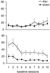
mPFC/AcbC disconnection lesions: impulsive responding. The average number of premature nose-pokes performed by animals with mPFC/AcbC disconnection lesions and their sham-operated controls, within each of 10 baseline sessions, after correct (top) or failed (bottom) trials is shown. For definition of trial outcomes, see Materials and Methods. disc, mPFC/AcbC disconnection lesioned group; sham, mPFC/AcbC disconnection sham-operated control group.
Figure 7.
Figure 7.
mPFC/AcbC disconnection lesions: compulsive responding. The average number of perseverative nose-pokes performed by animals with mPFC/AcbC disconnection lesions and their sham-operated controls, within each of 10 baseline sessions, after correct (top) or failed (bottom) trials is shown. For definition of trial outcomes, see Materials and Methods. disc, mPFC/AcbC disconnection lesioned group; sham, mPFC/AcbC disconnection sham-operated control group.

References

    1. Amalric M, Koob GF (1987) Depletion of dopamine in the caudate–nucleus but not in nucleus–accumbens impairs reaction-time performance in rats. J Neurosci 7: 2129–2134. - PMC - PubMed
    1. Apicella P, Ljungberg T, Scarnati E, Schultz W (1991) Responses to reward in monkey dorsal and ventral striatum. Exp Brain Res 85: 491–500. - PubMed
    1. Baunez C, Robbins TW (1999) Effects of dopamine depletion of the dorsal striatum and further interaction with subthalamic nucleus lesions in an attentional task in the rat. Neuroscience 92: 1343–1356. - PubMed
    1. Bowman EM, Brown VJ (1998) Effects of excitotoxic lesions of the rat ventral striatum on the perception of reward cost. Exp Brain Res 123: 439–448. - PubMed
    1. Bowman EM, Aigner TG, Richmond BJ (1996) Neural signals in the monkey ventral striatum related to motivation for juice and cocaine rewards. J Neurophysiol 75: 1061–1073. - PubMed

Publication types

MeSH terms

LinkOut - more resources