. 2004 Jan 28;24(4):808-18.
doi: 10.1523/JNEUROSCI.4610-03.2004.

Repulsive guidance molecule (RGM) gene function is required for neural tube closure but not retinal topography in the mouse visual system

Affiliations

Repulsive guidance molecule (RGM) gene function is required for neural tube closure but not retinal topography in the mouse visual system

Vera Niederkofler et al. J Neurosci. .

Abstract

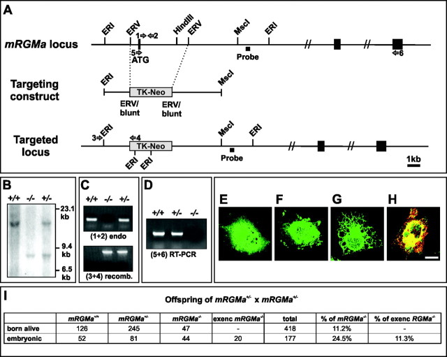
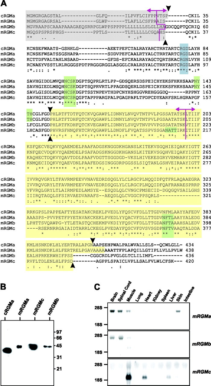
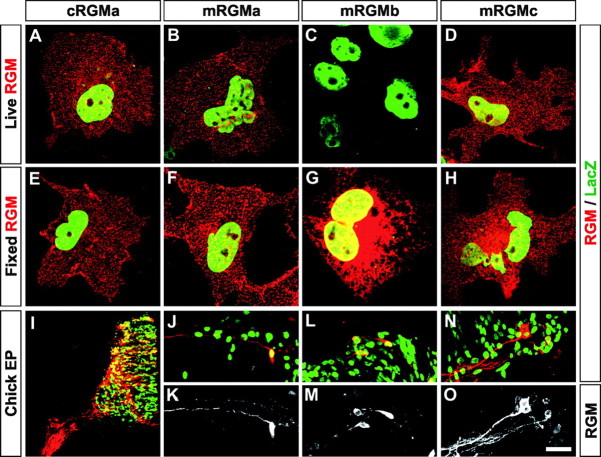
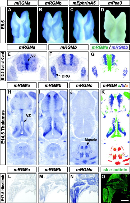
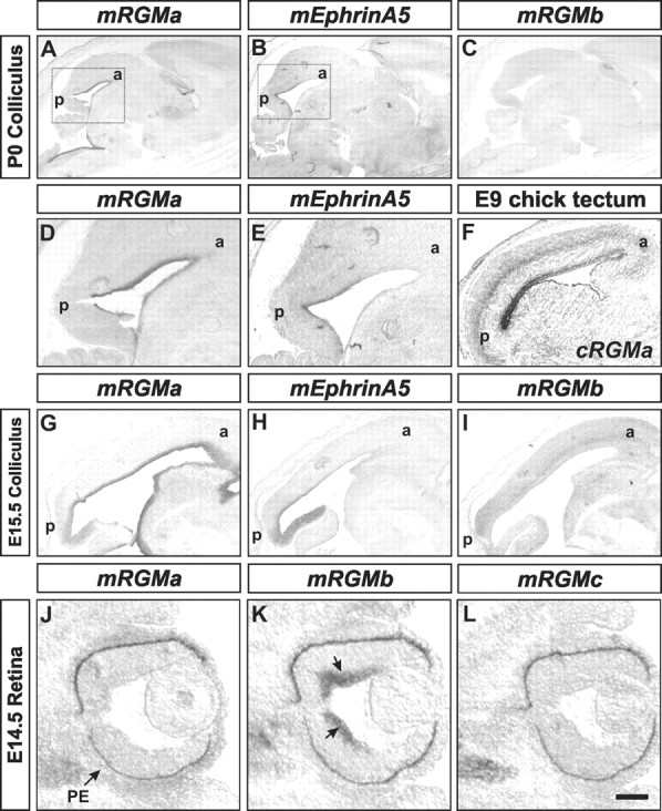
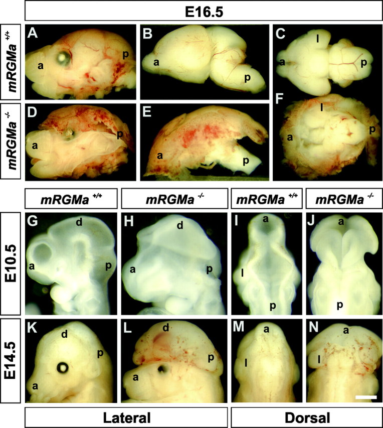
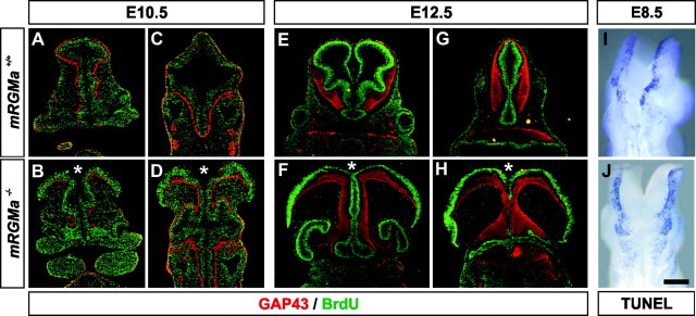
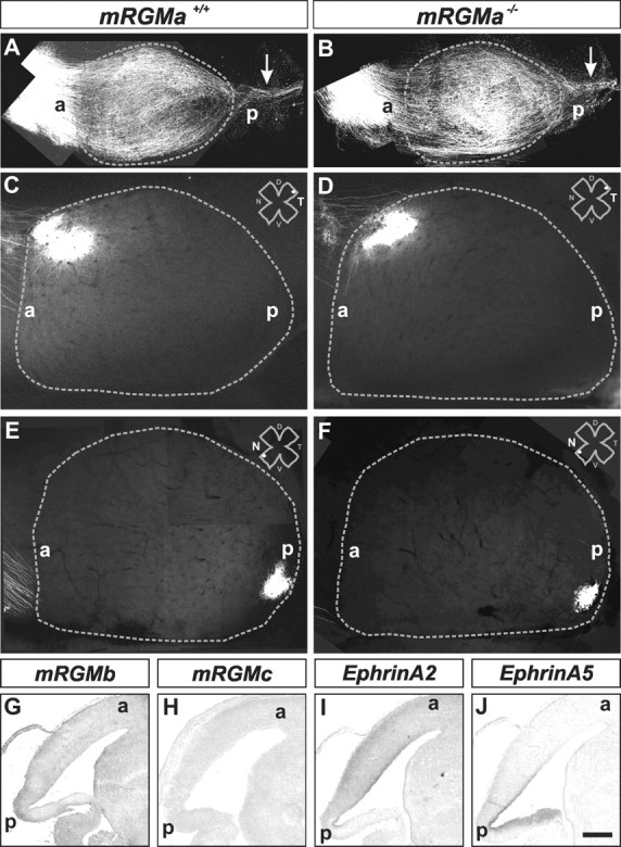
The establishment of topographic projections in the developing visual system depends on the spatially and temporally controlled expression of axon guidance molecules. In the developing chick tectum, the graded expression of the repulsive guidance molecule (RGM) has been proposed to be involved in controlling the topography of the retinal ganglion cell (RGC) axon termination zones along the anteroposterior axis of the tectum. We now show that there are three mouse proteins homologous to chick RGM displaying similar proteolytic processing but exhibiting differential cell-surface targeting by glycosyl phosphatidylinositol anchor addition. Two members of this gene family (mRGMa and mRGMb) are expressed in complementary patterns in the nervous system, and mRGMa is expressed prominently in the superior colliculus at the time of anteroposterior targeting of RGC axons. The third member of the family (mRGMc) is expressed almost exclusively in skeletal muscles. Functional studies in the mouse reveal a role for mRGMa in controlling cephalic neural tube closure, thus defining an unexpected role for mRGMa in early embryonic development. In contrast, mRGMa mutant mice did not exhibit defects in anteroposterior targeting of RGC axons to their stereotypic termination zones in the superior colliculus.


Figures

Figure 5.
Figure 5.
Generation of mRGMa mutant mice. A–D, Targeting strategy for homologous recombination in ES cells to eliminate mRGMa gene function. An EcoRV fragment including the exon containing the methionine of the signal peptide for targeting of mRGMa to the endoplasmic reticulum was replaced by a TK-neomycin resistant cassette (light gray). Coding exons for mRGMa and the probe used for genomic Southern analysis (B) are indicated by black boxes. Oligonucleotides to determine the absence of the methionine-containing exon are indicated by arrows 1 and 2 (C), oligonucleotides to verify 5′ homologous recombination by arrows 3 and 4 (C), and oligonucleotides used for RT-PCR by arrows 5 and 6 (D). RT-PCR analysis (D) was used to verify the absence of the exon containing the signal peptide on mRGMa mRNA in mRGMa mutant mice (arrows 5 and 6 in A). E–H, Immunocytochemistry of COS-7 cells transfected with a cDNA construct containing mRGMa exons 3′ of the targeted exon and eGFP on a bicistronic plasmid (E, F) or a cDNA construct containing an additional artificial in-frame N-terminal methionine and an eGFP containing a signal peptide for targeting to the endoplasmic reticulum (G, H). Cells were stained for eGFP (green) and mRGMa (red). In E and G, the incubation of cells with antibody to mRGMa was performed before the fixation and permeabilization of cells to label cell-surface-associated mRGMa. In F and H, the fixation and permeabilization of cells was performed before incubation with antibodies. Note that even the addition of an artificial N-terminal methionine in frame with the C-terminal mRGMa exons did not result in cell-surface-exposed mRGMa after transfection (G, H). Scale bar, 15 μm. I, Statistical analysis of the offspring recovered from matings of mRGMa+/– breeder pairs. exenc, Exencephalic mRGMa–/– embryos.
Figure 1.
Figure 1.
Characterization of the murine RGM protein family. A, Protein sequence alignment of cRGMa, mRGMa, mRGMb, and mRGMc. Asterisks indicate identical amino acids; pink lines, intron–exon junctions; the gray box, predicted signal peptides; the blue box, potential integrin binding sites (RGD); green boxes, predicted N-glycosylation sites; the yellow box, mature C-terminal RGM fragments after full proteolytic cleavage and C-terminal GPI anchor addition (proteolytic cleavage sites indicated by arrowheads). B, Western blot analysis of supernatant collected from COS-7 cells transfected with C-terminally truncated histidine–Myc-labeled cRGMa, mRGMa, mRGMc, and mRGMb detected with an anti-Myc antibody. Molecular weight standards in kilodaltons are indicated on the right. C, Northern blot analysis on total RNA from a variety of P3 mouse tissues as indicated, using mRGMa, mRGMb, and mRGMc as probes.
Figure 2.
Figure 2.
Differential cell-surface targeting of mRGMs. A–H, Expression of full-length cDNAs coding for cRGMa (A, E), mRGMa (B, F), mRGMb (C, G), and mRGMc (D, H) andβ-galactosidase on the same plasmid using an internal ribosome entry site in COS-7 cells. Transfected COS-7 cells are identified by staining forβ-galactosidase after the fixation and permeabilization of cells (green). RGMs (red) are detected either before fixation and permeabilization of cells (A–D) to detect cell-surface-accumulated RGM or after fixation and permeabilization of cells (E–H) to detect all RGM in transfected cells. I–O, Chick spinal cords electroporated with cDNAs coding for cRGMa (I), mRGMa (J, K), mRGMb (L, M), and mRGMc (N, O) and β-galactosidase on the same plasmid using an IRES. Sections were stained for RGM (red:I,J,L,N;white:K,M,O) and β-galactosidase (green:I,J,L,N). Note the extensive labeling of axonal processes in I,J,K,N, and O and the predominant cell-body and proximal axonal labeling in L and M. Scale bar: (in O) A–H, 15 μm; I, 150 μm; J–O, 60 μm.
Figure 3.
Figure 3.
Embryonic expression of mRGMs in complementary patterns. A–D, Whole-mount in situ hybridization of E8.5 mouse embryos using mRGMa (A), mRGMb (B), mEphrinA5 (C), and mPea3 (D) as probes. Note the absence of mPea3 expression from the tips of neural folds. E–G, Expression of mRGMa (E) and mRGMb (F) in E12.5 mouse spinal cord. Arrows point to the ventricular zone (VZ) in E and the DRG in F, detected by in situ hybridization on transverse sections. G, Artificial overlay (mRGMa in green, mRGMb in blue) to demonstrate complementarity of expression patterns. H–K, Expression of mRGMa (H, VZ), mRGMb (I), and mRGMc (J) in E14.5 mouse thalamus by in situ hybridization on coronal brain sections. The arrow in J points to the signal of mRGMc in skeletal muscles. K shows an artificial overlay (mRGMa in green, mRGMb in blue, and mRGMc in red) to demonstrate the complementarity of expression patterns. L–O, Expression of mRGMa (L), mRGMb (M), and mRGMc (N) by in situ hybridization and skeletal α-actinin (O) by immunohistochemistry on E17.5 mouse hindlimbs. Scale bar: (in O) A–D, 0.15 mm; E–G, 0.35 mm; H–K, 0.32 mm; L–O, 1.8 mm.
Figure 4.
Figure 4.
Expression of mRGMs in the developing retinocollicular system. A–E, Expression of mRGMa (A, D), mEphrinA5 (B, E), and mRGMb (C) in the superior colliculus of P0 sagittal brain sections detected by in situ hybridization. Boxes in A and B indicate regions shown in higher magnification in D and E. a, Anterior; p, posterior. F, Expression of cRGMa in E9 chick tectum detected by in situ hybridization. G–I, Expression of mRGMa (G), mEphrinA5 (H), and mRGMb (long exposure to detect faint expression; I) in the superior colliculus of E15.5 sagittal brain sections detected by in situ hybridization. J–L, Expression of mRGMa (J), mRGMb (K), and mRGMc (L) in E14.5 retina detected by in situ hybridization. Note the expression of mRGMb in RGCs (K, arrows). Pigment epithelium is marked by an arrow in J. Scale bar: (in L) A–C, 1 mm; D, E, 0.4 mm; F, 0.85 mm; G–I, 0.25 mm; J–L, 0.13 mm.
Figure 6.
Figure 6.
mRGMa mutant mice show an exencephalic phenotype. A–F, Lateral (A, B, D, E) or ventral (C, F) view of E16.5 heads (A, D) or dissected brains (B, C, E, F) from mRGMa+/+ (A–C) and exencephalic mRGMa–/– (D–F) embryos. G–J, Lateral (G, H) or dorsal (I, J) head view of E10.5 mRGMa+/+ (G, I) and mRGMa–/– (H, J) embryos. K–N, Lateral (K, L) or dorsal (M, N) head view of E14.5 mRGMa+/+ (K, M) and mRGMa–/– (L, N) embryos. a, Anterior; p, posterior; d, dorsal; l, lateral. Scale bar: (in N) A, D, 1.5 mm; B, E, 1 mm; C, F, 1.1 mm; G–J, 0.6 mm; K–N, 1.2 mm.
(Figure 7.
(Figure 7.
Exencephalic mRGMa mutant embryos do not show defects in proliferation in the brain. A–H, Immunohistochemical analysis (GAP-43: red; BrdU: green) of coronal brain sections from E10.5 (A–D) and E12.5 (E–H) BrdU pulse-labeled mRGMa+/+ (A, C, E, G) and exencephalic mRGMa–/– (B, D, F, H) embryos. *Open ventricle. I, J, Whole-mount TUNEL of E8.5 mRGMa+/+ (I) and exencephalic mRGMa–/– (J) embryos in dorsal view. Scale bar: (in J) A, B, E, F, 0.38 mm; C, D, G, H, 0.5 mm; I, J, 0.13 mm.
Figure 8.
Figure 8.
Lack of retinocollicular projection phenotype in mRGMa mutant mice. A, B, Dorsal views of superior and inferior colliculi from mRGMa+/+ (A) and mRGMa–/– (B) mice after anterograde Dil labeling from the retina at P0 to label many RGC axons. The gray dashed line outlines the superior colliculus; arrows point to RGC axons projecting to the inferior colliculus. C–F, Anterograde Dil labeling from the temporal (C, D) or nasal (E, F) retina to the superior colliculus (outlined by gray dashed lines) after focal Dil injection into the retina of mRGMa+/+ (C, E) and mRGMa–/– (D, F) mice at P10 (injection point in the retina indicated by the white dot the top right corner: T, Temporal; N, nasal; D, dorsal; V, ventral). GJ, Expression of mRGMb (G), mRGMc (H), mEphrinA2(I), and mEphrinA5 (J) in superior colliculi of E16.5 sagittal brain sections from mRGMa/ mice detected by in situ hybridization. a, Anterior; p, posterior. Scale bar: (in J) A, B, 0.5 mm; C–F, 0.33 mm; G–J, 0.3 mm.

References

    1. Arber S, Han B, Mendelsohn M, Smith M, Jessell TM, Sockanathan S (1999) Requirement for the homeobox gene Hb9 in the consolidation of motor neuron identity. Neuron 23: 659–674. - PubMed
    1. Briscoe J, Pierani A, Jessell TM, Ericson J (2000) A homeodomain protein code specifies progenitor cell identity and neuronal fate in the ventral neural tube. Cell 101: 435–445. - PubMed
    1. Brittis PA, Flanagan JG (2001) Nogo domains and a Nogo receptor: implications for axon regeneration. Neuron 30: 11–14. - PubMed
    1. Chen MS, Huber AB, van der Haar ME, Frank M, Schnell L, Spillmann AA, Christ F, Schwab ME (2000) Nogo-A is a myelin-associated neurite outgrowth inhibitor and an antigen for monoclonal antibody IN-1. Nature 403: 434–439. - PubMed
    1. Cheng HJ, Nakamoto M, Bergemann AD, Flanagan JG (1995) Complementary gradients in expression and binding of ELF-1 and Mek4 in development of the topographic retinotectal projection map. Cell 82: 371–381. - PubMed

Publication types

MeSH terms