. 2004 Jan 28;24(4):991-8.
doi: 10.1523/JNEUROSCI.4792-03.2004.

Presynaptic localization of neprilysin contributes to efficient clearance of amyloid-beta peptide in mouse brain

Affiliations

Presynaptic localization of neprilysin contributes to efficient clearance of amyloid-beta peptide in mouse brain

Nobuhisa Iwata et al. J Neurosci. .

Abstract

A local increase in amyloid-beta peptide (Abeta) is closely associated with synaptic dysfunction in the brain in Alzheimer's disease. Here, we report on the catabolic mechanism of Abeta at the presynaptic sites. Neprilysin, an Abeta-degrading enzyme, expressed by recombinant adeno-associated viral vector-mediated gene transfer, was axonally transported to presynaptic sites through afferent projections of neuronal circuits. This gene transfer abolished the increase in Abeta levels in the hippocampal formations of neprilysin-deficient mice and also reduced the increase in young mutant amyloid precursor protein transgenic mice. In the latter case, Abeta levels in the hippocampal formation contralateral to the vector-injected side were also significantly reduced as a result of transport of neprilysin from the ipsilateral side, and in both sides soluble Abeta was degraded more efficiently than insoluble Abeta. Furthermore, amyloid deposition in aged mutant amyloid precursor protein transgenic mice was remarkably decelerated. Thus, presynaptic neprilysin has been demonstrated to degrade Abeta efficiently and to retard development of amyloid pathology.


Figures

Figure 1.
Figure 1.
Expression profiles of neprilysin in brain after the vector injection. Immunohistochemical staining for neprilysin (A–E,G–I) or β-galactosidase (F) was performed 4 weeks after the injection of rAAV vectors (1.3 × 10¹⁰ genome copies). rAAV-NEP (C) or rAAV-NEPinactive (E) was unilaterally injected into the dentate gyrus of neprilysin–/– mouse. C, E, and F show the injection (ipsilateral) sides; D is the contralateral side to C. Three immunostained sections from anterior to posterior are shown in C and D. F shows the expression pattern of β-galactosidase in the dentate gyrus of wild-type mouse injected with rAAV-LacZ. A, I, and B show typical immunostaining patterns for neprilysin+/+ and neprilysin–/– mouse brains, respectively. rAAV-NEP was injected into the frontal isocortex (G) and lateral entorhinal cortex (H) of neprilysin–/– mice. Arrows, arrowheads, and asterisks indicate the injection sites, the outer molecular layer of the dentate gyrus, and the lateral entorhinal cortex, respectively. Boxed areas were analyzed for subcellular localization of neprilysin as shown in Figure 2. Scale bars, 500 μm.
Figure 2.
Figure 2.
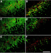
Subcellular localization of neprilysin expressed in the hippocampal formation after gene transfer. The subcellular localization of neprilysin expressed in the dentate gyrus of the contralateral hippocampal formation of neprilysin–/– mice by rAAV-mediated gene transfer was analyzed by double immunofluorescence staining for neprilysin (red) and several marker proteins (green). The merged images are shown here. A, SV2; B, GAP-43; C, tau; D, GluR1; E, MAP2; F, GFAP. Scale bar, 20 μm.
Figure 3.
Figure 3.
Functional expression of neprilysin after gene transfer. A, Levels of neprilysin-dependent endopeptidase activities in ipsilateral hippocampal formations of intact, buffer-injected, rAAV-NEP-injected, or rAAV-NEPinactive-injected neprilysin+/+ and neprilysin–/– mice. Each column with a bar represents the mean ± SE of three mice. B, Duration of neprilysin gene expression. The endopeptidase activities in ipsilateral hippocampal formations were measured 1, 2, 4, 8, 16, and 24 weeks after unilateral injection of rAAV-NEP, rAAV-NEPinactive, or buffer into the hippocampal formation of neprilysin+/+ mice. Each column with a bar represents the mean ± SE of three mice.
Figure 4.
Figure 4.
Reduction in intrahippocampal Aβ levels in neprilysin-deficient and wild-type mice after gene transfer. A, Aβ levels in both soluble and insoluble fractions of bilateral hippocampal formations were determined by sandwich ELISA 4 weeks after bilateral injection of rAAV-LacZ (1.3 × 10¹⁰ genome copies), rAAV-NEPinactive (1.3 × 10¹⁰), rAAV-NEP (1.3 × 10¹⁰), or buffer into the hippocampal formations of 10-week-old neprilysin–/– mice. Each column with a bar represents the mean ± SE of 5–7 mice. B,x-40 levels were determined 4 weeks after bilateral injection of rAAV vectors or buffer into the hippocampal formations of 10-week-old neprilysin+/+ mice under the same conditions as for A. The amount of amyloid precursor protein in all groups remained unchanged as analyzed by Western blotting (data not shown). Each column with a bar represents the mean ± SE of 6 or 7 mice. *p < 0.05, significantly different from rAAV-NEP-injected group.
Figure 5.
Figure 5.
Changes in Aβ levels or endopeptidase activity in young Tg2576 mice after neprilysin gene transfer. Aβ levels in both soluble and insoluble fractions (A, B, D, E) and endopeptidase activities (C) of the ipsilateral (A) and contralateral (B) hippocampal formations, and the ipsilateral (D) and contralateral and (E) cerebral cortices were determined 8 weeks after unilateral injection of rAAV-NEPinactive (open column) and rAAV-NEP (shadowed column) (1.3 × 10¹⁰ genome copies) into the hippocampal formation of 10-week-old Tg2576 mice, respectively. Each column with a bar represents the mean ± SE of 6 or 7 mice. *p < 0.05, significantly different from rAAV-NEPinactive-injected group.
Figure 6.
Figure 6.
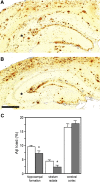
Deceleration of Aβ deposition in aged Tg2576 brains by neprilysin gene transfer. Twelve weeks after unilateral injection of rAAV-NEPinactive (A) or rAAV-NEP (B) (1.3 × 10¹⁰ genome copies) into the hippocampal formation of 18-month-old Tg2576 mice, brain sections were immunostained with anti-Aβ antibody, which recognized the C-terminal of Aβ42. Asterisks, stratum radiata. Scale bar, 500 μm. C, Aβ deposits in various areas of sections from the rAAV-NEPinactive-injected (shadowed column, n = 7) or rAAV-NEP-injected (open column, n = 5) brains were quantitated as described in Materials and Methods. Each column with a bar represents the mean ± SE. *p < 0.05, significantly different from rAAV-NEPinactive-injected group.
Figure 7.
Figure 7.
Detection of immunoreactive neprilysin from neuritic components surrounding amyloid plaques. Sections from rAAV-NEPinactive-injected brains of Tg2576 mice were immunolabeled with anti-SNAP-25 (A; green), anti-neprilysin (B; red), and anti-Aβ antibodies (C; blue). D, Merged images of sections labeled with anti-SNAP-25 and anti-neprilysin antibodies. E, Merged images of triply labeled section. Experimental conditions are the same as in the case of Figure 6. Scale bar, 20 μm.

References

    1. Akiyama H, Kondo H, Ikeda K, Kato M, McGeer PL (2001) Immunohistochemical localization of neprilysin in the human cerebral cortex: inverse association with vulnerability to amyloid β-protein (Aβ) deposition. Brain Res 902: 277–281. - PubMed
    1. Amaral DG, Witter MP (1995) Hippocampal formation. In: The rat nervous system (Paxinos G, ed), pp 443–493. San Diego: Academic.
    1. Barnes K, Turner AJ, Kenny AJ (1988) Electronmicroscopic immunocytochemistry of pig brain shows that endopeptidase-24.11 is localized in neuronal membranes. Neurosci Lett 94: 64–69. - PubMed
    1. Barnes K, Turner AJ, Kenny AJ (1992) Membrane localization of endopeptidase-24.11 and peptidyl dipeptidase A (angiotensin converting enzyme) in the pig brain: a study using subcellular fractionation and electron microscopic immunocytochemistry. J Neurochem 58: 2088–2096. - PubMed
    1. Buttini M, Yu GQ, Shockley K, Huang Y, Jones B, Masliah E, Mallory M, Yeo T, Longo FM, Mucke L (2002) Modulation of Alzheimer-like synaptic and cholinergic deficits in transgenic mice by human apolipoprotein E depends on isoform, aging, and overexpression of amyloid β peptides but not on plaque formation. J Neurosci 22: 10539–10548. - PMC - PubMed

Publication types

MeSH terms