. 2004 Feb;5(2):213-8.
doi: 10.1038/sj.embor.7400074. Epub 2004 Jan 23.

DJ-1 has a role in antioxidative stress to prevent cell death

Affiliations

DJ-1 has a role in antioxidative stress to prevent cell death

Takahiro Taira et al. EMBO Rep. 2004 Feb.

Erratum in

  • EMBO Rep. 2004 Apr;5(4):430

Abstract

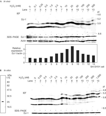
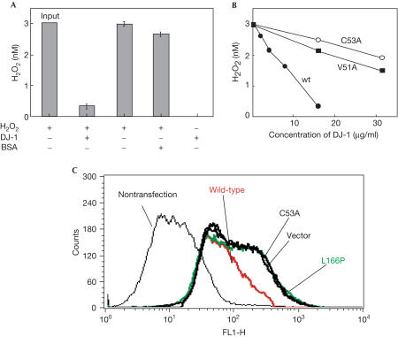
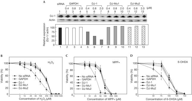
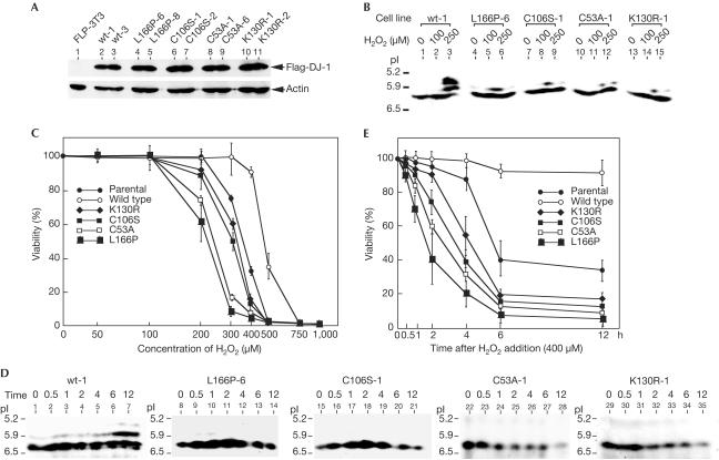
Deletion and point (L166P) mutations of DJ-1 have recently been shown to be responsible for the onset of familial Parkinson's disease (PD, PARK7). The aim of this study was to determine the role of DJ-1 in PD. We first found that DJ-1 eliminated hydrogen peroxide in vitro by oxidizing itself. We then found that DJ-1 knockdown by short interfering RNA rendered SH-SY5Y neuroblastoma cells susceptible to hydrogen peroxide-, MPP+- or 6-hydroxydopamine-induced cell death and that cells harbouring mutant forms of DJ-1, including L166P, became susceptible to death in parallel with the loss of oxidized forms of DJ-1. These results clearly showed that DJ-1 has a role in the antioxidative stress reaction and that mutations of DJ-1 lead to cell death, which is observed in PD.


Figures

Figure 1
Figure 1
pI shift of DJ-1 after its treatment with hydrogen peroxide. (A) SH-SY5Y cells were treated with various concentrations of hydrogen peroxide for 12 h, and proteins in the extracts were then analysed by isoelectric focusing phoresis gel (IEF, upper panel) or PAGE containing SDS (SDS–PAGE in the lower panel) as described in Methods. Intensities of the bands of DJ-1 and actin were measured and are shown as ‘relative expression'. (B) In all, 1 μg of recombinant DJ-1 was reacted with various concentrations of hydrogen peroxide for 30 min at room temperature and subjected to isoelectric focusing Diet phoresis and SDS–PAGE electrophoresis as described in A (right panel). The recombinant DJ-1 used in this experiment was separated on PAGE containing SDS and stained with Coomassie brilliant blue R-250 (left panel).
Figure 2
Figure 2
Elimination of hydrogen peroxide by DJ-1. (A) In all, 3 nM of hydrogen peroxide was reacted with 0.5 nM of DJ-1 for 1 h at 30°C, and the concentration of hydrogen peroxide was measured as described in Methods. (B) In all, 3 nM of hydrogen peroxide was reacted with various amounts of wild-type, V51A and C53A DJ-1 for 1 h at 30°C, and the concentrations of hydrogen peroxide were measured as described in Methods. (C) SHSY-5Y cells in 10-cm dishes were transfected with 5 μg each of plasmids used for the establishment of cell lines by the lipofectamine method. At 36 h after transfection, cells were treated with 10 μM hydrogen peroxide for 60 min and then with 5 μM DCFH-DA for 30 min, and analysed by flow cytometry. BSA, bovine serum albumin.
Figure 3
Figure 3
Acceleration of hydrogen-peroxide-induced cell death in DJ-1-knockdown cells. (A) SH-SY5Y cells were transfected with siRNAs targeting GAPDH and DJ-1. At 36 h after transfection, the proteins in the extracts were blotted with an anti-DJ-1 antibody or anti-actin antibody as described in Methods. The intensities of the bands in western blotting shown in A (upper panel) were measured by an Odyssey system, and the intensity of DJ-1 was normalized to that of actin (lower panel). SH-SY5Y cells were transfected with 2 μM of siRNAs targeting GAPDH and DJ-1. At 36 h after transfection, cells were reacted with various concentrations of hydrogen peroxide (B), MPP+ (C) or 6-OHDA (D) for 12 h, and their viabilities were measured by an MTT assay.
Figure 4
Figure 4
Acceleration of hydrogen-peroxide-induced cell death in cells harbouring various mutants of DJ-1, including L166P, which is observed in PARK7 patients. (A) Expression levels of Flag-DJ-1 and actin in NIH3T3 cell lines harbouring Flag-tagged wild-type DJ-1 and various mutants of DJ-1 were examined by western blotting. Two different cell lines were used for each construct. (B) NIH3T3 cell lines were reacted with 100 and 250 μM of hydrogen peroxide for 12 h. Proteins in cells were then subjected to isoelectric focusing phoresis and blotted with an anti-DJ-1 antibody. (C) NIH3T3 cell lines were reacted with various concentrations of hydrogen peroxide for 24 h and their viabilities were measured by an MTT assay. (D) NIH3T3 cell lines were reacted with 400 μM of hydrogen peroxide. Proteins in cells at various times after treatment with hydrogen peroxide were then subjected to isoelectric focusing phoresis and blotted with an anti-DJ-1 antibody. (E) NIH3T3 cell lines were reacted with 400 μM hydrogen peroxide, and cell viabilities were measured by an MTT assay at various times after treatment with hydrogen peroxide.

References

    1. Bonifati V et al. (2003) Mutations in the DJ-1 gene associated with autosomal recessive early-onset Parkinsonism. Science 299: 256–259 - PubMed
    1. Heikkila R, Cohen G (1971) Inhibition of biogenic amine uptake by hydrogen peroxide: a mechanism for toxic effects of 6-hydroxydopamine. Science 172: 1257–1258 - PubMed
    1. Honbou K et al. (2003) The crystal structure of DJ-1, a protein related to male fertility and Parkinson's disease. J Biol Chem 278: 31380–31384 - PubMed
    1. Huai Q et al. (2003) Crystal structure of DJ-1/RS and implication on familial Parkinson's disease. FEBS Lett 549: 171–175 - PubMed
    1. Jacquot JP et al. (2002) Thioredoxins and related proteins in photosynthetic organisms: molecular basis for thiol dependent regulation. Biochem Pharmacol 64: 1065–1069 - PubMed

Publication types

MeSH terms