. 2004 Feb 11;23(3):650-8.
doi: 10.1038/sj.emboj.7600090. Epub 2004 Jan 29.

A glycosylated type I membrane protein becomes cytosolic when peptide: N-glycanase is compromised

Affiliations

A glycosylated type I membrane protein becomes cytosolic when peptide: N-glycanase is compromised

Daniël Blom et al. EMBO J. .

Abstract

The human cytomegalovirus-encoded glycoprotein US2 catalyzes proteasomal degradation of Class I major histocompatibility complex (MHC) heavy chains (HCs) through dislocation of the latter from the endoplasmic reticulum (ER) to the cytosol. During this process, the Class I MHC HCs are deglycosylated by an N-glycanase-type activity. siRNA molecules designed to inhibit the expression of the light chain, beta(2)-microglobulin, block the dislocation of Class I MHC molecules, which implies that US2-dependent dislocation utilizes correctly folded Class I MHC molecules as a substrate. Here we demonstrate it is peptide: N-glycanase (PNGase or PNG1) that deglycosylates dislocated Class I MHC HCs. Reduction of PNGase activity by siRNA expression in US2-expressing cells inhibits deglycosylation of Class I MHC HC molecules. In PNGase siRNA-treated cells, glycosylated HCs appear in the cytosol, providing the first evidence for the presence of an intact N-linked type I membrane glycoprotein in the cytosol. N-glycanase activity is therefore not required for dislocation of glycosylated Class I MHC molecules from the ER.


Figures

Figure 1
Figure 1
Attenuation of β2m expression levels in U373 astrocytoma cells. (A) Western blot analyses of U373 control cells and β2m siRNA cells. Total extracts (30 μg per lane) of U373 control cells (wt) and U373 cells infected with two independent β2m siRNA constructs (I and II, respectively) were separated by SDS–PAGE, blotted onto PVDF and tested for the presence of β2m. (B) Same as in (A); extracts were tested for the presence of Class I MHC HC. (C) Same as in (A); extracts were tested for the presence of PDI. Note that extracts from two independent cell lines harboring the β2m siRNA construct II were tested, since the complete nucleotide sequence of this construct could not be determined.
Figure 2
Figure 2
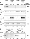
US2-mediated breakdown of Class I MHC HC can be disrupted by attenuation of β2m expression levels. (A) Control cells (control; lanes 1–3), US2-expressing cells (control+US2; lanes 4–6), β2m siRNA cells (β2mRNAi; lanes 7–9) and β2m siRNA plus US2-expressing cells (β2mRNAi+US2; lanes 10–12) were pulse-labeled with 35S-methionine for 10 min and chased for the indicated times. Cells were lysed in NP-40 lysis buffer, followed by immunoprecipitation for properly folded Class I MHC (W6/32; panel (A)), Class I MHC HC (αHC; panel (B)), US2 (αUS2; panel (C)) and the TfR (panel (D)). Indicated on the left-hand side are the migration of the glycosylated (+CHO) and deglycosylated (−CHO) form of Class I MHC HC, respectively, or glycosylated and deglycosylated US2 (panel C). TfRHM and TfRC indicate the migration of the TfR containing high-mannose- and complex-type sugars, respectively (panel (D)). (E) Cell extracts of US2-expressing cells (control+US2; lanes 1–6) and β2m siRNA plus US2-expressing cells (β2mRNAi+US2; lanes 7–12) were labeled as described in (A) and subjected to subcellular fractionation by ultracentrifugation at 100 000 g (described in Tortorella et al, 1998). The obtained fractions were subsequently used to recover Class I MHC HC by immunoprecipitation with the HC70 antibody. The migration of the glycosylated (+CHO) and deglycosylated (−CHO) form of Class I MHC HC is indicated on the left-hand side.
Figure 3
Figure 3
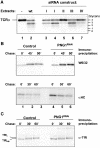
PNGase siRNA cells display normal glycoprotein turnover. (A) PNGase siRNA cell lines were generated as described in Materials and methods. NP-40 cell extracts (575 μg) were tested for PNGase activity in a deglycosylation assay using radiolabeled TCRα as a substrate (Hirsch et al, 2003). Lane 1: TCRα incubated without cell extracts (−). Lane 2: TCRα incubated with NP-40 extracts from U373 control cells (wt). Lanes 3–7: TCRα incubated with NP-40 extracts from cells infected with the indicated PNGase siRNA constructs (I–IV). Note that extracts from two independent cell lines harboring the PNGase siRNA construct I were tested, since the complete nucleotide sequence of this construct could not be determined. The number of N-linked glycans of TCRα is indicated on the right-hand side. Note that the doublet observed for TCRα carrying two residual N-linked glycans is most probably due to heterogeneous deglycosylation of the polypeptide, since the four N-linked attachment sites of TCRα allow six different glycosylation patterns. (B) Control cells (control; lanes 1–3) and PNGase siRNA cells (PNG1RNAi; lanes 4–6) were pulse-labeled with 35S-methionine for 10 min and chased for the indicated times. Cells were lysed in NP-40 lysis buffer followed by immunoprecipitation for properly folded Class I MHC (W6/32; upper panel) or Class I MHC HC (αHC; lower panel). (C) Same procedure as in (B); NP-40 cell extracts were used for immunoprecipitation of the TfR (α-TfR).
Figure 4
Figure 4
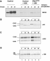
PNGase is responsible for deglycosylation of Class I MHC HCs in US2-mediated dislocation. (A) US2-expressing cells (control+US2; lanes 1–3) and US2-expressing PNGase siRNA cells (PNG1RNAi+US2; lanes 4–6) were pulse-labeled with 35S-methionine for 10 min and chased for the indicated times. NP-40 cell extracts were used for the immunoprecipitation of correctly folded Class I MHC (W6/32). To demonstrate downregulation of Class I MHC molecules, control cells were run in parallel (control; left-hand side). (B) Same as in (A); NP-40 cell extracts were used for the immunoprecipitation of free Class I MHC HC (αHC). The migration of glycosylated (+CHO) and deglycosylated (−CHO) HCs, respectively, is indicated on the left-hand side. (C) Same as in (A); NP-40 extracts were used for the immunoprecipitation of US2 (αUS2), which migrates as a doublet. (D) Same as in (A); NP-40 cell extracts were used for the immunoprecipitation of the TfR glycoprotein. Slower migration of the TfR at later chase points is due to the processing of high-mannose N-linked sugars.
Figure 5
Figure 5
Glycosylated Class I MHC HCs accumulate in the cytosol after dislocation in PNGase knockdown cells. (A) US2-expressing cells (control+US2; lanes 1–3, 7–9 and 13–15) and US2-expressing PNGase siRNA cells (PNG1RNAi+US2; lanes 4–6, 10–12 and 16–18) were pulse-labeled with 35S-methionine for 10 min and chased for 0, 20 and 40 min. Cells were resuspended in 250 mM sucrose containing 10 mM Tris, pH 7.4 and 50 mM NaCl and passed through a ball bearing homogenizer 14 times. One quarter of this homogenate was used to immunoprecipitate Class I MHC HC (total; lanes 1–3 and 4–6). The remaining three quarters were spun at 100 000 g for 1 h. The supernatant fraction was then adjusted to 0.5% NP-40 and 125 mM sucrose; the pellet fraction was resuspended in NP-40 lysis buffer. Both fractions were used for the immunoprecipitation of Class I MHC HC. (B) Same as in (A), fractions were used for the immunoprecipitation of the ER resident protein calnexin. (C) Same as in (A), fractions were used for the immunoprecipitation of fully assembled Class I MHC by use of the W6/32 monoclonal antibody. Note that part (C) was exposed 10 times longer than part (A), in order to detect W6/32-reactive Class I MHC, which is downregulated in US2-expressing cells. Note that lanes 1–6 contain one-third of the amount used in lanes 7–18 for each immunoprecipitation.
Figure 6
Figure 6
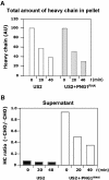
Quantitation of dislocated Class I MHC HC in US2- and US2 plus PNGRNAi-expressing cells. (A) Radioactive signal of glycosylated (+CHO) and deglycosylated (−CHO) HC shown in Figure 5 was quantitated by use of a Phosphorimager for both US2- and US2 plus PNG1RNAi-expressing cells. Signals were expressed as the percent HC recovered at the indicated time points relative to the 0-min time point of each cell line. The sum of the amount of glycosylated HC and deglycosylated HC in the 100 000 g pellet fraction at 0, 20 and 40 min is depicted. White bars represent the total amount of Class I HC in US2-expressing cells, and light gray bars represent that in US2 plus PNG1RNAi-expressing cells. (AU=arbitrary units). (B) Depiction of the ratio of glycosylated versus deglycosylated HCs in the 100 000 g supernatant fraction in US2-expressing cells (black bars) and US2 plus PNG1RNAi-expressing cells (white bars).

References

    1. Bebok Z, Mazzochi C, King SA, Hong JS, Sorscher EJ (1998) The mechanism underlying cystic fibrosis transmembrane conductance regulator transport from the endoplasmic reticulum to the proteasome includes Sec61beta and a cytosolic, deglycosylated intermediary. J Biol Chem 273: 29873–29878 - PubMed
    1. Beckmann R, Spahn CM, Eswar N, Helmers J, Penczek PA, Sali A, Frank J, Blobel G (2001) Architecture of the protein-conducting channel associated with the translating 80S ribosome. Cell 107: 361–372 - PubMed
    1. Bogyo M, McMaster JS, Gaczynska M, Tortorella D, Goldberg AL, Ploegh H (1997) Covalent modification of the active site threonine of proteasomal beta subunits and the Escherichia coli homolog HslV by a new class of inhibitors. Proc Natl Acad Sci USA 94: 6629–6634 - PMC - PubMed
    1. Brodsky FM, Bodmer WF, Parham P (1979) Characterization of a monoclonal anti-beta 2-microglobulin antibody and its use in the genetic and biochemical analysis of major histocompatibility antigens. Eur J Immunol 9: 536–545 - PubMed
    1. Brummelkamp TR, Bernards R, Agami R (2002) Stable suppression of tumorigenicity by virus-mediated RNA interference. Cancer Cell 2: 243–247 - PubMed

Publication types

MeSH terms

Substances