Review
. 2004 Mar;14 Suppl 3(3):E2-20.
doi: 10.1007/s00330-003-2162-7.

Community-acquired and nosocomial pneumonia

Affiliations
Review

Community-acquired and nosocomial pneumonia

Christian J Herold et al. Eur Radiol. 2004 Mar.

Abstract

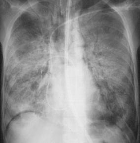
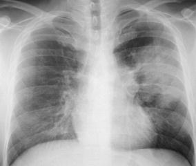
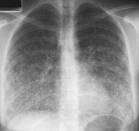
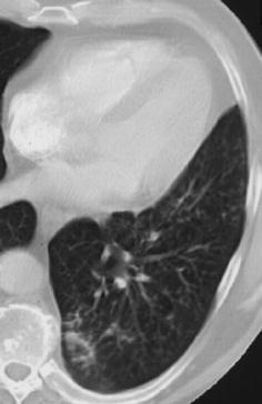
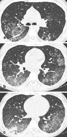
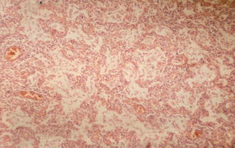
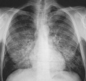
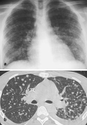
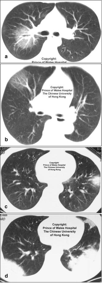
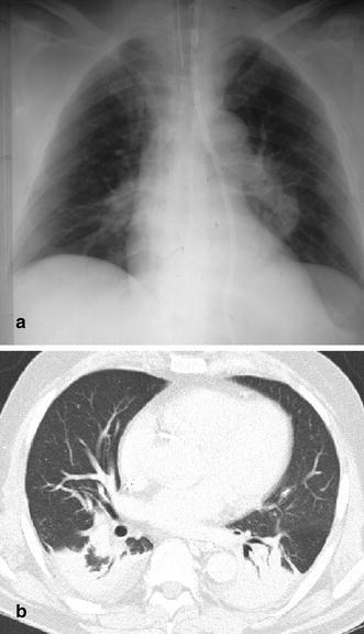
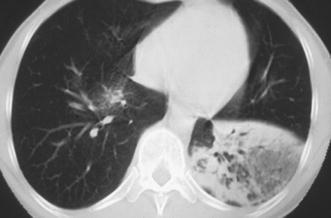
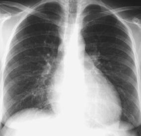
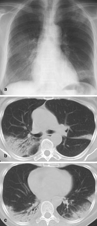
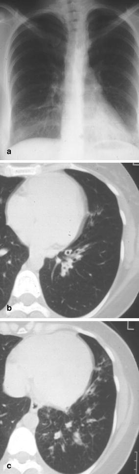
Pneumonia is one of the leading causes of morbidity, hospitalization, and mortality in both industrialized and developing countries. In particular, pulmonary infections acquired in the community, and pneumonias arising in the hospital setting, represent a major medical and economic problem and thus a continuous challenge to health care. For the radiologist, it is important to understand that community-acquired pneumonia (CAP) and nosocomial pneumonia (NP) share a number of characteristics, but should, in many respects be regarded as separate entities. CAP and NP arise in different populations, host different spectra of causative pathogens, and pose different challenges to both the clinician and the radiologist. CAP is generally seen in outpatients, is most frequently caused by Streptococcus pneumoniae, Mycoplasma pneumoniae, Haemophilus influenzae, and Chlamydia, and its radiologic diagnosis is relatively straightforward. NP, in contrast, develops in the hospital setting, is commonly caused by gram-negative bacteria, and may generate substantial problems for the radiologist. Overall, both for CAP and NP, imaging is an integral component of the diagnosis, important for classification and differential diagnosis, and helpful for follow-up.
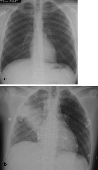
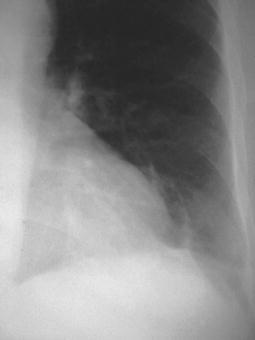
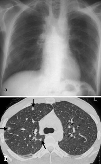
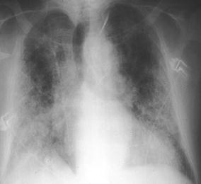
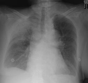
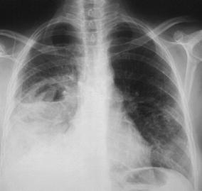
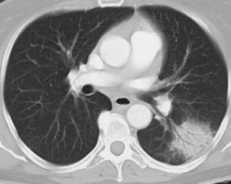
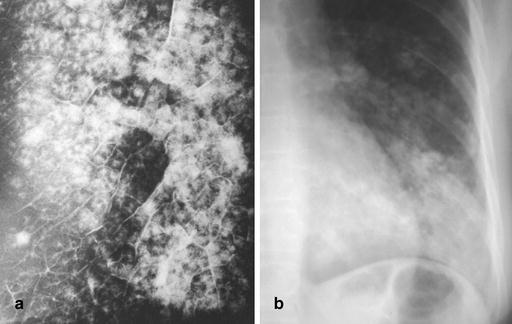
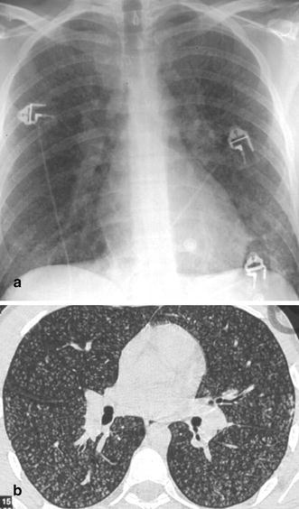
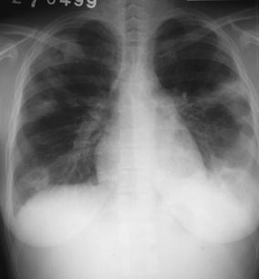
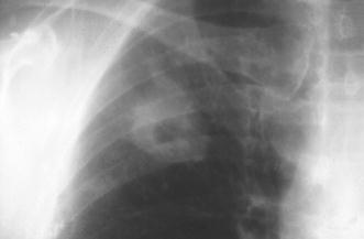
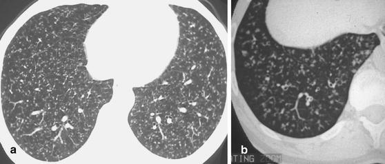

Figures

Fig. 1a,b
Fig. 1a,b
Rapid development of pneumonia in a 39-year-old man with a hematologic disorder. a When the patient developed fever, the first chest radiogram was taken 5 h after the onset of symptoms. b Follow-up 20 h after the onset of symptoms demonstrates an extensive alveolar infiltrate in the right upper lung representing lobar pneumonia due to Staphylococcus aureus infection
Fig. 2
Fig. 2
A 54-year-old diabetic inpatient with fever, non-productive cough, and elevated white cell count. The chest radiograph shows a faint segmental density in the left lower lobe representing delayed and incomplete development of left lower lobe pneumonia. At bronchoscopy, E. coli was identified as the causative organism
Fig. 3a,b
Fig. 3a,b
A 38-year-old patient with relapsing Hodgkin’s disease. In the neutropenic phase following chemotherapy the patient developed fever. a The chest radiograph demonstrates paramediastinal fibrosis as a consequence of the initial radiotherapy but reveals no signs of infection. b In contrast, the CT scan in the same patient 2 days later shows three small focal lesions with a halo phenomenon (arrows). Based on the CT findings, the patient was diagnosed to suffer from invasive aspergillosis and was treated successfully
Fig. 4
Fig. 4
A 62-year-old inpatient with previous smoking history and proven idiopathic pulmonary fibrosis. Clinical symptoms included cough, fever, dyspnea, and malaise: CRP was 26 mg/dl and white cell count was 23.6 g/l. Chest radiography does not allow identification of a pneumonic infiltrate, although the patient was diagnosed as suffering from Pseudomonas aeruginosa infection (using protected catheter brushing)
Fig. 5
Fig. 5
A 28-year-old patient with adult respiratory distress syndrome (ARDS) and sepsis. Supine portable chest radiogram displays extensive consolidation in both lungs, characteristic of ARDS, but does not allow either identification of pneumonic abnormalities or differentiation between ARDS and pneumonia. Protected catheter brushing revealed Pseudomonas aeruginosa infection
Fig. 6
Fig. 6
Acute air-space pneumonia in a male outpatient with symptoms suggestive of pneumonia. A chest radiogram shows a large area of consolidation with relatively well-defined borders and a central discrete air bronchogram in the left lung. This acute air-space pneumonia was caused by Streptococcus pneumoniae
Fig. 7
Fig. 7
A 27-year-old woman with high-grade fever, dyspnea, and maculo-papular rash. Varicella zoster pneumonia in this patient is characterized by diffuse bilateral patchy and confluent nodular densities, a common pattern in pulmonary VZV infection
Fig. 8
Fig. 8
Pathohistologic correlate of acute air-space pneumonia (hematoxylin–eosin stain). An abundance of neutrophils fill the alveolar spaces and are accompanied by a few macrophages and fibrin accumulation
Fig. 9
Fig. 9
Typical upper lobe pneumonia in a 63-year-old outpatient. Note that acute air-space disease may involve predominantly the periphery of the lungs while the perihilar area is relatively spared. The aerobronchogram is indicative of an air-space process
Fig. 10
Fig. 10
Typical radiographic characteristics of acute lobar pneumonia in a 53-year-old inpatient. The chest radiogram displays an extensive, dense alveolar infiltrate in the right mid- and lower lung fields with loss of the silhouettes of the lower right heart border and right hemidiaphragm. These features suggest involvement of the right lower lobe and middle lobe by the pneumonic infiltrate. In addition, an air–fluid level indicates cavitation. Note that the infection has spread transbronchially to the left lung. The patient was diagnosed to have E. coli pneumonia
Fig. 11
Fig. 11
Computed tomography signs of acute lobar pneumonia due to Streptococcus pneumoniae. At CT, the air-space consolidation in the left lower lobe displays a round shape and an air bronchogram, and is surrounded by a halo of ground-glass attenuation (which is occasionally seen in acute air-space pneumonia)
Fig. 12a,b
Fig. 12a,b
Radiographic appearance of lobular (broncho) pneumonia. a Specimen radiogram of lobular (broncho) pneumonia. This radiogram, obtained from a post-mortem examination of a patient with Staphylococcus aureus pneumonia, demonstrates the centrilobular origin of this particular form of pneumonia. As disease spreads from the walls of terminal and respiratory bronchioles, large portions of secondary lobules are involved, and confluent disease may cause frank lobar consolidation. [By courtesy of Saunders, Philadelphia. Potchen J, Grainger R, Greene R (eds) Pulmonary radiology.] b Radiographic appearance of lobular (broncho) pneumonia caused by Staphylococcus aureus and shows patchy and partly confluent densities in the left lower lobe. This pattern is distinctly different from the one seen in acute lobar (air-space) pneumonia
Fig. 13
Fig. 13
Computed tomography findings in early bronchopneumonia. The CT section through the left lower lobe in a patient with Haemophilus influenzae pneumonia demonstrates small nodular and patchy acinar lesions with a tendency towards confluence in the posterior basal segment of the left lower lobe
Fig. 14a–c
Fig. 14a–c
Computed tomography features of Mycoplasma pneumonia. The CT scans in a 31-year-old woman with fever, incessant cough, malaise, and fatigue demonstrates excessive areas of ground-glass attenuation in all pulmonary lobes. Certain ground-glass densities show a centrilobular location (a), whereas others have formed widespread ground-glass infiltrates (b,c). Areas of ground-glass attenuation in a lobular distribution are a distinct feature of Mycoplasma pneumonia
Fig. 15
Fig. 15
Histopathologic correlate of interstitial pneumonia. This histopathologic specimen (hematoxylin–eosin stain) demonstrates moderate lymphocytic infiltrates surrounding a blood vessel and extending into the neighboring alveolar walls. The presence of macrophages and pneumocytes suggest the beginning of acute alveolar damage
Fig. 16
Fig. 16
Interstitial pneumonia. In a 36-year-old HIV-positive patient with Pneumocystis carinii pneumonia the disease is characteristized by reticular, reticulo-nodular, and alveolar patchy densities predominately located in the central and basal portions of the lungs. The pattern may occasionally be confused with hydrostatic lung edema
Fig. 17a,b
Fig. 17a,b
Acute viral broncholitis and pneumonia in a 19-year-old recruit. a At chest radiography, the viral infection is characterized by a myriad of tiny nodules scattered throughout the lung parenchyma bilaterally. b Thin-section CT reveals unimorphous centrilobular densities demarcating the peripheral terminal and respiratory bronchioles
Fig. 18
Fig. 18
Nodular lesions in a young female drug addict with endocarditis. The chest radiogram demonstrates peripheral densities in both lungs showing a nodular aspect in a right upper and lower lobes. The peripheral location, nodular aspect, and central cavitation is characteristic of septic emboli in this patient setting. Note the enlargement of the cardiac silhouette and the left pleural effusion
Fig. 19
Fig. 19
A 58-year-old patient 2 years after renal transplant surgery. The isolated, centrally cavitating macronodular lesion in the right upper lobe is caused by Mycobacterium avium intracellulare infection
Fig. 20a,b
Fig. 20a,b
Patterns of micronodular disease caused by tuberculous infection. a Miliary pattern with small, well-defined interstitial nodules scattered randomly throughout the lung parenchyma. b Micronodular disease originating from tuberculous infection of the small airways. Note that the tree-in-bud phenomena, seen in the lung periphery, are characteristic of infectious small airways disease
Fig. 21a,b
Fig. 21a,b
Micronodular disease caused by Candida infection. The a chest radiogram and b CT scan demonstrate well-defined nodules scattered throughout the parenchyma of both lungs. In patients with long-standing granulocytopenia and symptoms of infection, this pattern is indicative of Candida infection with small Candida abscesses. Occasionally, these abscesses are also seen in the liver and the spleen
Fig. 22a–d
Fig. 22a–d
Different patterns of disease caused by the same organism in severe acute respiratory syndrome (SARS). Images from a patient with proven SARS. (From the Princess of Wales Hospital, The Chinese University of Hong Kong) demonstrate different patterns of disease including a,b ground-glass densities, c focal acute air-space disease with halo phenomenon, and d bilateral areas of consolidation. The SARS has been shown to cause several different radiographic patterns at chest radiography and CT
Fig. 23a,b
Fig. 23a,b
Partial atelectasis of the left lower lobe mimicking pneumonia. a In this 64-year-old stroke patient with arterial hypertension, an alveolar opacity in the medial aspects of the left lower lobe was misdiagnosed as pneumonia. b At CT, this density was identified as partial atelectasis of the left lower lobe
Fig. 24
Fig. 24
Lobar lymphoma mimicking lobar pneumonia. A 28-year-old outpatient with subfebrile temperatures and a chronic left lower lobe infiltrate, non-responsive to standard antibiotic treatment. The patient was biopsied and a low-grade B-cell lymphoma was identified histopathologically
Fig. 25
Fig. 25
Lupus pneumonitis mimicking pneumonia. A young woman with known systemic lupus erythematosus, presenting with an episode of fever and cough. The chest radiogram demonstrates bilateral diffuse interstitial abnormalities and an increased background density of the lung parenchyma. No causative organism could be identified. The enlargement of the cardiac silhouette, due to a pericardial effusion and the amount of circulating anti-DSDNA antibodies, pointed towards acute damage to the alveolar capillary unit (acute lupus pneumonitis)
Fig. 26a–c
Fig. 26a–c
Bronchiolitis obliterans with organizing pneumonia (BOOP) mimicking pneumonia. A 62-year-old woman with a 3 month history of subfebrile temperatures, cough, and failure of response to several antibiotic regimens. a The plain chest radiogram demonstrates “ground-glass” opacities in the right middle and lower lung fields as well as in the left upper lobe and left lower lobe. b,c At CT, alveolar consolidation is seen in the periphery of both upper lobes and both lower lobes. The distinct peripheral location of the abnormalities and the discretely ectatic airways raised the suspicion of BOOP. The diagnosis was confirmed through video-assisted fluoroscopic biopsy
Fig. 27a–c
Fig. 27a–c
Recurrent left lower lobe pneumonia in a young woman with bronchiectasis. a The left lower lobe pneumonia due to Pseudomonas aeruginosa infection is well documented at chest radiography. b,c After successful treatment, the patient underwent CT scanning which showed dilated airways in the left lower lobe with wall thickening and peribronchial and peribronchiolar inflammation

References

    1. Vincent J Am Med Assoc. 1995;274:634. doi: 10.1001/jama.274.8.639. - DOI
    1. National Monthly Vital Stat Rep. 1993;42:1.
    1. Mandell Chest. 1995;108:35S. - PubMed
    1. Halm N Engl J Med. 2002;347:2039. doi: 10.1056/NEJMcp020499. - DOI - PubMed
    1. Jokinen Am J Epidemiol. 1993;137:977. - PubMed

MeSH terms