. 2004 Jan 15;18(2):210-22.
doi: 10.1101/gad.1163204.

Splicing enhances translation in mammalian cells: an additional function of the exon junction complex

Affiliations

Splicing enhances translation in mammalian cells: an additional function of the exon junction complex

Ajit Nott et al. Genes Dev. .

Abstract

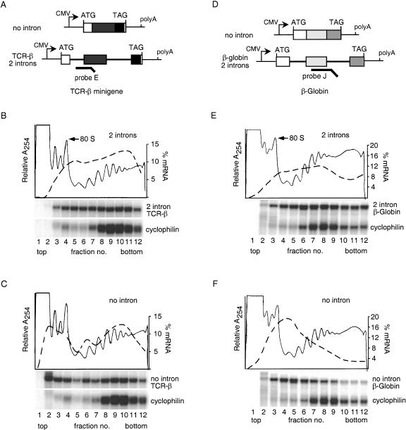
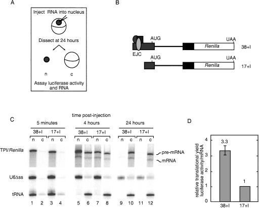
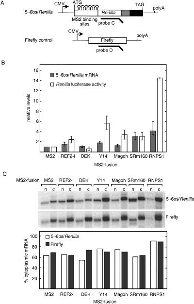
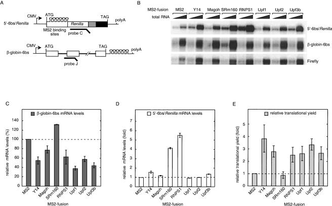
In mammalian cells, spliced mRNAs yield greater quantities of protein per mRNA molecule than do otherwise identical mRNAs not made by splicing. This increased translational yield correlates with enhanced cytoplasmic polysome association of spliced mRNAs, and is attributable to deposition of exon junction complexes (EJCs). Translational stimulation can be replicated by tethering the EJC proteins Y14, Magoh, and RNPS1 or the nonsense-mediated decay (NMD) factors Upf1, Upf2, and Upf3b to an intronless reporter mRNA. Thus, in addition to its previously characterized role in NMD, the EJC also promotes mRNA polysome association. Furthermore, the ability to stimulate translation when bound inside an open reading frame appears to be a general feature of factors required for NMD.


Figures

Figure 1.
Figure 1.
Splicing enhances mRNA polysome association. (A,D) Schematic representation of TCR-β and β-globin constructs. Boxes represent exons, and lines connecting them denote introns. RPA probes are indicated by heavy lines. (B,C,E,F) Sucrose gradient fractionation of cytoplasmic extracts from cells expressing no-intron or intron-containing versions of TCR-β and β-globin. RNA extracted from each fraction (see Materials and Methods) was subject to RPA with probe E or J (A,D) and a probe specific to endogenous cyclophilin mRNA. Protected fragments were separated on a 10% denaturing polyacrylamide gel. Relative absorbance at 254 nm is depicted by thin lines, and the percent total mRNA in each fraction is shown by the dashed lines (scale on right).
Figure 2.
Figure 2.
EJC-dependent stimulation of translational yield in Xenopus oocytes. (A) Overview of experimental procedure. (B) Schematic representation of in vitro transcribed, radiolabeled RNAs injected into Xenopus oocytes. TPI intron 6 (thick line) and flanking TPI exons (gray and black boxes) and Renilla luciferase ORF (white box) are indicated. First exons were either 38 or 17 nt in length. (C) Denaturing polyacrylamide gel electrophoresis of RNA extracted from nucleus (n) and cytoplasm (c) at 5 min, 4 h, and 24 h post-injection. (Top panel) 6% gel for Renilla RNAs; (bottom panel) identical samples loaded on a 10% gel to separate the U6Δss RNA and tRNA. (D) Translational efficiencies at 24 h post-injection relative to 17 no I mRNA. Translational efficiencies from two independent experiments were calculated by first normalizing Renilla luciferase activity to Firefly luciferase activity and then dividing by cytoplasmic mRNA levels; error bars represent the range.
Figure 3.
Figure 3.
Effects of tethering individual EJC proteins inside the Renilla luciferase ORF. (A) Schematic representation of 5′-6bs/Renilla reporter construct and Firefly control. Six MS2-binding sites were inserted 49-nt downstream of the start codon to be in-frame with the Renilla luciferase gene. (B) Levels of 5′-6bs/Renilla reporter mRNA and luciferase activity normalized to Firefly control and represented relative to MS2 alone. The data shown are the average of three to four independent experiments; error bars represent standard deviation. (C) Nuclear and cytoplasmic distribution of 5′-6bs/Renilla reporter and Firefly control mRNA in the presence of various MS2 fusion proteins. RNA was extracted from fractionated nuclear (n) and cytoplasmic (c) compartments and analyzed by RPA (top panel). The percent of total mRNA in the cytoplasm (lower panel).
Figure 4.
Figure 4.
Upf1, Upf2, and Upf3b also enhance the translational yield of 5′-6bs/Renilla mRNA. (A) Schematic representation of 5′-6bs/Renilla and β-globin-6bs constructs carrying identical MS2-binding sites. RPA probes are indicated with thick lines. (B) RPA of 2 and 8 μg of total mRNA extracted from transfected cells. (C) β-globin-6bs mRNA levels in the presence of MS2-fusion proteins relative to MS2 alone. The data reflect the average of two independent experiments; error bars represent the range. (D) Renilla reporter mRNA levels in the presence of MS2-fusion proteins relative to MS2 alone. The data reflect the average of two independent experiments; error bars represent the range. (E) Relative translational yields of Renilla luciferase. Luciferase activities were normalized to mRNA levels and are represented relative to MS2 alone. The data reflect the average of three independent experiments; errors were propagated using standard algorithms.
Figure 5.
Figure 5.
MS2-fusions of RNPS1, Y14. Magoh and Upf proteins enhance polysome association of Renilla reporter mRNA. (A) Sucrose gradient fractionation of cytoplasmic extracts from cells expressing 5′-6bs/Renilla reporter mRNA and individual MS2-fusion proteins (indicated on left). The absorbance profile is representative of all three gradients. RNA extracted from each fraction was subjected to RPA with probe C (Fig. 3A) and a probe specific to endogenous cyclophilin mRNA. Protected fragments were separated on a 10% denaturing polyacrylamide gel. (B) MS2–RNPS1 cotransfected cells were treated with Puromycin prior to lysis and fractionation. (C) Sucrose gradient fractionation of cytoplasmic extracts from cells expressing 5′-6bs/Renilla reporter mRNA plus MS2 alone, MS2–Y14, MS2–Magoh, MS2–Upf1, MS2–Upf2, or MS2–Upf3b. The absorbance profile is representative of all gradients. (D) Quantitative representation of 5′-6bs/Renilla mRNA distribution in polysome gradients in the presence of indicated MS2-fusion proteins. Relative mRNA levels in each fraction were calculated as a percent of the total, and amounts in fractions 1–4 and 5–12 were pooled to represent mRNAs cofractionating with monosomes and ribosomal subunits and polysomes, respectively. The data represent the averages of two independent experiments, and error bars reflect the range.

Similar articles

Cited by

References

    1. Aravind L. and Koonin, E.V. 2000. Eukaryote-specific domains in translation initiation factors: Implications for translation regulation and evolution of the translation system. Genome Res. 10: 1172-1184. - PMC - PubMed
    1. Badolato J., Gardiner, E., Morrison, N., and Eisman, J. 1995. Identification and characterization of a novel human RNA-binding protein. Gene 166: 323-327. - PubMed
    1. Bonneau A.M., Darveau, A., and Sonenberg, N. 1985. Effect of viral infection on host protein synthesis and mRNA association with the cytoplasmic cytoskeletal structure. J. Cell Biol. 100: 1209-1218. - PMC - PubMed
    1. Bouvet P. and Wolffe, A.P. 1994. A role for transcription and FRGY2 in masking maternal mRNA within Xenopus oocytes. Cell 77: 931-941. - PubMed
    1. Braddock M., Muckenthaler, M., White, M.R., Thorburn, A.M., Sommerville, J., Kingsman, A.J., and Kingsman, S.M. 1994. Intron-less RNA injected into the nucleus of Xenopus oocytes accesses a regulated translation control pathway. Nucleic Acids Res. 22: 5255-5264. - PMC - PubMed

Publication types