. 2004 Apr 23;279(17):17650-9.
doi: 10.1074/jbc.M313837200. Epub 2004 Jan 29.

Cytoplasmic polyadenylation element (CPE)- and CPE-binding protein (CPEB)-independent mechanisms regulate early class maternal mRNA translational activation in Xenopus oocytes

Affiliations

Cytoplasmic polyadenylation element (CPE)- and CPE-binding protein (CPEB)-independent mechanisms regulate early class maternal mRNA translational activation in Xenopus oocytes

Amanda Charlesworth et al. J Biol Chem. .

Abstract

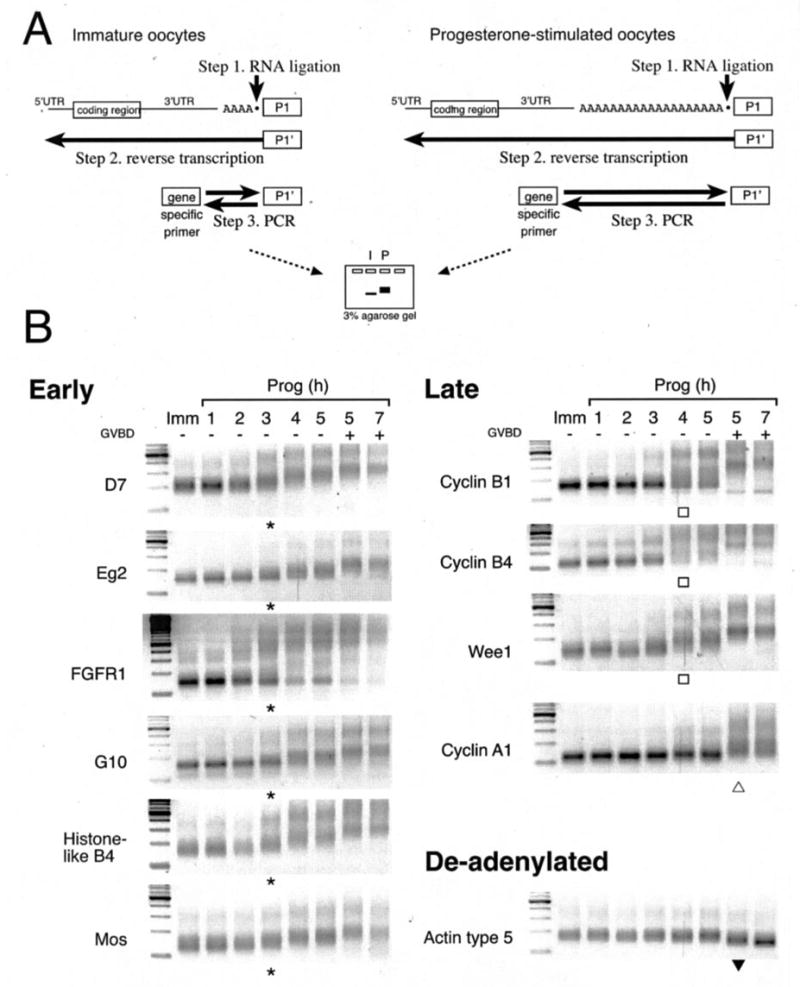
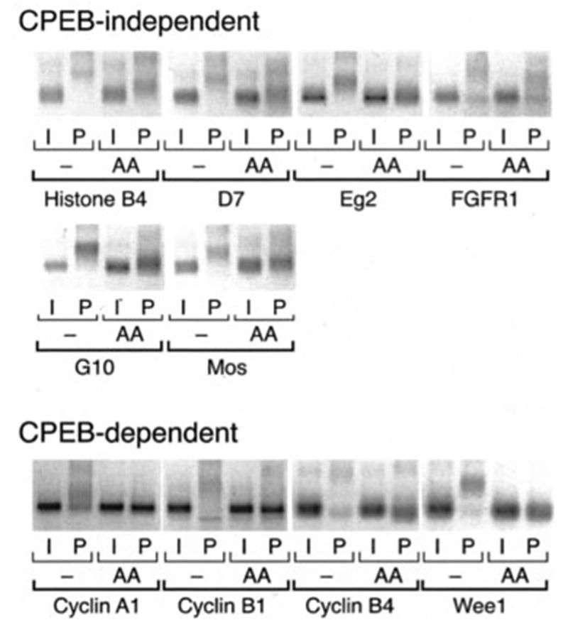
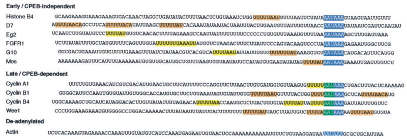
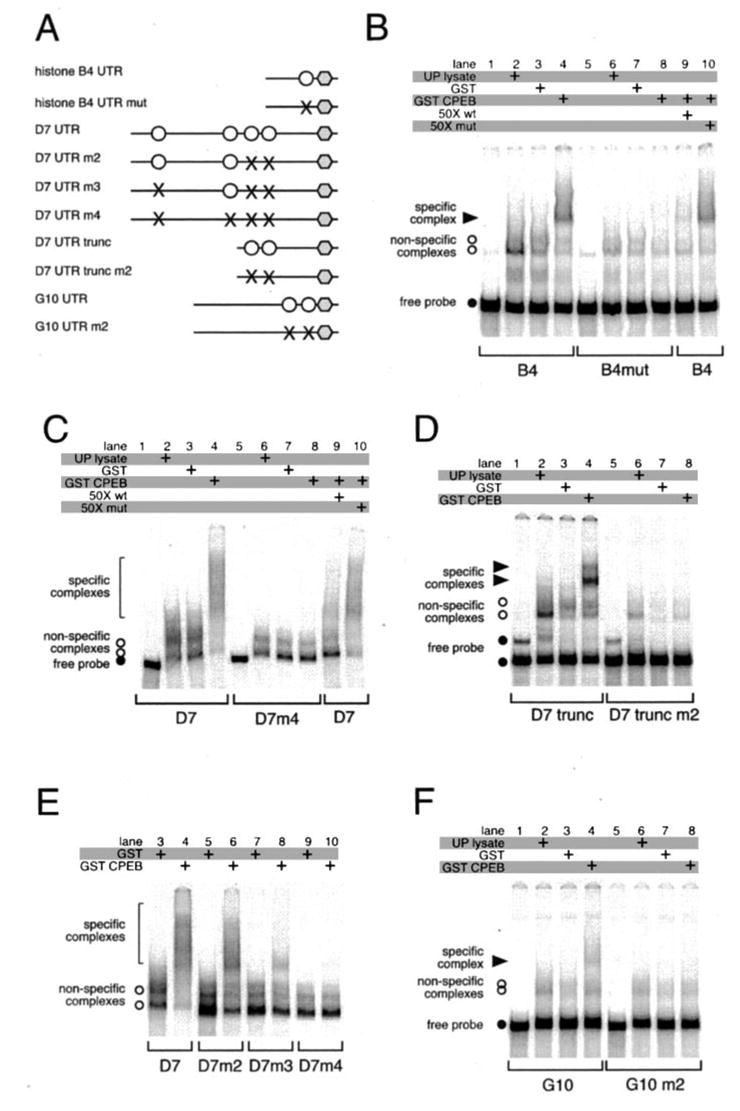
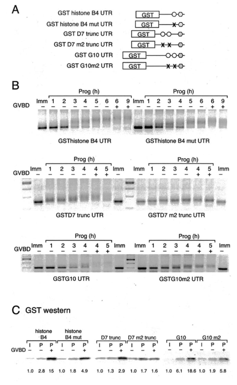
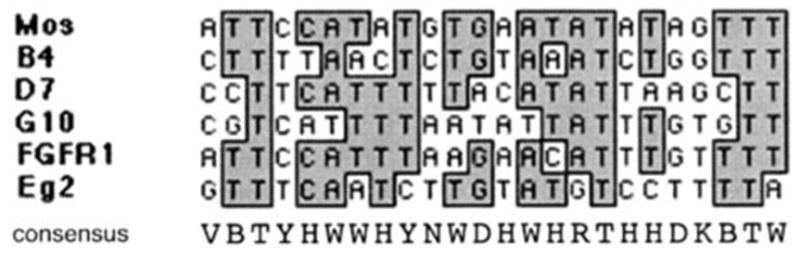
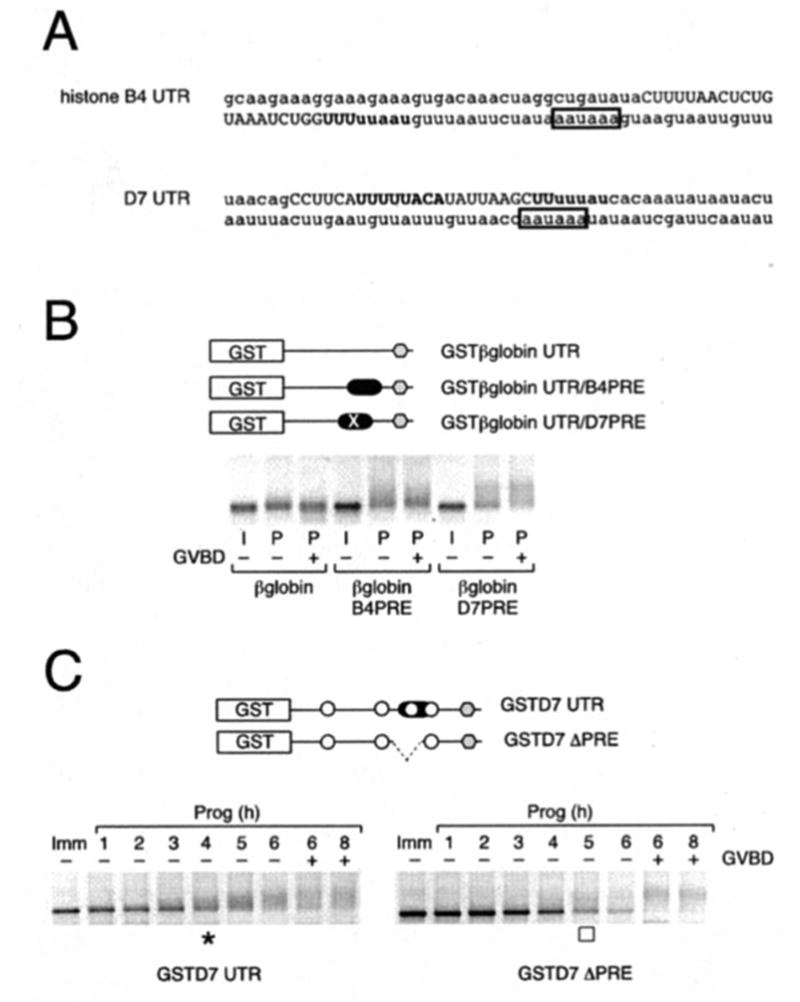
Meiotic cell cycle progression during vertebrate oocyte maturation requires the correct temporal translation of maternal mRNAs encoding key regulatory proteins. The mechanism by which specific mRNAs are temporally activated is unknown, although both cytoplasmic polyadenylation elements (CPE) within the 3'-untranslated region (3'-UTR) of mRNAs and the CPE-binding protein (CPEB) have been implicated. We report that in progesterone-stimulated Xenopus oocytes, the early cytoplasmic polyadenylation and translational activation of multiple maternal mRNAs occur in a CPE- and CPEB-independent manner. We demonstrate that polyadenylation response elements, originally identified in the 3'-UTR of the mRNA encoding the Mos proto-oncogene, direct CPE- and CPEB-independent polyadenylation of an early class of Xenopus maternal mRNAs. Our findings refute the hypothesis that CPE sequences alone account for the range of temporal inductions of maternal mRNAs observed during Xenopus oocyte maturation. Rather, our data indicate that the sequential action of distinct 3'-UTR-directed translational control mechanisms coordinates the complex temporal patterns and extent of protein synthesis during vertebrate meiotic cell cycle progression.


Figures

Fig. 1
Fig. 1. Temporal classification of endogenous mRNA cytoplasmic polyadenylation during oocyte maturation
A, schematic of the RNA ligation-coupled RT-PCR technique (35) utilized in this study to assess poly(A) tail length. Retarded migration indicates polyadenylation. B, time course of polyadenylation of endogenous mRNAs using RT-PCR with different gene-specific primers from the same cDNA preparation. On the left of each gel is the 100-bp ladder (New England Biolabs). GVBD50 (see “Experimental Procedures”) occurred at 5 h in this experiment. Asterisks and open squares indicate initiation of early and late polyadenylation, respectively. Within the late class, initiation of cyclin A1 polyadenylation occurred predominantly after GVDB (open triangle). Deadenylation of the actin mRNA is indicated by a closed triangle. Similar results were obtained in eight separate experiments using oocytes derived from different animals. In all cases the polyadenylation of the early mRNAs was consistently initiated 2–3 h prior to GVBD, no matter how long it took the oocytes to reach GVBD. Polyadenylation of the late class was initiated up to 1 h prior GVBD, and typically 1 h after initiation of the early class mRNAs.
Fig. 2
Fig. 2. Progesterone-stimulated cytoplasmic polyadenylation of multiple maternal mRNAs is CPEB-independent
Where indicated, immature oocytes were injected with RNA encoding CPEB-AA (AA) and left 20 h at 21 °C to express the protein prior to stimulation with progesterone. CPEB-AA significantly delayed oocyte maturation as expected (29, 30). When all the control oocytes had matured (6 h in this experiment), samples were prepared from control and CPEB-AA expressing oocytes (with (P) or without (I) progesterone stimulation), and polyadenylation was assessed by RT-PCR. The mRNAs are categorized based on whether expression of CPEB-AA reduced but did not eliminate polyadenylation (CPEB-independent) or ablated polyadenyl-ation (CPEB-dependent). Of the latter class, CPEB-AA expression prevented cyclin A1 and cyclin B1 polyadenylation in progesterone-stimulated oocytes and led to a slight deadenylation of the cyclin B4 and Wee1 mRNAs relative to the poly(A) tail present in immature oocytes.
Fig. 3
Fig. 3. Temporal control of cytoplasmic polyadenylation does not correlate with CPE number or sequence
The last 100 nucleotides of the mRNAs used in this study are shown. Polyadenylation hexanucleotides are shown in blue boxes; putative CPEs are shown in yellow boxes, and CPEs that have been experimentally determined by mutational analysis are shown in orange boxes: histone-like B4 (14, 43); D7 (this study); G10 ((13) this study); Mos (30, 44); cyclin B1 (18, 20, 22); and Wee1 (9). The overlap of a CPE with the polyadenylation hexanucleotide is noted in green. It should be noted that the D7 RT-PCR product contained 21 bp of extra 3′ sequence not reported previously (45), including a canonical polyadenylation hexanucleotide.
Fig. 4
Fig. 4. Xenopus CPEB does not bind to CPE-disrupted histone-like B4, D7, or G10 3′-UTRs
A, schematic showing constructs used in the study. Polyadenyl-ation hexanucleotides are represented by gray hexagons, CPEs by open circles, and disrupted CPEs by ×. The D7 3′-UTR has another CPE-like sequence 121 nt back from the polyadenylation hexanucleotide, and so was not noted in Fig. 3. B–F, RNA EMSAs. The indicated probes were incubated in the presence of unprogrammed lysate, or GST, or GST-XeCPEB expressing reticulocyte lysates, and specific complex formation was assessed. B, either wild-type (lanes 1–4, , and ) or CPE-disrupted (lanes 5–8) B4 3′-UTR probes were used. Where indicated, 50-fold molar excess of unlabeled wild-type (wt) (specific) or CPE-disrupted (mut) (non-specific) competitor B4 probe was included in the binding reactions. C, wild-type D7 3′-UTR probe (lanes 1–4, , and ) or a mutant D7 probe that had all four CPEs disrupted (lanes 5–8) were used. Where indicated, 50-fold molar excess of unlabeled wild-type or CPE-disrupted competitor D7 probe was included. D, two complexes are formed on the truncated D7 UTR demonstrating that there are at least two CPEs present. E, identification of all four CPEs in the D7 3′-UTR. F, either wild-type (lanes 1–4) or CPE-disrupted (lanes 5–8) G10 3′-UTR probes were used.
Fig. 5
Fig. 5. The initiation of CPEB-independent class mRNA polyadenylation and translational activation occurs in a CPE-independent manner
A, schematic of the reporters used in this study. The histone-like B4, D7, and G10 3′-UTRs from Fig. 4 were fused to the GST coding region. B and C, wild-type and mutant histone-like B4, D7, or G10 reporter constructs were injected into immature oocytes. B, polyadenylation of reporter constructs after progesterone stimulation was assessed by RT-PCR using a GST forward primer. In this experiment, the truncated D7 UTR was used because the PCR product from the GST forward primer is considerably smaller than that obtained with the full-length D7 UTR, and hence the change in poly(A) tail length is more readily observed. C, accumulation of GST protein after progester-one stimulation was assessed by Western blot with anti-GST antibodies. Samples were prepared at GVBD50 (see “Experimental Procedures”), segregated based on GVBD status (− or +) and shown relative to a time-matched sample prepared from immature oocytes. The numbers below the panels indicate the fold increase in GST accumulation over that seen in the immature oocyte control sample for each RNA reporter construct.
Fig. 6
Fig. 6. Sequence alignment comparison of PRE-like sequences in the 3′-UTRs of early class mRNAs
Below the alignment, the derivative consensus PRE sequence is shown using the single letter code: V, A or C or G; N, A or G or C or T; Y, C or T; H, A or C or T; W, A or T; D, A or G or T; R, A or G; B, C or G or T; K, G or T.
Fig. 7
Fig. 7. PRE sequences in 3′-UTRs direct temporally early, progesterone-stimulated cytoplasmic poly-adenylation
The indicated GST reporter RNA constructs were injected into immature oocytes. A, the last 100 nt of the histone-like B4 and D7 mRNAs are shown, with the PRE in uppercase, the CPEs in boldface, and the polyadenylation hexanucleotide in a box. B, histone-like B4 and D7 PRE sequences were inserted into the heterologous β-globin 3′-UTR. Polyadenylation of reporter constructs was assessed by RT-PCR using a GST forward primer. Progesterone-stimulated (P) samples were prepared at GVBD50 (see “Experimental Procedures”), segregated based on GVBD status (− or +), and shown relative to a time-matched sample prepared from immature (I) oocytes. Reporter constructs are schematically represented with the position of the histone-like B4 and D7 PREs (filled oblong) indicated. The PRE in D7 spans a CPE. To eliminate any possible contribution from the CPE, it was disrupted in the reporter RNA (denoted by ×). C, GST reporter RNA constructs fused to either the wild-type or PRE-deleted D7 3′-UTR were injected into immature oocytes. Poly-adenylation of reporter constructs was assessed by RT-PCR using a GST forward primer at the indicated times. GVBD50 (see “Experimental Procedures”) occurred at 6 h. Initiation of wild-type (asterisk, left panel) and PRE-deleted (open square, right panel) D7 3′-UTR-directed poly-adenylation is indicated. Reporter constructs are schematically represented with the position of CPEs (open circle), PRE (filled oblong), and deleted PRE (dashed line) indicated. − or + indicates GVBD status of harvested oocyte samples at the indicated times.

References

    1. Hochegger H, Klotzbucher A, Kirk J, Howell M, le Guellec K, Fletcher K, Duncan T, Sohail M, Hunt T. Development. 2001;128:3795–3807. - PubMed
    1. Nakajo N, Yoshitome S, Iwashita J, Iida M, Uto K, Ueno S, Okamoto K, Sagata N. Genes Dev. 2000;14:328–338. - PMC - PubMed
    1. Howard EL, Charlesworth A, Welk J, MacNicol AM. Mol Cell Biol. 1999;19:1990–1999. - PMC - PubMed
    1. Sheets MD, Wu M, Wickens M. Nature. 1995;374:511–516. - PubMed
    1. Gross SD, Schwab MS, Taieb FE, Lewellyn AL, Qian YW, Maller JL. Curr Biol. 2000;10:430–438. - PubMed

Publication types

MeSH terms