. 2004 Mar 16;101(11):3729-36.
doi: 10.1073/pnas.0308082100. Epub 2004 Jan 29.

Structures of the N-terminal modules imply large domain motions during catalysis by methionine synthase

Affiliations

Structures of the N-terminal modules imply large domain motions during catalysis by methionine synthase

John C Evans et al. Proc Natl Acad Sci U S A. .

Abstract

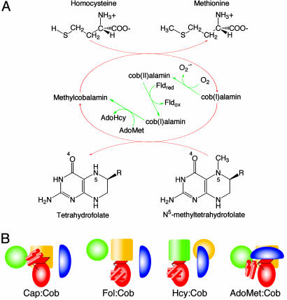
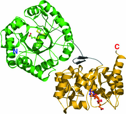
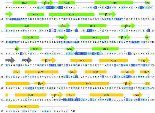
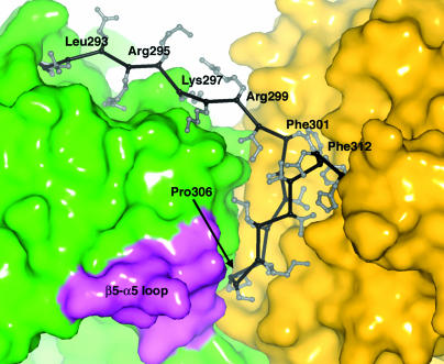
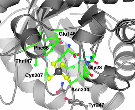
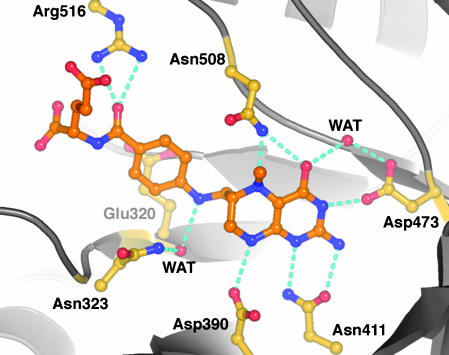
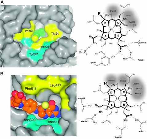
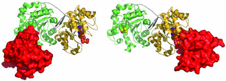
B(12)-dependent methionine synthase (MetH) is a large modular enzyme that utilizes the cobalamin cofactor as a methyl donor or acceptor in three separate reactions. Each methyl transfer occurs at a different substrate-binding domain and requires a different arrangement of modules. In the catalytic cycle, the cobalamin-binding domain carries methylcobalamin to the homocysteine (Hcy) domain to form methionine and returns cob(I)alamin to the folate (Fol) domain for remethylation by methyltetrahydrofolate (CH(3)-H(4)folate). Here, we describe crystal structures of a fragment of MetH from Thermotoga maritima comprising the domains that bind Hcy and CH(3)-H(4)folate. These substrate-binding domains are (beta alpha)(8) barrels packed tightly against one another with their barrel axes perpendicular. The properties of the domain interface suggest that the two barrels remain associated during catalysis. The Hcy and CH(3)-H(4)folate substrates are bound at the C termini of their respective barrels in orientations that position them for reaction with cobalamin, but the two active sites are separated by approximately 50 A. To complete the catalytic cycle, the cobalamin-binding domain must travel back and forth between these distant active sites.


Figures

Fig. 1.
Fig. 1.
The reactions catalyzed by MetH (A) and the conformational states of MetH (B). (A) During primary turnover (red arrows), a methyl group is transferred from CH3-H4folate to the enzyme-bound cob(I)alamin cofactor, forming H4-folate and methylcobalamin. Methylcobalamin is then demethylated by Hcy, forming methionine (Met) and regenerating cob(I)alamin (1). In reactivation (green arrows), cob(II)alamin is reduced by one-electron transfer from flavodoxin and the resulting cob(I)alamin is methylated by AdoMet (1). (B) Cartoons depicting the arrangements of the modules of MetH. The diagrams designate four states in which the cobalamin-binding domain interacts with different partners: the four-helix bundle or Cap domain, the Fol domain, the Hcy domain, or the AdoMet domain. The Hcy (green) and Fol (gold) barrels are represented by large cylinders, the red oval corresponds to the α/β Rossmann domain (Cob) that binds cobalamin, and the blue oval is the activation (AdoMet) domain. The helical Cap domain is represented as a group of small red cylinders.
Fig. 2.
Fig. 2.
A ribbon drawing of the substrate-binding domains of MetH from T. maritima. The Hcy barrel is shown in green, the linker is shown in gray, and the Fol barrel is shown in gold. Substrates are shown as ball-and-stick models. The view is approximately along the axis of the Hcy barrel and is oriented to show the hairpin component of the domain interface.
Fig. 3.
Fig. 3.
The sequence of the substrate-binding modules of MetH from T. maritima. Invariant and conserved residues, framed in dark and light blue, are assigned from a multiple alignment performed in clustalw (19) using 19 sequences from the NCBI database (see Fig. 10). The secondary structure assignments for T. maritima MetH are displayed above the alignments and are labeled H or F to designate Hcy or Fol domains.
Fig. 4.
Fig. 4.
The interface between the Hcy and Fol domains. Residues 292-300 comprise the extended part of the interdomain linker. The hairpin segment of the linker that packs against both barrels begins at Phe-301 (see text). Residues in a conserved motif (180G-R/K-S/T-L-S/T-G) from the loop β5H-α5H (lavender) contact the conserved Pro-306 of the hairpin.
Fig. 5.
Fig. 5.
Substrate interactions in the Hcy domain. The sulfur of Hcy coordinates the metal, the amino group is hydrogen-bonded to Glu-146, and the carboxyl group interacts with backbone amides following the fingerprint sequence, 19D-G-A. Phe-66 stacks against the substrate, and Thr-147 is hydrogen-bonded to the sulfur of Hcy.
Fig. 6.
Fig. 6.
CH3-H4folate and its interactions with conserved residues from the Fol barrel. The extended arrangement of the folate is determined by the interactions of the p-aminobenzoic acid (PABA) side chain: the ring stacks with Glu-321, N10 makes a through-water interaction with the conserved Asn-323, and Arg-516 binds the PABA carbonyl oxygen. In the saturated pyrazine ring of the pterin, the C6 side chain substituent is axial and the N5-methyl group is equatorial, pointing toward the reader. Hydrogen bonds are indicated for donor-acceptor pairs that are closer than 3.2 Å. The side chain amide of Asn-508 may reorient in the ternary complex to allow the oxygen to interact with a protonated pterin N5 (see text). The viewpoint is approximately along the expected approach of the corrin ring.
Fig. 7.
Fig. 7.
Models of cobalamin binding to each of the substrate domains. (A) (Left) The surface of the cobalamin binding site of the Hcy barrel, with Hcy represented as van der Waals spheres; the Hcy sulfur is yellow. Hydrophobic patches are yellow, and hydrophilic regions are cyan. (Right) A schematic diagram of the modeled orientation of the corrin macrocycle, showing the possible interactions between cobalamin and residues of the Hcy domain. Cys-207 and -273 of the (Cys)3 Hcy-metal cluster are positioned behind the B and C rings of the corrin. Invariant residues that interact with cobalamin side chains are labeled, and hydrophobic contacts are shaded. Preliminary characterization of the Tyr247Phe mutant shows an 8-fold decrease in the rate constant for the reaction of Hcy with methylcobalamin (D. Touw, L. Paul, and R.G.M., unpublished data). (B) (Left) The surface of the cobalamin binding site of the Fol barrel, with the atoms of CH3-H4folate represented as van der Waals spheres; the 5N-methyl group is pale yellow. Hydrophobic patches are yellow, and hydrophilic regions are cyan. (Right) A schematic diagram showing the orientation and interactions of cobalamin in the model complex with the Fol domain. Invariant residues that interact with cobalamin side chains are labeled; Asp-390 and Asn-411 also interact with the pterin ring of CH3-H4folate (Fig. 6).
Fig. 8.
Fig. 8.
Models of the complexes of the cobalamin-binding domain with the Hcy and Fol barrels. The α/β (Cob) domain, represented in surface mode (red), interacts with the Hcy domain (Left) or with the Fol domain (Right). The Hcy:Cob and Fol:Cob conformations are shown side by side to illustrate the large displacement of the Cob domain that occurs during the reaction cycle; the centers of the Cob domains are separated by ≈70 Å. During turnover, exchange of product for substrate is assumed to occur only in “open” accessible substrate barrels, as proposed earlier (46).

Similar articles

Cited by

References

    1. Ludwig, M. L. & Matthews, R. G. (1997) Annu. Rev. Biochem. 66, 269-313. - PubMed
    1. Chen, L. H., Liu, M.-L., Hwang, H.-Y., Chen, L.-S., Korenberg, J. & Shane, B. (1997) J. Biol. Chem. 272, 3628-3634. - PubMed
    1. van der Put, N. M., van der Molen, E. F., Kluijtmans, L. A., Heil, S. G., Trijbels, J. M., Eskes, T. K., van Oppenraaij-Emmerzaal, D., Banerjee, R. & Blom, H. J. (1997) Q. J. Med. 90, 511-517.
    1. LeClerc, D., Campeau, E., Goyette, P., Adjalla, C. E., Christensen, B., Ross, M., Eydoux, P., Rosenblatt, D. S., Rozen, R. & Gravel, R. A. (1996) Hum. Mol. Genet. 5, 1867-1874. - PubMed
    1. Peariso, K., Goulding, C. W., Huang, S., Matthews, R. G. & Penner-Hahn, J. E. (1998) J. Am. Chem. Soc. 120, 8410-8416.

Publication types

MeSH terms

Substances

LinkOut - more resources