

Multivariate statistical model for 3D image segmentation with application to medical images
- PMID: 14752607
- PMCID: PMC3044072
- DOI: 10.1007/s10278-003-1664-9
Multivariate statistical model for 3D image segmentation with application to medical images
Abstract
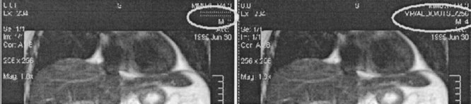
In this article we describe a statistical model that was developed to segment brain magnetic resonance images. The statistical segmentation algorithm was applied after a pre-processing stage involving the use of a 3D anisotropic filter along with histogram equalization techniques. The segmentation algorithm makes use of prior knowledge and a probability-based multivariate model designed to semi-automate the process of segmentation. The algorithm was applied to images obtained from the Center for Morphometric Analysis at Massachusetts General Hospital as part of the Internet Brain Segmentation Repository (IBSR). The developed algorithm showed improved accuracy over the k-means, adaptive Maximum Apriori Probability (MAP), biased MAP, and other algorithms. Experimental results showing the segmentation and the results of comparisons with other algorithms are provided. Results are based on an overlap criterion against expertly segmented images from the IBSR. The algorithm produced average results of approximately 80% overlap with the expertly segmented images (compared with 85% for manual segmentation and 55% for other algorithms).
Figures
Similar articles
-
A framework for quantification and visualization of segmentation accuracy and variability in 3D lateral ventricle ultrasound images of preterm neonates.Med Phys. 2015 Nov;42(11):6387-405. doi: 10.1118/1.4932366. Med Phys. 2015. PMID: 26520730
-
Three-dimensional lung tumor segmentation from x-ray computed tomography using sparse field active models.Med Phys. 2012 Feb;39(2):851-65. doi: 10.1118/1.3676687. Med Phys. 2012. PMID: 22320795
-
A 3D global-to-local deformable mesh model based registration and anatomy-constrained segmentation method for image guided prostate radiotherapy.Med Phys. 2010 Mar;37(3):1298-308. doi: 10.1118/1.3298374. Med Phys. 2010. PMID: 20384267
-
Magnetic resonance image segmentation using pattern recognition, and applied to image registration and quantitation.NMR Biomed. 1998 Jun-Aug;11(4-5):157-67. doi: 10.1002/(sici)1099-1492(199806/08)11:4/5<157::aid-nbm528>3.0.co;2-l. NMR Biomed. 1998. PMID: 9719570 Review.
-
Image processing and radiotherapy.Cancer Radiother. 2004 Apr;8(2):120-9. doi: 10.1016/j.canrad.2003.10.002. Cancer Radiother. 2004. PMID: 15132145 Review. English, French.
Cited by
-
An artificial immune-activated neural network applied to brain 3D MRI segmentation.J Digit Imaging. 2008 Oct;21 Suppl 1(Suppl 1):S69-88. doi: 10.1007/s10278-007-9081-0. Epub 2007 Dec 11. J Digit Imaging. 2008. PMID: 18071820 Free PMC article. Review.
-
Evaluation of automated brain MR image segmentation and volumetry methods.Hum Brain Mapp. 2009 Apr;30(4):1310-27. doi: 10.1002/hbm.20599. Hum Brain Mapp. 2009. PMID: 18537111 Free PMC article.
-
Automated detection of intercellular signaling in astrocyte networks using the converging squares algorithm.J Neurosci Methods. 2008 May 30;170(2):294-9. doi: 10.1016/j.jneumeth.2008.01.013. Epub 2008 Jan 29. J Neurosci Methods. 2008. PMID: 18328570 Free PMC article.
References
-
- Bloch RH, Udupa JK. Application of computerized tomography to radiation therapy and surgical planning. Proc IEEE. 1983;71:351–355.
-
- Brewster LJ, Trivedi S, Tut H., et al. Interactive surgical planning. IEEE Computer Graphics Appl. 1984;4:31–40.
-
- Lorensen W, Cline H. Marching cubes: a high-resolution 3D surface construction algorithm. Computer Graphics. 1987;21:163–169.
-
- Burk D, Mears D, Kennedy W, et al. Three-dimensional computed tomography of acetabula fractures. Radiology. 1985;155:33–43. - PubMed
-
- Hemmy DC, Tessier PL. Three-dimensional reconstruction of craniofacial deformity using computed tomography. Neurosurgery. 1985;13:534–541. - PubMed
Publication types
MeSH terms
LinkOut - more resources
Full Text Sources
