. 2004 Apr 15;556(Pt 2):521-30.
doi: 10.1113/jphysiol.2003.058792. Epub 2004 Jan 30.

Interstitial cells of Cajal are functionally innervated by excitatory motor neurones in the murine intestine

Affiliations

Interstitial cells of Cajal are functionally innervated by excitatory motor neurones in the murine intestine

Satoshi Iino et al. J Physiol. .

Abstract

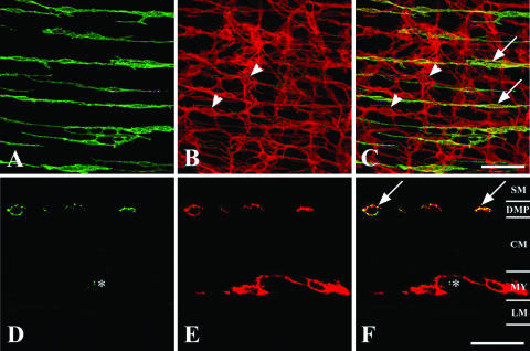
Recent studies have demonstrated that intramuscular interstitial cells of Cajal (ICC) are preferential targets for neurotransmission in the stomach. Terminals of enteric motor neurones also form tight, synaptic-like contacts with ICC in the small intestine and colon, but little is known about the role of these cells in neurotransmission. ICC at the deep muscular plexus (ICC-DMP) of the small intestine express neurokinin 1 receptors (NK1R) and internalize these receptors in response to exogenous substance P. We used NK1R internalization as an assay of functional innervation of ICC-DMP in the murine small intestine. Under basal conditions NK1R-like immunoreactivity (NK1R-LI) was mainly observed in ICC-DMP (519 cells counted, 100% were positive) and myenteric neurones. ICC-DMP were closely apposed to substance P-containing nerve fibres. Of 338 ICC-DMP examined, 65% were closely associated with at least one substance P-positive nerve fibre, 32% were associated with at least two, 2% were associated with more than two nerve fibres and 1% with none. After electrical field stimulation (EFS, 10 Hz; 1 min) NK1R-LI was internalized in more than 80% of ICC-DMP, as compared to 10% of cells before EFS. Internalization of NK1R was not observed in myenteric ICC or smooth muscle cells in response to nerve stimulation. Internalization of NK1R-LI was blocked by the specific NK1 receptor antagonist WIN 62577 (1 microm) and by tetrodotoxin (0.3 microm), suggesting that internalization resulted from stimulation of receptors with neurally released neurokinins. These data suggest that ICC-DMP are primary targets for neurokinins released from enteric motor neurones in the intestine.


Figures

Figure 1
Figure 1. Distribution of neurokinin 1 receptor-like immunoreactivity (NK1R-LI) in the murine small intestine and colocalization with Kit-LI in ICC-DMP
In whole mount preparations a dense network of NK1R-LI cells were observed running parallel to the circular muscle fibres (A, green). Double labelling with Kit-LI (B, red) showed that NK1R-LI was located in ICC-DMP near the inner aspect of the circular muscle layer (C, yellow, see arrows). NK1R-LI was not resolved in ICC at the level of the myenteric plexus (ICC-MY; red, see arrowheads in B and C). In cryostat sections (D–F), NK1R-LI (D, green) and Kit-LI (E, red) were colocalized in the plasma membranes of ICC-DMP (F, yellow, see arrows). NK1R-LI was not observed in ICC-MY; however, some enteric neurones displayed NK1R-LI (*). A–C are digital reconstructions from 8 sections, total thickness of reconstructed stack = 7 μm; D–F are from a single optical section 0.9 μm thickness. In D–F, SM = submucosa; DMP = deep muscular plexus; CM = circular muscle; MY = myenteric plexus; LM = longitudinal muscle. Scale bar in C = 50 μm and applies to A–C and scale bar in F = 20 μm and applies to D–F.
Figure 2
Figure 2. NK1R-LI and Kit-LI at the level of the myenteric plexus in the small intestine
A shows NK1R-LI in enteric neurones at the level of the myenteric plexus (green). Myenteric neurones had a morphological appearance typical of Dogiel type II neurones. B shows Kit-LI in ICC-MY (red) and C shows a merged image of NK1R-LI and Kit-LI. ICC-MY did not possess NK1R-LI. A–C are digital reconstructions from 5 sections, total thickness of reconstructed stack = 3 μm. Scale bar in C = 50 μm and applies to all panels.
Figure 3
Figure 3. Whole mount and cryostat sections of NK1R-LI and substance P-LI
A–C show double labelling of a whole mount preparation (single optical section, 1.3 μm thickness) revealing NK1R-LI (A, green) and substance P-LI nerve fibres (B, red) at the level of the deep muscular plexus. Even in single optical sections substance P-containing nerve fibres and varicosities were closely associated with ICC-DMP with NK1R-LI (C). Of 338 ICC-DMP examined, 64.8% of cells were associated with at least one substance P-LI nerve fibre, 32.3% were associated with at least two substance P-LI nerve fibres, 2.1% were associated with more than two nerve fibres. Clear morphological associations between substance P-containing nerve fibres and ICC-DMP were not resolved in 0.8% of the cells examined. Cryostat sections (single optical section 0.9 μm thickness) confirmed the close association of substance P-containing nerve fibres and ICC-DMP at the level of the deep muscular plexus (D–F, arrows). There was no obvious relationship between ICC and substance P-containing neurones in the myenteric region (F, *). Scale bar in C = 50 μm and applies to A–C and scale bar in F = 20 μm and applies to D–F.
Figure 4
Figure 4. Exogenous substance P causes NK1R-LI internalization
Diffuse labelling of NK1R-LI was observed on ICC-DMP under control conditions (A, arrows). After exposure to Substance P (1 μm; 60 min at 4°C; followed by 20 min at 37°C), NK1-LI was concentrated into small granular structures within the cytoplasm of ICC-DMP (B, arrows). Granular labelling of NK1R-LI was also observed in circular smooth muscle cells (arrowheads in B and C) and myenteric neurones (arrowheads in D) after substance P exposure. Even after exposure to exogenous substance P, we could not resolve NK1R-LI in ICC-MY. A and B are digital reconstructions from 10 sections, total thickness of reconstructed stack = 10 μm; C and D are from a single optical section 1.0 μm in thickness. Scale bars in all panels = 20 μm.
Figure 5
Figure 5. Distribution of NK1R-LI in ICC-DMP
A is a wholemount preparation of ICC-DMP labelled with NK1R-LI. In unstimulated muscles NK1R-LI was diffusely distributed around the peripheries of ICC-DMP. A is a single optical section (0.8 μm thickness). B shows high power single optical section (0.5 μm thickness) of the area outlined by a square in A. Scale bars = 20 μm in both panels.
Figure 6
Figure 6. NK1R-LI is internalized by ICC-DMP after electrical field stimulation (EFS)
A and B show NK1R-LI on ICC-DMP after EFS (10 Hz for 1 min, 0.5 ms pulse duration). After stimulation, NK1R-LI was concentrated into small granular structures (possibly endosomes; see arrows) in ICC-DMP. Stimulation did not produce resolution of NK1R-LI in ICC-MY. A is a single optical section (0.8 μm thickness). B is a high power single optical section (0.5 μm thickness) of the area outlined by a square in A. Scale bars = 20 μm in both panels.
Figure 7
Figure 7. Summary of NK1R internalization on ICC-DMP after EFS
A shows images prior to (Control) and after electric field stimulation (EFS) demonstrating internalization of NK1R-LI in ICC-DMP. B quantifies the degree of NK1R-LI internalization under control conditions and after EFS (10 Hz for 1 min, 0.5 ms pulse duration). NK1R-LI were internalized into 80.1 ± 1.3% of ICC-DMP after EFS as compared to 10.4 ± 1.0% of ICC-DMP prior to EFS (*P < 0.01). The data are expressed as mean ±s.e.m. (n = 10; each experiment on muscles of different animals). Scale bars in A = 10 μm.
Figure 8
Figure 8. NK1R-LI internalization was due to specific stimulation of NK1R
A shows distribution of NK1R-LI before (Control) and after EFS (EFS). Left panel in A shows that NK1R-LI internalization in response to EFS (10 Hz for 1 min, 0.5 ms pulse duration) was blocked by the selective NK1R antagonist WIN 62577 (1 μm). B summarizes the percentage of ICC-DMP that displayed NK1R-LI internalization under control conditions and after EFS in the presence of WIN 62577. Prior to EFS 11.3 ± 1.4% ICC-DMP displayed internalized NK1R-LI. After EFS in the presence of WIN 62577, 15.3 ± 1.1% of ICC-DMP showed internalization. The data are expressed as the mean ±s.e.m. (n = 3 animals). Scale bars in A = 10 μm.
Figure 9
Figure 9. NK1R-LI internalization in response to EFS was blocked by tetrodotoxin (TTX)
A shows the normal distribution of NK1R-LI before (Control) and after EFS (EFS) in the presence of TTX (0.3 μm). Prior to EFS, intestinal muscles were exposed to TTX (20 min). Under these conditions EFS (10 Hz for 1 min, 0.5 ms pulse duration) caused no significant increase in NK1R-LI internalization (A). B summarizes the degree of internalization before stimulation and during stimulation in the presence of TTX. After TTX, only 14.2 ± 1.5% of ICC-DMP showed internalization compared to 12.2 ± 1.1% prior to stimulation. The data are expressed as the mean ±s.e.m. (n = 3 animals). Scale bars in A = 10 μm.

Similar articles

Cited by

References

    1. Beckett EA, Horiguchi K, Khoyi M, Sanders KM, Ward SM. Loss of enteric motor neurotransmission in the gastric fundus of Sl/Sl (d) mice. J Physiol. 2002;543:871–887. - PMC - PubMed
    1. Beckett EA, McGeough CA, Sanders KM, Ward SM. Pacing of interstitial cells of Cajal in the murine gastric antrum : neurally mediated and direct stimulation. J Physiol. 2003;553:545–559. - PMC - PubMed
    1. Bowden JJ, Garland AM, Baluk P, Lefevre P, Grady EF, Vigna SR, Bunnett NW, McDonald DM. Direct observation of substance P-induced internalization of neurokinin 1 (NK1) receptors at sites of inflammation. Proc Natl Acad Sci U S A. 1994;91:8964–8968. - PMC - PubMed
    1. Burns AJ, Lomax AE, Torihashi S, Sanders KM, Ward SM. Interstitial cells of Cajal mediate inhibitory neurotransmission in the stomach. Proc Natl Acad Sci U S A. 1996;93:12008–12013. - PMC - PubMed
    1. Daniel EE, Posey-Daniel V. Neuromuscular structures in opossum esophagus : role of interstitial cells of Cajal. Am J Physiol. 1984;246:G305–G315. - PubMed

Publication types

LinkOut - more resources