. 2004 Feb 10;101(6):1714-9.
doi: 10.1073/pnas.0308102100. Epub 2004 Jan 30.

STAT1 is overexpressed in tumors selected for radioresistance and confers protection from radiation in transduced sensitive cells

Affiliations

STAT1 is overexpressed in tumors selected for radioresistance and confers protection from radiation in transduced sensitive cells

Nikolai N Khodarev et al. Proc Natl Acad Sci U S A. .

Abstract

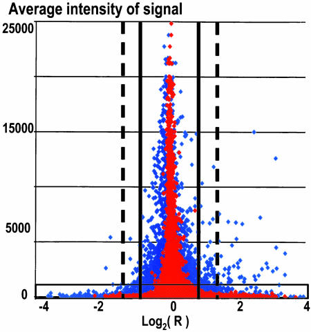
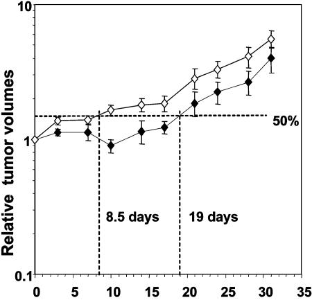
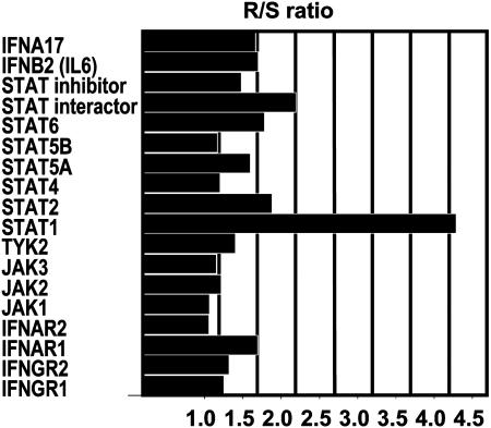
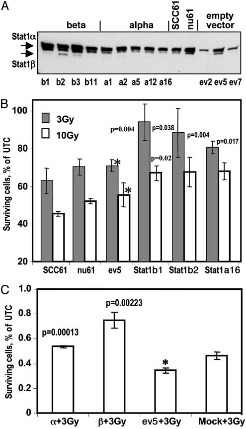
Nu61, a radiation-resistant human tumor xenograft, was selected from a parental radiosensitive tumor SCC-61 by eight serial cycles of passage in athymic nude mice and in vivo irradiation. Replicate DNA array experiments identified 52 genes differentially expressed in nu61 tumors compared with SCC-61 tumors. Of these, 19 genes were in the IFN-signaling pathway and moreover, 25 of the 52 genes were inducible by IFN in the nu61 cell line. Among the genes involved in IFN signaling, STAT1alpha and STAT1beta were the most highly overexpressed in nu61 compared to SCC-61. STAT1alpha and STAT1beta cDNAs were cloned and stably transfected into SCC-61 tumor cells. Clones of SCC-61 tumor cells transfected with vectors expressing STAT1alpha and STAT1beta demonstrated radioprotection after exposure to 3 Gy (P < 0.038). The results indicate that radioresistance acquired during radiotherapy treatment may account for some treatment failures and demonstrate an association of acquired tumor radioresistance with up-regulation of components of the IFN-related signaling pathway.


Figures

Fig. 1.
Fig. 1.
Scheme of multistep filtration (MSF) of DNA array data. Red diamonds corresponds to the same-to-same hybridizations (nu61/nu61 and SCC-61/SCC-61), and blue diamonds correspond to the same-to-different hybridizations (nu61/SCC-61). Light blue region on the bottom of the histogram corresponds to the genes with intensity signals below cutoff level, estimated by the receiver operating characteristic (ROC) analysis. Note, that genes in this region possess the highest ratios of response producing false positive data (see ref. 12). Solid vertical bars, range of ±2 SD of the reference set, corresponding to ratios of ± 1.919. Dashed vertical bars, range of ± 3 SD of the reference set, corresponding to the ratios of ±2.657. Shown are data not filtrated by P values and CV.
Fig. 2.
Fig. 2.
Relative increase in SCC-61 and nu61 tumors as a function of time after irradiation. Tumors were permitted to grow to a volume of 250-300 mm3. At that time (day 0) the tumors were subjected to six 500-cGy irradiations as described in Materials and Methods. Measurements of relative tumor volumes were performed during 32 days. Growth delay was defined as the time, necessary for irradiated tumor to increase relative volume by 50%, compared to the volume on day 0. Error bars are SEM.
Fig. 3.
Fig. 3.
Stat1 is overexpressed in the nu61 compared with SCC-61 more than other genes, involved in the IFN signaling and presented on U133A DNA Chip. Data were taken from the first experiment with pooled RNA samples. Databases, obtained with U133A arrays were screened manually to identify genes, involved in IFN signaling. Shown are the ratios of the gene expression in the nu61 cells relative to the SCC-61 parental cells. Stat1 (β isoform, target ID 209969_s_at) is overexpressed more than other IFN-signaling genes.
Fig. 4.
Fig. 4.
Stable transfection of SCC-61 parental cell line by Stat1α and Stat1β cDNAs leads to increased radioprotection of the transfected clones. (A) Protein expression of Stat1 in stably transfected clones. pAttBStat1α, pAttBStat1β, or pAttB were cotransfected into SCC-61 with pCMV-INT, kindly provided by Michele Calos. Cells were selected in the Hyg-containing media, and selected monoclones were further propagated for experiments (see Materials and Methods). Several clones, transfected by pAttBStat1β, pAttBStat1α, or pAttB (empty vector), were checked by PCR of genomic DNA for appropriate integration and checked by Western analysis for expression of α and β isoforms of Stat1. b1, b2, b3, and b11 correspond to the clones stably transfected by pAttBStat1β. a1, a2, a5, a12, and a16 correspond to the clones stably transfected by pAttBStat1α. ev2, ev5, and ev7 correspond to the clones, stably transfected by pAttB and related as “empty vector.” For the further experiments we choose b1, b2, and a16 clones as Stat1 stable transfectants and ev5 clone as the “empty vector” control. (B) Stably transfected α and β clones protect parental cell line from ionizing radiation. SCC-61, nu61, and b1, b2, a16, and ev5 clones were plated in the 96-well plates and either irradiated at 3 or 10 Gy (see Materials and Methods) or left untreated. All experiments were made in triplicates. Viability of irradiated and untreated cells was assessed by the MTS assay 72 h after irradiation (see Materials and Methods). Fraction of surviving cells was calculated as percent of viable cells after irradiation to unirradiated control for the each type of the cells. For the stable transfectants of the SCC-61, significance of survival was calculated relative to the ev5 clone, marked by asterisks. P values were calculated as the two-tailed, unpaired t test and are indicated in B above each corresponding bar. Error bars are standard deviations of triplicate experiments. (C) Transient transfection of the parental cell line SCC-61 by plasmids, carrying Stat1β and Stat1α leads to the increased radioprotection. SCC-61 cells were transfected with the pAttBStat1α, pAttBStat1β or pAttB plasmids (see above and Materials and Methods). As the mock we used SCC-61, treated in the same way as transfected cells but without exogenous plasmids. Twenty-four hours after transfection, cells were irradiated at 3 Gy, and 24 h after irradiation viable cells were scored by the Trypan Blue assay (see Materials and Methods). Surviving fraction was calculated as the percent of viable cells after irradiation to unirradiated control for the each type of the cells. Significance was calculated relative to the ev5-transfected cells (marked by asterisk), using two-tailed unpaired t test. P values are indicated above the appropriate columns. Error bars are standard deviations of the triplicate experiments.

References

    1. Hall, E. J. (2000) Radiobiology for Radiologist (Lippincott Williams and Wilkins, Philadelphia), 5th Ed., pp 5-17.
    1. Harris, A. L. (2002) Nat. Rev. Cancer 2, 38-47. - PubMed
    1. Taylor, A. M. (1998) Int. J. Radiat. Biol. 73, 365-371. - PubMed
    1. Young, B. R. & Painter, R. B. (1989) Hum. Genet. 82, 113-117. - PubMed
    1. Valerie, K. & Povirk, L. F. (2003) Oncogene 22, 5792-5812. - PubMed

Publication types