. 2004 Feb 10;101(6):1491-5.
doi: 10.1073/pnas.2637011100. Epub 2004 Jan 30.

Molecular dissection of the roles of nucleotide binding and hydrolysis in dynein's AAA domains in Saccharomyces cerevisiae

Affiliations

Molecular dissection of the roles of nucleotide binding and hydrolysis in dynein's AAA domains in Saccharomyces cerevisiae

Samara L Reck-Peterson et al. Proc Natl Acad Sci U S A. .

Retraction in

Abstract

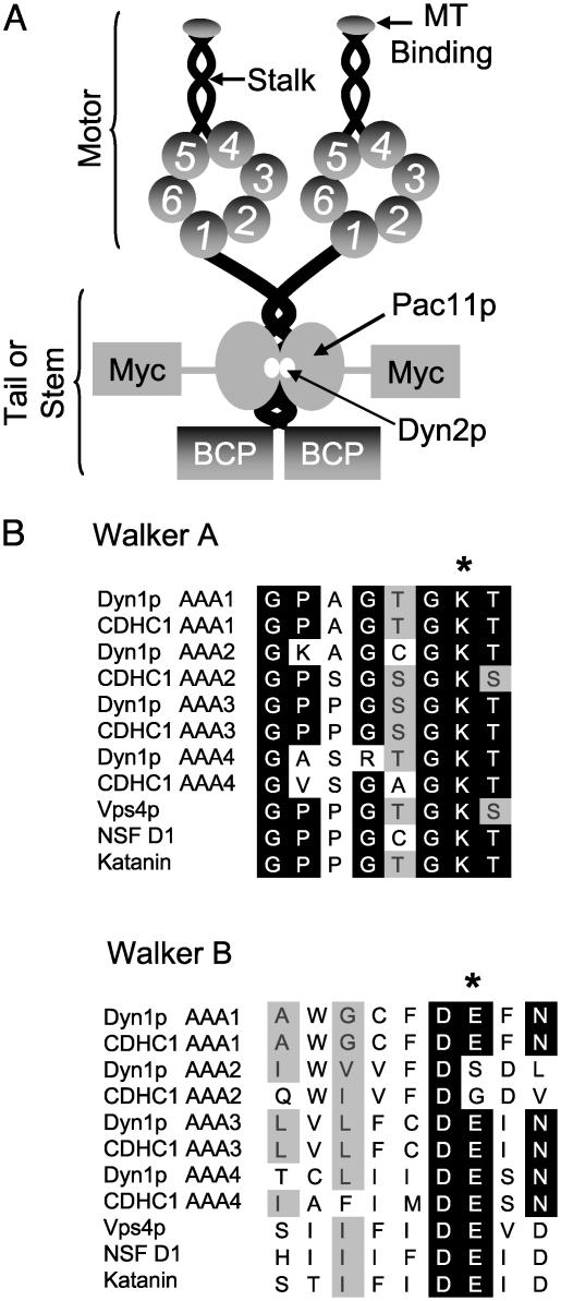
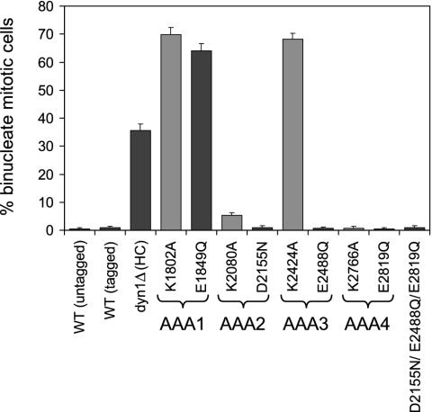
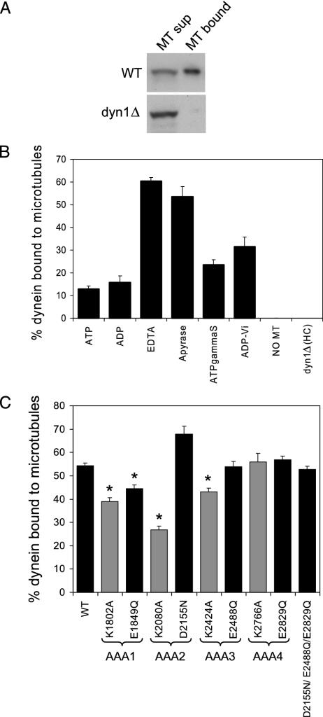
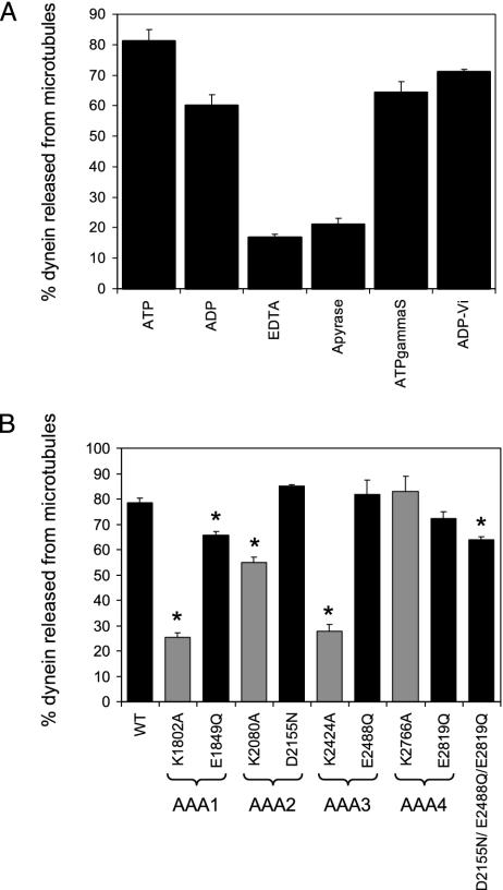
The motor protein cytoplasmic dynein is responsible for most of the minus-end-directed microtubule traffic within cells. Dynein contains four evolutionarily conserved AAA (ATPase associated with various cellular activities) domains that are thought to bind nucleotide; the role of nucleotide binding and hydrolysis in each of these four AAA domains has constituted an important and unresolved question in understanding dynein's mechanism. Using Saccharomyces cerevisiae cytoplasmic dynein as a model system, we mutagenized residues involved in nucleotide binding or hydrolysis in the four AAA domains and examined the ability of the mutant dyneins to mediate nuclear segregation in vivo and to bind microtubules in vitro. Our analysis shows that an AAA1 hydrolysis mutant blocks dynein function, whereas a triple AAA2/3/4 hydrolysis mutant does not, suggesting that nucleotide binding is required at only one site. We also show that nucleotide binding at AAA3, but not hydrolysis, is essential for motor activity in vivo and ATP-induced dissociation of dynein from microtubules, suggesting that this domain acts as a critical allosteric site. In contrast, mutations in AAA2 cause subtle defects in dynein function, whereas mutation in AAA4 produce no obvious defects. These results show that the four conserved dynein AAA domains have distinct functions in dynein's mechanochemical cycle.


Figures

Fig. 1.
Fig. 1.
Overall dynein structure and mutations generated in the AAA domains. (A) Predicted domain structure of S. cerevisiae cytoplasmic dynein and the introduction of tags. Cytoplasmic dynein is composed of two heavy chains that contain six AAA domains within the motor domain, the first four of which are predicted to bind nucleotide. The light (Dyn2p) and intermediate chain (Pac11p) are associated with the tail domain. Dyn1p was tagged with a 96-aa biotinylatable domain from the P. pastoris Pyc1p protein. Pacllp was tagged with 13 copies of the Myc epitope. (B) Sequence alignment of the Walker A and Walker B motifs from S. cerevisiae (Dyn1p) and rat (CDHC1) cytoplasmic dynein and other members of the AAA superfamily (Vps4p, NSF, and katanin). The starred (*) amino acids are the amino acids that were mutated in Dyn1p (K changed to A; E changed to Q). In the AAA2 Walker B box, DSDL was mutated to NSDL.
Fig. 2.
Fig. 2.
Mutations in AAA1 and AAA3 cause defects in nuclear segregation. To monitor nuclear segregation, the number of binucleate mitotic cells was determined after growth at 16°C for 16 h. The mean and standard error of the proportion are shown (n > 200). Walker A mutants (predicted to block nucleotide binding) are shaded with gray, whereas Walker B mutants (predicted to block nucleotide hydrolysis) are shaded with black.
Fig. 3.
Fig. 3.
Mutations in dynein AAA domains minimally affect dynein microtubule (MT) binding. (A) Anti-myc immunoblots of WT and dyn1Δ microtubule supernatant and pellet fractions loaded stoichiometrically on the gel, showing that the dynein intermediate chain does not associate with microtubules in the absence of the heavy chain. (B) Yeast extract containing WT dynein or no dynein (extract from dyn1Δ) was mixed with taxol-stabilized bovine brain microtubules in the presence or absence of the indicated Mg-nucleotides (5 mM). The dynein intermediate chain present in the microtubule pellet fractions was plotted as the percentage of total dynein (supernatant plus pellet fraction), and the mean and SEM are shown (n = 4). Because these experiments were performed in extracts containing adenylate kinase, we cannot exclude conversion of some ADP to ATP. (C) Yeast extract from WT or AAA mutant cells was mixed with bovine brain microtubule in the presence of apyrase. The percentage of dynein intermediate chain present in the microtubule pellet fractions was plotted as the percentage of total dynein, and the mean and SEM are shown (n = 4-14). The starred (*) means are statistically different from the WT mean (P < 0.005, t test). Walker A mutants (predicted to block nucleotide binding) are shaded with gray, whereas Walker B mutants (predicted to block nucleotide hydrolysis) are shaded with black.
Fig. 4.
Fig. 4.
Nucleotide binding at AAA1 and AAA3 is required for ATP-dependent dynein microtubule release. (A) Yeast extract containing WT dynein was bound to microtubules in the presence of apyrase and then released from microtubules by using different nucleotide conditions. The dynein intermediate chain released from microtubules was plotted as the percentage of total dynein (supernatant plus pellet fraction), and the mean and the SEM are shown (n = 4). (B) Yeast extract containing WT or mutant dynein protein was first bound to microtubules in the presence of apyrase and then released from microtubules with ATP. The dynein intermediate chain released from microtubules was plotted as the percentage of total dynein (supernatant plus pellet fraction), and the mean and the SEM are shown (n = 3-17). The starred (*) means are statistically different from the WT mean (P < 0.005, t test). Walker A mutants (predicted to block nucleotide binding) are shaded with gray, whereas Walker B mutants (predicted to block nucleotide hydrolysis) are shaded with black.

References

    1. Asai, D. J. & Koonce, M. P. (2001) Trends Cell Biol. 11, 196-202. - PubMed
    1. Vale, R. D. (2003) Cell 112, 467-480. - PubMed
    1. Vale, R. D. (2000) J. Cell Biol. 150, F13-F19. - PMC - PubMed
    1. Ogura, T. & Wilkinson, A. J. (2001) Genes Cells 6, 575-597. - PubMed
    1. Mocz, G. & Gibbons, I. R. (2001) Structure (London) 9, 93-103. - PubMed

Publication types