. 2004 Feb;113(3):357-69.
doi: 10.1172/JCI19448.

Defects in nuclear structure and function promote dilated cardiomyopathy in lamin A/C-deficient mice

Affiliations

Defects in nuclear structure and function promote dilated cardiomyopathy in lamin A/C-deficient mice

Vesna Nikolova et al. J Clin Invest. 2004 Feb.

Abstract

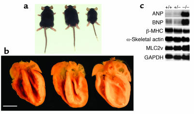
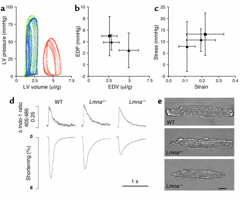
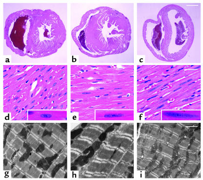
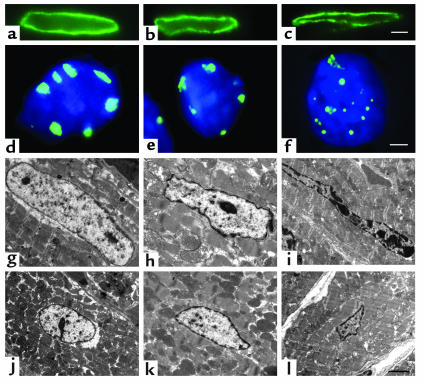
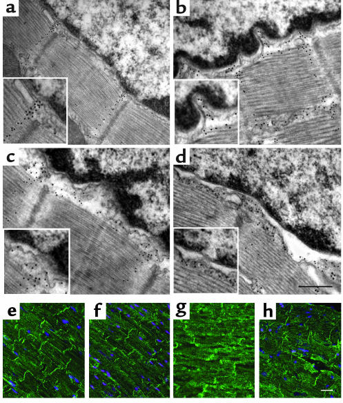
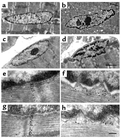
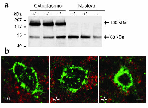
Laminopathies are a group of disorders caused by mutations in the LMNA gene that encodes the nuclear lamina proteins, lamin A and lamin C; their pathophysiological basis is unknown. We report that lamin A/C-deficient (Lmna(-/-)) mice develop rapidly progressive dilated cardiomyopathy (DCM) characterized by left ventricular (LV) dilation and reduced systolic contraction. Isolated Lmna(-/-) myocytes show reduced shortening with normal baseline and peak amplitude of Ca(2+) transients. Lmna(-/-) LV myocyte nuclei have marked alterations of shape and size with central displacement and fragmentation of heterochromatin; these changes are present but less severe in left atrial nuclei. Electron microscopy of Lmna(-/-) cardiomyocytes shows disorganization and detachment of desmin filaments from the nuclear surface with progressive disruption of the cytoskeletal desmin network. Alterations in nuclear architecture are associated with defective nuclear function evidenced by decreased SREBP1 import, reduced PPARgamma expression, and a lack of hypertrophic gene activation. These findings suggest a model in which the primary pathophysiological mechanism in Lmna(-/-) mice is defective force transmission resulting from disruption of lamin interactions with the muscle-specific desmin network and loss of cytoskeletal tension. Despite severe DCM, defects in nuclear function prevent Lmna(-/-) cardiomyocytes from developing compensatory hypertrophy and accelerate disease progression.


Figures

Figure 1
Figure 1
Comparative morphometry in mice aged 4–6 weeks. (a) When compared with WT (left) and Lmna+/– littermates (center), Lmna–/– (right) mice exhibit growth retardation with 50% reduction in body weight. (b) Despite marked differences in body size, longitudinal sections show that heart sizes are similar in WT (left), Lmna+/– (center), and Lmna–/– (right) mice. Lmna–/– hearts show LV and LA dilation with wall thinning. Scale bar: 2.5 mm. (c) The phenotype of severe DCM without compensatory hypertrophy in 4- to 6-week-old Lmna–/– mice is reflected by Northern blot analyses that show appropriate increases in LV expression of ANP and BNP but no induction of β-MHC or α-skeletal actin. +/+, WT; +/–, Lmna+/–; –/–, Lmna–/–.
Figure 2
Figure 2
In vivo analyses of cardiac function with micromanometry and sonomicrometry in mice aged 4–6 weeks. (a) Representative LV pressure-volume loops recorded at steady state are shown for WT (blue), Lmna+/– (green), and Lmna–/– (red) mice. Steady-state LV end-diastolic pressure (EDP) plotted against LV end-diastolic volume (EDV) (b) and stress-strain relationships (c) for WT (diamonds, n = 9) Lmna+/– mice (squares, n = 11), and Lmna–/– mice (triangles, n = 6). Volume data are normalized to body weight; data are shown as mean ± SD. Functional and morphologic characteristics of cardiac myocytes isolated from mice aged 4–6 weeks were also assessed. (d) Representative recordings of Ca2+ transients (top panel) and shortening (lower panel) in single myocytes. Intracellular Ca2+ concentration was measured as the change in the 405:485 nm emission ratio for Indo-1. 0.25 represents the magnitude of the Ca2+ transient. Lmna–/– myocytes have similar baseline and peak intracellular Ca2+ concentrations to WT and Lmna+/– myocytes but significantly reduced shortening. (e) Lmna–/– myocytes are shorter and thinner than WT and Lmna+/– myocytes but the length/width ratios are similar. Scale bar: 20 μm.
Figure 3
Figure 3
Myocardial histology in mice aged 4–6 weeks. Heart tissue from WT (a, d, and g), Lmna+/– (b, e, and h), and Lmna–/– (c, f, and i) mice was evaluated by light microscopy after staining with H&E (af) and electron microscopy (gi). Cross-sections at the midventricular level (ac) show LV and RV dilation in Lmna–/– hearts. Scale bar: 1 mm. Higher magnification (df) shows overall myocardial architecture in Lmna–/– LV myofibrils is relatively preserved. Nuclear elongation and chromatin dispersion are evident in Lmna+/– and Lmna–/– cardiomyocytes (insets). Scale bar: 50 μm. Sarcomere organization (gi) appears normal in all mice. Scale bar: 2 μm.
Figure 4
Figure 4
Nuclear morphology in LV cardiomyocytes from WT (a, d, g, and j), Lmna+/– (b, e, h, and k) and Lmna–/– (c, f, i, and l) mice aged 4–6 weeks. Immunostaining with a lamin B antibody (ac) shows the nuclear lamina intact but altered in shape in Lmna+/– and Lmna–/– mice. Scale bar: 2 μm. Immunostaining with a pancentromeric heterochromatin probe (green) and DAPI (blue) (df) shows that centromeric heterochromatin forms discrete peripherally located blocks in WT nuclei with some irregularities of shape in Lmna+/– nuclei. In Lmna–/– nuclei, centromeric heterochromatin appears as small round foci at the periphery and within the nuclear interior. Confluent masses with a “beaded” appearance and larger, less dense masses with indistinct irregular borders are present. Scale bar: 1 μm. Longitudinal (gi) and cross-sectional (jl) electron microscopy images also demonstrate changes in nuclear morphology and heterochromatin organization. WT nuclei are characteristically oval-shaped with a thin rim of densely staining heterochromatin and a few small central deposits (g and j). Lmna+/– nuclei have some irregularities of shape and peripheral heterochromatin clumping (h and k). Abnormalities of shape and heterochromatin distribution are evident in Lmna–/– myocyte nuclei at 2 weeks of age (data not shown). By 4–6 weeks, the majority of Lmna–/– myocyte nuclei demonstrate morphologic changes, including elongation and bizarre shapes with clumping of peripheral heterochromatin (i and l). In approximately 50% of the nuclei, heterochromatin clumps of varying size and shape are observed within the nuclear interior. Scale bar: 2 μm.
Figure 5
Figure 5
Disruption of the lamin-desmin intermediate filament network in lamin A/C–deficient mice. Attachment sites of desmin filaments to the LV cardiomyocyte nuclear surface were identified using immunogold-labeled desmin Ab’s and electron microscopy. Approximately 30 nuclei from WT mice (n = 3), Lmna+/– mice (n = 3), and Lmna–/– mice (n = 3) aged 2 weeks and 4–6 weeks were evaluated. In WT mice (a), desmin filaments (black dots) generally appear as well-defined bands connecting the cytoskeleton with the nuclear surface through nuclear pores (insets). Disorganization of the desmin filaments with detachment from the nuclear surface (with or without widening of the gap between the nuclear and myofibril borders) was observed in 4 (13%) Lmna+/– nuclei (b) and 16 (59%) Lmna–/– nuclei (d), but in none of the WT nuclei at 4–6 weeks. These changes were evident in 14 (50%) Lmna–/– nuclei (c) at 2 weeks; scale bar = 0.25 μm. Immunostaining with a desmin Ab (eh) shows normal Z disc striations and intercalated discs in WT and Lmna+/– mice, but progressive disorganization of the desmin filament network in 2-week-old (g) and 4- to 6-week-old (h) Lmna–/– mice. Scale bar: 5 μm.
Figure 6
Figure 6
Nuclear morphology and nuclear-desmin attachments in LA and skeletal myocytes in mice aged 4–6 weeks. Electron microscopic images of myocyte nuclei in the LA (a, b, e, and f) and gastrocnemius (c, d, g, and h) show normal nuclear morphology and nuclear-desmin attachments in WT mice (a, c, e, and g). Lmna–/– nuclei (b, d, f, and h) show changes in shape and heterochromatin clumping in the LA and skeletal muscle, although to a lesser extent than in the LV, with disruption and detachment of desmin filaments from the nuclear surface in both tissues. Scale bars: 2 μm (ad) and 0.1 μm (eh).
Figure 7
Figure 7
SREBP1 localization in LV cardiomyocytes of mice aged 4–6 weeks. (a) Western blot analysis of cytoplasmic and nuclear fractions of LV tissue hybridized with an SREBP1 Ab. Representative data for one WT (+/+), Lmna+/– (+/–), and Lmna–/– (–/–) mouse are shown. Experiments were repeated with six mice from each group. When compared with WT cardiomyocytes, Lmna–/– cardiomyocytes show a mean decrease (35%) in uncleaved (130 kDa) SREBP1 and a mean increase (55%) in cleaved (60 kDa) SREBP1 in the cytoplasmic fraction, with a mean reduction (40%) of cleaved SREBP1 in the nuclear fraction. (b) Immunofluorescence microscopy shows a perinuclear and intranuclear distribution of SREBP1 in WT and Lmna+/– nuclei, but predominant perinuclear SREBP1 staining in Lmna–/– nuclei. Scale bar: 2 μm.

Comment in

References

    1. Hozak P, Sasseville AM, Raymond Y, Cook PR. Lamin proteins form an internal nucleoskeleton as well as a peripheral lamina in human cells. J. Cell Sci. 1995;108:635–644. - PubMed
    1. Barboro P, et al. Unravelling the organization of the internal nuclear matrix: RNA-dependent anchoring of NuMA to a lamin scaffold. Exp. Cell Res. 2002;279:202–218. - PubMed
    1. Manilal S, et al. Distribution of emerin and lamins in the heart and implications for Emery-Dreifuss muscular dystrophy. Hum. Mol. Genet. 1999;8:353–359. - PubMed
    1. Stuurman N, Heins S, Aebi U. Nuclear lamins: their structure, assembly and interactions. J. Struct. Biol. 1998;122:42–66. - PubMed
    1. Burke B, Stewart CL. Life at the edge: the nuclear envelope and human disease. Nat. Rev. Mol. Cell Biol. 2002;3:575–585. - PubMed

Publication types