. 2004 Feb;21(2):75-85.
doi: 10.1002/hbm.10153.

Functional brain mapping during free viewing of natural scenes

Affiliations

Functional brain mapping during free viewing of natural scenes

Andreas Bartels et al. Hum Brain Mapp. 2004 Feb.

Abstract

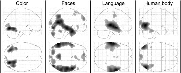
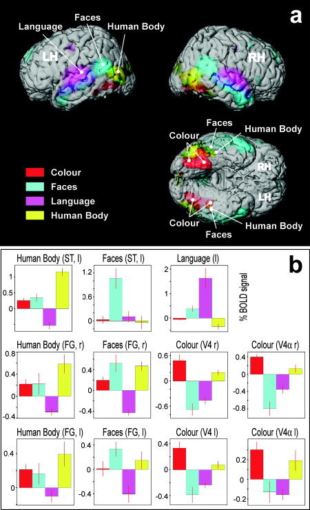
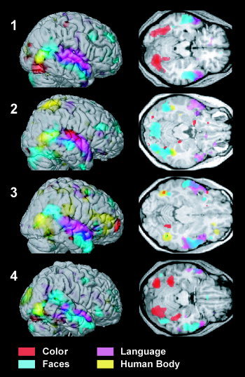
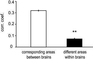
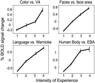
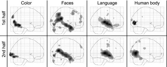
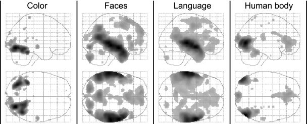
Previous imaging studies have used mostly perceptually abstracted, idealized, or static stimuli to show segregation of function in the cerebral cortex. We wanted to learn whether functional segregation is maintained during more natural, complex, and dynamic conditions when many features have to be processed simultaneously, and identify regions whose activity correlates with the perception of specific features. To achieve this, we used functional magnetic resonance imaging (fMRI) to measure brain activity when human observers viewed freely dynamic natural scenes (a James Bond movie). The intensity with which they perceived different features (color, faces, language, and human bodies) was assessed psychometrically in separate sessions. In all subjects different features were perceived with a high degree of independence over time. We found that the perception of each feature correlated with activity in separate, specialized areas whose activity also varied independently. We conclude that even in natural conditions, when many features have to be processed simultaneously, functional specialization is preserved. Our method thus opens a new way of brain mapping, which allows the localization of a multitude of brain areas based on a single experiment using uncontrolled, natural stimuli. Furthermore, our results show that the intensity of activity in a specialized area is linearly correlated with the intensity of its perceptual experience. This leads us to suggest that each specialized area is directly responsible for the creation of a feature-specific conscious percept (a microconsciousness). Hum. Brain Mapp. 21:75-83, 2004.


Figures

Figure 1
Figure 1
Brain activity that correlated with the intensity of experience of color, faces, language, and human bodies during free viewing of the action movie. Shown are glass‐brain views for each of the four feature regressors obtained from a group analysis (P < 0.05, corrected).
Figure 2
Figure 2
Overview showing the spatial segregation of feature‐specific activity (A), and random‐effects parameter estimates from regions of interest (B). A: Rendered views of a template brain (after removal of the cerebellum), with superimposed group‐activity related to the four features (same data as in Fig. 1, P < 0.05 corrected). B: Parameter estimates of the four feature regressors of subjective experience, expressed as mean percent change in blood oxygen level dependent (BOLD) signal ± standard error (across eight subjects), for the group‐peak voxels indicated by white markers in (A). l, left; r, right; RH/LH, right/left hemisphere; FG, fusiform gyrus; ST, superior temporal cortex.
Figure 3
Figure 3
Neural correlates of perception of different features during free viewing on the example of four subjects. Significant voxels (P < 0.001 uncorrected) were color‐coded for each of the four feature regressors and shown on renderings of the right hemisphere (left) and on transverse slices (right) for each individual. Note that slices do not reflect the full extent of activity for all features, as this varied from plane to plane. Slices were taken at z = −19, −18, −16, −20 mm.
Figure 4
Figure 4
Corresponding areas of different brains have higher correlations of activity than different areas within the same brain. Data include all corresponding pairwise correlations across all eight subjects (see also text and Table II). nbetween brains = 140, nwithin brains = 80, Wilcoxon rank sum test and two‐sample t‐test: P < e−10, error bars = SEM.
Figure 5
Figure 5
Linear correlation between the intensity of subjective experience of a given feature with the activity in the areas specialized for it. The perceived feature‐intensities (as rated on a scale from 1–4) for color, faces, language and human bodies are plotted against the BOLD signal taken from V4, the face‐selective region in the ST cortex, Wernicke's area, and EBA from all subjects. Corresponding graphs for V4α and FFA were virtually identical and omitted for graphical simplicity. Error bars = SEM.
Figure 6
Figure 6
Separate analyses of data from the first (top) and the second (bottom) halves of the movie provided consistent results, suggesting that functional mapping during natural free viewing conditions does not depend on the precise nature of the stimuli used (group, P < 0.05, corrected).
Figure 7
Figure 7
Spatial specificity of feature‐specific activation. Results from four different analyses, using only one of the four feature regressors in each to maximize their power and shown at a low threshold of P < 0.001 uncorrected to illustrate that activity is contained to the functionally specialized regions identified in the original analysis shown in Figure 1.

Similar articles

Cited by

References

    1. Bar M, Tootell RB, Schacter DL, Greve DN, Fischl B, Mendola JD, Rosen BR, Dale AM (2001): Cortical mechanisms specific to explicit visual object recognition. Neuron 29: 529–535. - PubMed
    1. Bartels A, Zeki S (1998): The theory of multistage integration in the visual brain. Proc R Soc Lond B Biol Sci 265: 2327–2332. - PMC - PubMed
    1. Bartels A, Zeki S (2000): The architecture of the colour centre in the human visual brain: new results and a review. Eur J Neurosci 12: 172–193. - PubMed
    1. Bartels A, Zeki S (2003): The chronoarchitecture of the human brain—functional anatomy based on natural brain dynamics and on the principle of functional independence In: Frackowiak R, Friston K, Frith C, Dolan R, Zeki S, editors. Human brain function, 2nd edition UK: Academic Press; p 201–229.
    1. Beauchamp MS, Haxby JV, Jennings JE, DeYoe EA (1999): An fMRI version of the Farnsworth‐Munsell 100‐Hue test reveals multiple color‐selective areas in human ventral occipitotemporal cortex. Cereb Cortex 9: 257–263. - PubMed

Publication types