

Characterizing the diffusion/perfusion mismatch in experimental focal cerebral ischemia
- PMID: 14755724
- PMCID: PMC2949945
- DOI: 10.1002/ana.10803
Characterizing the diffusion/perfusion mismatch in experimental focal cerebral ischemia
Abstract
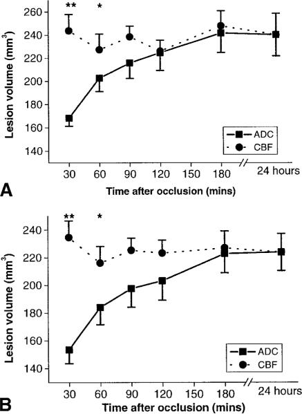
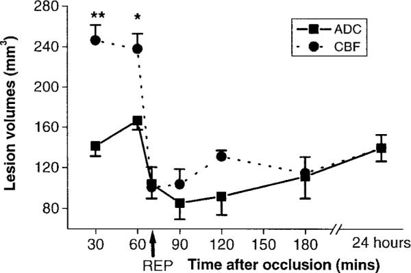
Diffusion-weighted imaging (DWI) and perfusion-weighted imaging (PWI) can rapidly detect lesions in acute ischemic stroke patients. The PWI volume is typically substantially larger than the DWI volume shortly after onset, that is, a diffusion/ perfusion mismatch. The aims of this study were to follow the evolution of the diffusion/ perfusion mismatch in permanent and 60- minute temporary focal experimental ischemia models in Sprague-Dawley rats using the intraluminal middle cerebral artery occlusion (MCAO) method. DWI and arterial spin-labeled PWI were performed at 30, 60, 90, 120, and 180 minutes after occlusion and lesion volumes (mm(3)) calculated At 24 hours after MCAO, and infarct volume was determined using triphenyltetrazolium chloride staining. In the permanent MCAO group, the lesion volume on the ADC maps was significantly smaller than that on the cerebral blood flow maps through the first 60 minutes after MCAO; but not after 90 minutes of occlusion. With 60 minutes of transient ischemia, the diffusion/perfusion mismatch was similar, but after reperfusion, the lesion volumes on ADC and cerebral blood flow maps became much smaller. There was a significant difference in 24- hour infarct volumes between the permanent and temporary occlusion groups.
Figures
Similar articles
-
Differences in ischemic lesion evolution in different rat strains using diffusion and perfusion imaging.Stroke. 2005 Sep;36(9):2000-5. doi: 10.1161/01.STR.0000177486.85508.4d. Epub 2005 Jul 21. Stroke. 2005. PMID: 16040589 Free PMC article.
-
SB 234551 selective ET(A) receptor antagonism: perfusion/diffusion MRI used to define treatable stroke model, time to treatment and mechanism of protection.Exp Neurol. 2008 Jul;212(1):53-62. doi: 10.1016/j.expneurol.2008.03.011. Epub 2008 Mar 25. Exp Neurol. 2008. PMID: 18462720
-
Spontaneous hyperthermia and its mechanism in the intraluminal suture middle cerebral artery occlusion model of rats.Stroke. 1999 Nov;30(11):2464-70; discussion 2470-1. doi: 10.1161/01.str.30.11.2464. Stroke. 1999. PMID: 10548685
-
[Application of diffusion-weighted and perfusion magnetic resonance imaging in definition of the ischemic penumbra in hyperacute cerebral infarction].Zhonghua Yi Xue Za Zhi. 2003 Jun 10;83(11):952-7. Zhonghua Yi Xue Za Zhi. 2003. PMID: 12899795 Chinese.
-
The role of diffusion- and perfusion-weighted magnetic resonance imaging in drug development for ischemic stroke: from laboratory to clinics.Curr Vasc Pharmacol. 2004 Oct;2(4):343-55. doi: 10.2174/1570161043385493. Curr Vasc Pharmacol. 2004. PMID: 15320814 Review.
Cited by
-
Improving standardization and accuracy of in vivo omega plot exchange parameter determination using rotating-frame model-based fitting of quasi-steady-state Z-spectra.Magn Reson Med. 2025 Jan;93(1):151-165. doi: 10.1002/mrm.30259. Epub 2024 Sep 2. Magn Reson Med. 2025. PMID: 39221563
-
Artificial neural network prediction of ischemic tissue fate in acute stroke imaging.J Cereb Blood Flow Metab. 2010 Sep;30(9):1661-70. doi: 10.1038/jcbfm.2010.56. Epub 2010 Apr 28. J Cereb Blood Flow Metab. 2010. PMID: 20424631 Free PMC article.
-
Animal models of focal brain ischemia.Exp Transl Stroke Med. 2009 Nov 13;1:7. doi: 10.1186/2040-7378-1-7. Exp Transl Stroke Med. 2009. PMID: 20150985 Free PMC article.
-
A review of current imaging methods used in stroke research.Neurol Res. 2013 Dec;35(10):1092-102. doi: 10.1179/1743132813Y.0000000250. Epub 2013 Aug 16. Neurol Res. 2013. PMID: 24070268 Free PMC article. Review.
-
Perfluorocarbon Enhanced Glasgow Oxygen Level Dependent (GOLD) Magnetic Resonance Metabolic Imaging Identifies the Penumbra Following Acute Ischemic Stroke.Theranostics. 2018 Feb 12;8(6):1706-1722. doi: 10.7150/thno.21685. eCollection 2018. Theranostics. 2018. PMID: 29556351 Free PMC article.
References
-
- Minematsu K, Li L, Fisher M, et al. Diffusion-weighted magnetic resonance imaging: rapid and quantitative detection of focal brain ischemia. Neurology. 1992;42:235–240. - PubMed
-
- Moseley ME, Cohen Y, Mintorovitch J, et al. Early detection of regional cerebral ischemia in cats: comparison of diffusion- and T2-weighted MRI and spectroscopy. Magn Res Med. 1990;14:330–346. - PubMed
-
- Fisher M, Albers GW. Applications of diffusion-perfusion magnetic resonance imaging in acute ischemic stroke. Neurology. 1999;52:1750–1756. - PubMed
-
- Fisher M, Prichard JW, Warach S. New magnetic resonance techniques for acute ischemic stroke. JAMA. 1995;274:908–911. - PubMed
-
- Kidwell CS, Saver JL, Mattiello J, et al. Thrombolytic reversal of acute human cerebral ischemic injury shown by diffusion/perfusion magnetic resonance imaging. Ann Neurol. 2000;47:462–469. - PubMed
Publication types
MeSH terms
Grants and funding
LinkOut - more resources
Full Text Sources
Other Literature Sources
