Neural mechanisms underlying reaching for remembered targets cued kinesthetically or visually in left or right hemispace
- PMID: 14755836
- PMCID: PMC6871955
- DOI: 10.1002/hbm.20001
Neural mechanisms underlying reaching for remembered targets cued kinesthetically or visually in left or right hemispace
Abstract
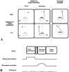
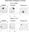
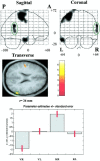
Reaching for a target involves integrative coordinate transformation processes between the representation of the target location, the sensorimotor information of limb of reach, and body space. Although right hemisphere dominance for visuospatial information processing is well established, corresponding right hemisphere dominance for kinesthetic spatial information processing remains to be demonstrated. We explored neural mechanisms of encoding target locations using 15O-butanol positron emission tomography (PET) in normal volunteers in a factorial experiment, where modality (visual/kinesthetic) and hemispace of target presentation (left/right of midsagittal plane) were varied systematically. After target presentation, subjects reached to the encoded target location. PET data analysis using SPM99 showed increased neural activity (P < 0.05, corrected) associated with left hemispace target presentation in right hemisphere areas (sensorimotor, anterior cingulate, insular, and temporo-occipital cortex) only. By contrast, right hemispace target presentation activated bilateral temporo-occipital cortex, which extended into the right temporo-parietal cortex and left sensorimotor cortex. A significant interaction of hemispace and modality of target presentation observed in right temporo-parietal cortex resulted from an increase in neural activity with kinesthetic target presentation in right hemispace. The data support an important role for the right temporo-parietal area in visuospatial processing and suggest a specific role of the right hemisphere in kinesthetic spatial processing.
Copyright 2004 Wiley-Liss, Inc.
Figures


Similar articles
-
Visual cortex activation in kinesthetic guidance of reaching.Exp Brain Res. 2007 Jun;179(4):607-19. doi: 10.1007/s00221-006-0815-x. Epub 2006 Dec 15. Exp Brain Res. 2007. PMID: 17171536
-
Pointing in 3D space to remembered targets. I. Kinesthetic versus visual target presentation.J Neurophysiol. 1998 Jun;79(6):2833-46. doi: 10.1152/jn.1998.79.6.2833. J Neurophysiol. 1998. PMID: 9636090 Clinical Trial.
-
Visuomotor transformations for reaching to memorized targets: a PET study.Neuroimage. 1997 Feb;5(2):129-46. doi: 10.1006/nimg.1996.0254. Neuroimage. 1997. PMID: 9345543 Clinical Trial.
-
Human cortical areas underlying the perception of optic flow: brain imaging studies.Int Rev Neurobiol. 2000;44:269-92. doi: 10.1016/s0074-7742(08)60746-1. Int Rev Neurobiol. 2000. PMID: 10605650 Review.
-
The effects of practice on the functional anatomy of task performance.Proc Natl Acad Sci U S A. 1998 Feb 3;95(3):853-60. doi: 10.1073/pnas.95.3.853. Proc Natl Acad Sci U S A. 1998. PMID: 9448251 Free PMC article. Review.
Cited by
-
Multisensory Integration in Stroke Patients: A Theoretical Approach to Reinterpret Upper-Limb Proprioceptive Deficits and Visual Compensation.Front Neurosci. 2021 Apr 7;15:646698. doi: 10.3389/fnins.2021.646698. eCollection 2021. Front Neurosci. 2021. PMID: 33897359 Free PMC article.
-
Effects of Stimulus Type and Strategy on Mental Rotation Network: An Activation Likelihood Estimation Meta-Analysis.Front Hum Neurosci. 2016 Jan 7;9:693. doi: 10.3389/fnhum.2015.00693. eCollection 2015. Front Hum Neurosci. 2016. PMID: 26779003 Free PMC article.
-
Crossmodal interference in bimanual movements: effects of abrupt visuo-motor perturbation of one hand on the other.Exp Brain Res. 2015 Mar;233(3):839-49. doi: 10.1007/s00221-014-4159-7. Epub 2014 Dec 6. Exp Brain Res. 2015. PMID: 25479738
-
The contributions of vision and haptics to reaching and grasping.Front Psychol. 2015 Sep 16;6:1403. doi: 10.3389/fpsyg.2015.01403. eCollection 2015. Front Psychol. 2015. PMID: 26441777 Free PMC article. Review.
-
Task-dependent asymmetries in the utilization of proprioceptive feedback for goal-directed movement.Exp Brain Res. 2007 Jul;180(4):693-704. doi: 10.1007/s00221-007-0890-7. Epub 2007 Feb 13. Exp Brain Res. 2007. PMID: 17297548
References
-
- Andersen RA, Essick GK, Siegel RM (1985): Encoding of spatial location by posterior parietal neurons. Science 230: 456–458. - PubMed
-
- Binkofski F, Dohle C, Posse S, Stephan KM, Hefter H, Seitz RJ, Freund HJ (1998): Human anterior intraparietal area subserves prehension: a combined lesion and functional MRI activation study. Neurology 50: 1253–1259. - PubMed
-
- Bloedel JR, Bracha V (1995): On the cerebellum, cutaneomuscular reflexes, movement control and the elusive engrams of memory. Behav Brain Res 68: 1–44. - PubMed
Publication types
MeSH terms
LinkOut - more resources
Full Text Sources

