

Brain capillary endothelium and choroid plexus epithelium regulate transport of transferrin-bound and free iron into the rat brain
- PMID: 14756801
- PMCID: PMC3980859
- DOI: 10.1046/j.1471-4159.2003.02221.x
Brain capillary endothelium and choroid plexus epithelium regulate transport of transferrin-bound and free iron into the rat brain
Abstract
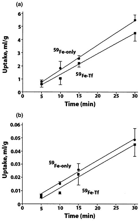
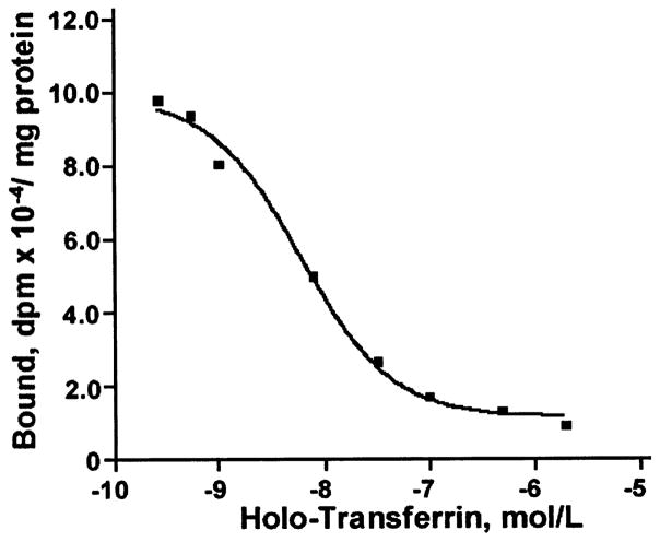
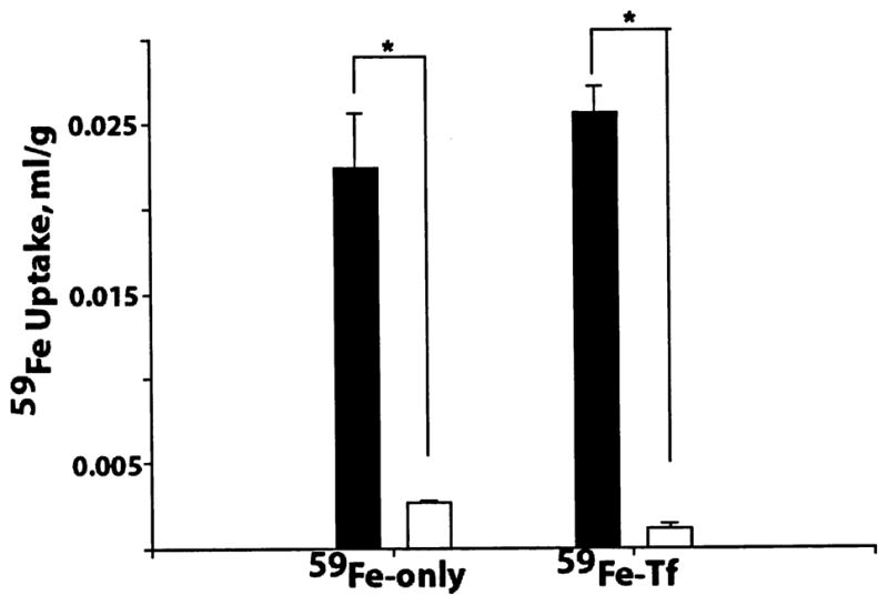
Iron transport into the CNS is still not completely understood. Using a brain perfusion technique in rats, we have shown a significant brain capillary uptake of circulating transferrin (Tf)-bound and free 59Fe (1 nm) at rates of 136 +/- 26 and 182 +/- 23 microL/g/min, respectively, while their respective transport rates into brain parenchyma were 1.68 +/- 0.56 and 1.52 +/- 0.48 microL/g/min. Regional Tf receptor density (Bmax) in brain endothelium determined with 125I-holo-Tf correlated well with 59Fe-Tf regional brain uptake rates reflecting significant vascular association of iron. Tf-bound and free circulating 59Fe were sequestered by the choroid plexus and transported into the CSF at low rates of 0.17 +/- 0.01 and 0.09 +/- 0.02 microL/min/g, respectively, consistent with a 10-fold brain-CSF concentration gradient for 59Fe, Tf-bound or free. We conclude that transport of circulating Tf-bound and free iron could be equally important for its delivery to the CNS. Moreover, data suggest that entry of Tf-bound and free iron into the CNS is determined by (i) its initial sequestration by brain capillaries and choroid plexus, and (ii) subsequent controlled and slow release from vascular structures into brain interstitial fluid and CSF.
Figures
Similar articles
-
Brain iron homeostasis.Dan Med Bull. 2002 Nov;49(4):279-301. Dan Med Bull. 2002. PMID: 12553165 Review.
-
Kinetics and distribution of [59Fe-125I]transferrin injected into the ventricular system of the rat.Brain Res. 1998 Apr 20;790(1-2):115-28. doi: 10.1016/s0006-8993(98)00055-9. Brain Res. 1998. PMID: 9593852
-
Transcytosis of protein through the mammalian cerebral epithelium and endothelium. III. Receptor-mediated transcytosis through the blood-brain barrier of blood-borne transferrin and antibody against the transferrin receptor.Exp Neurol. 1996 Nov;142(1):47-65. doi: 10.1006/exnr.1996.0178. Exp Neurol. 1996. PMID: 8912898
-
Evidence for low molecular weight, non-transferrin-bound iron in rat brain and cerebrospinal fluid.J Neurosci Res. 1998 Nov 15;54(4):486-94. doi: 10.1002/(SICI)1097-4547(19981115)54:4<486::AID-JNR6>3.0.CO;2-I. J Neurosci Res. 1998. PMID: 9822159
-
Transferrin and transferrin receptor function in brain barrier systems.Cell Mol Neurobiol. 2000 Feb;20(1):77-95. doi: 10.1023/a:1006948027674. Cell Mol Neurobiol. 2000. PMID: 10690503 Free PMC article. Review.
Cited by
-
Amyloid beta immunization worsens iron deposits in the choroid plexus and cerebral microbleeds.Neurobiol Aging. 2013 Nov;34(11):2613-22. doi: 10.1016/j.neurobiolaging.2013.05.013. Epub 2013 Jun 22. Neurobiol Aging. 2013. PMID: 23796662 Free PMC article.
-
Clinical Imaging of Choroid Plexus in Health and in Brain Disorders: A Mini-Review.Front Mol Neurosci. 2019 Feb 12;12:34. doi: 10.3389/fnmol.2019.00034. eCollection 2019. Front Mol Neurosci. 2019. PMID: 30809124 Free PMC article.
-
SARS-CoV-2: is there neuroinvasion?Fluids Barriers CNS. 2021 Jul 14;18(1):32. doi: 10.1186/s12987-021-00267-y. Fluids Barriers CNS. 2021. PMID: 34261487 Free PMC article. Review.
-
Molecular mechanism of distorted iron regulation in the blood-CSF barrier and regional blood-brain barrier following in vivo subchronic manganese exposure.Neurotoxicology. 2006 Sep;27(5):737-44. doi: 10.1016/j.neuro.2006.02.003. Epub 2006 Mar 20. Neurotoxicology. 2006. PMID: 16545456 Free PMC article.
-
Age-related changes in iron homeostasis and cell death in the cerebellum of ceruloplasmin-deficient mice.J Neurosci. 2006 Sep 20;26(38):9810-9. doi: 10.1523/JNEUROSCI.2922-06.2006. J Neurosci. 2006. PMID: 16988052 Free PMC article.
References
-
- Allen DD, Smith QR. Characterization of the blood–brain barrier choline transporter using the in situ rat brain perfusion technique. J Neurochem. 2001;76:1032–1041. - PubMed
-
- Andrews NC. The iron transporter, DMT1. Int J Biochem Cell Biol. 1999;31:991–994. - PubMed
-
- Bali PK, Zak O, Aisen P. A new role for the transferring receptor in the release of iron from transferrin. Biochemistry. 1991;30:324–328. - PubMed
-
- Beard JL, Dawson H, Pinero DJ. Iron metabolism: a comprehensive review. Nutr Rev. 1996;54:295–317. - PubMed
-
- Berg D, Gerlach M, Youdim MBH, Double KL, Zecca L, Riederer P, Becker G. Brain iron pathways and their relevance to Parkinson’s disease. J Neurochem. 2001;79:225–236. - PubMed
Publication types
MeSH terms
Substances
Grants and funding
LinkOut - more resources
Full Text Sources
Medical
Miscellaneous
